/
Exploring Demographics, Treatment, and Outcomes for Pediatric Bony Ulnar Collateral Ligament Exploring Demographics, Treatment, and Outcomes for Pediatric Bony Ulnar Collateral Ligament

Exploring Demographics, Treatment, and Outcomes for Pediatric Bony Ulnar Collateral Ligament - PowerPoint Presentation

ella
ella . @ella
Follow
4 views
Uploaded On 2024-03-15

Exploring Demographics, Treatment, and Outcomes for Pediatric Bony Ulnar Collateral Ligament - PPT Presentation

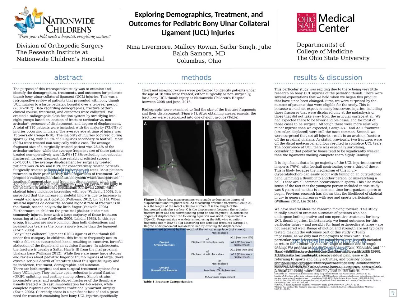
Nina Livermore Mallory Rowan Satbir Singh Julie Balch Samora MD Division of Orthopedic Surgery The Research Institute at Nationwide Childrens Hospital Columbus Ohio Departments of ID: 1048541

pediatric injuries ucl fractures injuries pediatric fractures ucl articular displacement patients thumb bony fracture injury hand study 2006 treated

Share:

Link:

Embed:

Download Presentation from below link

Download Presentation The PPT/PDF document "Exploring Demographics, Treatment, and O..." is the property of its rightful owner. Permission is granted to download and print the materials on this web site for personal, non-commercial use only, and to display it on your personal computer provided you do not modify the materials and that you retain all copyright notices contained in the materials. By downloading content from our website, you accept the terms of this agreement.


Presentation Transcript

1. Exploring Demographics, Treatment, and Outcomes for Pediatric Bony Ulnar Collateral Ligament (UCL) Injuries Nina Livermore, Mallory Rowan, Satbir Singh, Julie Balch Samora, MDDivision of Orthopedic SurgeryThe Research Institute atNationwide Children’s HospitalColumbus, OhioDepartment(s) ofCollege of MedicineThe Ohio State Universitymethodsresults & discussionabstractThe purpose of this retrospective study was to examine and identify the demographics, treatments, and outcomes for pediatric thumb bony ulnar collateral ligament (UCL) injuries. This was a retrospective review of patients that presented with bony thumb UCL injuries to a large pediatric hospital over a ten-year period (2007-2017). Data regarding demographics, fracture pattern, clinical course, treatment, and outcomes were collected. We created a radiographic classification system by stratifying into eight groups based on location of fracture (articular vs. non-articular), presence of displacement, and degree of displacement. A total of 110 patients were included, with the majority (73.6%) of injuries occurring in males. The average age at time of injury was 15 years old (range 8-18). The majority of injuries occurred during sports (70%), with 25.5% of all injuries secondary to football. Most (60%) were treated non-surgically with a cast. The average fragment size of a surgically treated patient was 28.4% of the articular surface, while the average fragment size of those patients treated non-operatively was 13.4% (17.8% excluding non-articular fractures). Larger fragment size reliably predicted surgery (p<0.001). The average displacement for surgically treated patients was 26.6% and 8.7% for conservatively treated patients. Surgically treated patients had higher healing rates. Most patients returned to their prior activity level, regardless of treatment. We propose a radiographic classification system which incorporates location, fragment size, and fragment displacement, which can help guide management of pediatric thumb bony UCL injuries.Chart and imaging reviews were performed to identify patients under the age of 18 who were treated, either surgically or non-surgically, for a bony UCL thumb injury at Nationwide Children’s Hospital between 2008 and June 2018. Radiographs were examined to find the size of the fracture fragments and their displacement (Figure 1). After obtaining measurements, the fractures were categorized into one of eight groups (Table). This particular study was exciting due to there being very little research on bony UCL injuries of the pediatric thumb. There were several expectations that we held when we began this product that have since been changed. First, we were surprised by the number of patients that were eligible for the study. This is because we did not expect so many less severe injuries, including those fractures that were displaced only at the metaphysis or those that did not take away from the articular surface at all. We had expected there to be fewer eligible cases, and for most of those cases to be surgical. Although there were more relatively minor injuries than we expected, Group A3.1 and A3.2 fractures (articular, displaced) were still the most common. Second, we were surprised that not all injuries result in an avulsion fracture off the proximal phalanx. As stated previously, two fractures were off the distal metacarpal and four resulted in complete UCL tears. The occurrence of UCL tears was especially surprising, considering that pediatric bones tend to be significantly weaker than the ligaments making complete tears highly unlikely.  It is significant that a large majority of the UCL injuries occurred in sports (70%), with football contributing over a third of those. This is likely because the mechanism of this injury (hyperabduction) can easily occur with falling on an outstretched hand, jamming a thumb into another person, or mis-catching a ball. These are all common occurrences in sports. This also makes sense of the fact that the youngest person included in this study was 8 years old, as that is a common time for organized sports to begin. Previous research has shown that the likelihood of skeletal injury in general increases with age and sports participation (Williams 2012, Liu 2014).   We have several ideas for research moving forward. This study initially aimed to examine outcomes of patients who had undergone both operative and non-operative treatment for bony UCL thumb injuries. Unfortunately, we found outcomes for this particular injury – and possibly for hand orthopedics at large – are not measured well. Range of motion and strength are not typically tested, making the outcomes part of this study virtually impossible, as we only had radiographs to work with. This particular research can be continued by asking patients included to return for a follow up visit for range of motion and strength testing. We propose using the Disabilities of Arm, Shoulder, and Hand (DASH) score to objectively gather this data (Hudak 1996). Additionally, we would ask about residual pain, ease with returning to sports and daily activities, and possibly obtain additional radiographs. This report has hopefully initiated more research on the topic of pediatric bony UCL injuries, and we look forward to seeing where this may lead in the future.backgroundThe hand is one of the most frequently injured parts of the body in the pediatric & adolescent populations (Cornwall 2006), with skeletal injury incidence increasing with age (Vadivelu 2006). It is suspected that the increase in skeletal injury is due to increase in weight and sports participation (Williams, 2012, Liu 2014). When skeletal injuries do occur the second highest rate of fracture is in the thumb, second only to the little finger (Vadivelu 2006). Furthermore, in children the proximal phalanx is the most commonly injured bone with a large majority of those fractures occurring at its base (Vadivelu 2006, Landin 1983). In this age group, fractures are more common than both dislocations and ligamentous tears as the bone is more fragile than the ligament (Kozin 2006).Bony ulnar collateral ligament (UCL) injuries of the thumb fall under this category. In children, this fracture frequently occurs with a fall on an outstretched hand, resulting in excessive, forceful abduction of the thumb and an avulsion fracture. In adolescents, the fracture is usually a Salter Harris III from the first proximal phalanx base (Williams 2012). While there are many large studies and reviews about pediatric finger or thumb injuries at large, there exists a serious dearth of literature about this specific injury and its incidence, treatment, demographic, and outcome. There are both surgical and non-surgical treatment options for a bony UCL injury. They include open reduction internal fixation (ORIF), splinting, and casting among others. Simple strains, incomplete tears, and nondisplaced fractures of the thumb are usually treated with cast immobilization for 4-6 weeks, while complete ruptures and fractures traditionally warrant surgery (Kozin 2006). Currently, there is a significant lack of and a great need for research examining how bony UCL injuries specifically are treated and the outcomes of these various treatment options.This study aims to evaluate the demographics, treatment, and outcomes of pediatric bony UCL injury patients at Nationwide Children’s Hospital. This will improve the way we assess and care for these patients, as well as elucidate the benefits and risks of all treatment options. acknowledgements & referencesNina would like to acknowledge the Samuel Roessler MDSR Scholarship for funding this workCornwall R., Ricchetti E.T. Pediatric phalanx fractures: Unique challenges and pitfalls. Clinical Orthopaedics and Related Research. 2006;445:146-156. Hudak PL, Amadio PC, Bombardier C. Development of an upper extremity outcome measure: the DASH (disabilities of the arm, shoulder and hand). The Upper Extremity Collaborative Group (UECG). Am J Ind Med. 1996 Jun;29(6):602-8. Kozin MD, SH. Fractures and dislocations along the pediatric thumb ray. Hand Clinics. 2006;22: 19-29. Landin LA. Fracture patterns in children. Analysis of 8682 fractures with special reference to incidence, etiology, and secular changes in a Swedish urban population 1950–1979. Acta Orthop Scand Suppl. 1983;202:1–109. Liu E.H., Borschel MD, G.H. A prospective study of pediatric hand fractures and review of the literature. Pediatric Emergency Care. 2015;30: 299-304. Vadivelu, R. Hand injuries in children: Prospective study. J Pediatric Ortho. 2006;26: 29-35. Williams AA, Lochner HV. Pediatric hand and wrist injuries. Current Reviews in Musculoskeletal Medicine. 2013;6(1):18-25. This “blue box” represents the margin that MUST be maintained around the logos at all times.Group AArticular fracturesA1Non-displacedA2Displaced at metaphysis onlyA2.1 (less than 15%)A2.2 (15% or more displacement)A3Displaced at articular surfaceA3.1 (less than 15%)A3.2 (15% or more displacement)Group BNon-articular fracturesB1Non-displacedB2Less than 15% displacementB315% or more displacementFigure 1 shows how measurements were made to determine degree of displacement and fragment size. A) Measuring articular fractures (Group A). A is the length of the intact articular surface. B is the length of the fragmented articular surface. C is the displacement between the articular fracture point and the corresponding point on the fragment. To determine degree of displacement the following equation was used: displacement = C/(A+B). Fragment size was determined using the following equation: fragment size = B/(A+B) B) Measuring non-articular fractures (Group B). Degree of displacement was determined by dividing the displacement measurement (shown) by the length of the articular surface (not shown). Table 1 Fracture Categorization