/
Spinal Injury    EMAN ALADLY                                           HUSAM ABU SUILIK Spinal Injury    EMAN ALADLY                                           HUSAM ABU SUILIK

Spinal Injury EMAN ALADLY HUSAM ABU SUILIK - PowerPoint Presentation

ella
ella . @ella
Follow
64 views
Uploaded On 2024-01-03

Spinal Injury EMAN ALADLY HUSAM ABU SUILIK - PPT Presentation

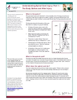
Anatomy of Spinal Cord The spinal cord extends from the foramen magnum where it is continuous with the medulla olbangata in brainstem and continues through to the  conus medullaris  near the second  ID: 1037455

disc spinal cord injury spinal disc injury cord cervical lumbar common spine fracture level prolapse motor nerve compression posterior

Share:

Link:

Embed:

Download Presentation from below link

Download Presentation The PPT/PDF document "Spinal Injury EMAN ALADLY ..." is the property of its rightful owner. Permission is granted to download and print the materials on this web site for personal, non-commercial use only, and to display it on your personal computer provided you do not modify the materials and that you retain all copyright notices contained in the materials. By downloading content from our website, you accept the terms of this agreement.


Presentation Transcript

1. Spinal Injury EMAN ALADLY HUSAM ABU SUILIK

2. Anatomy of Spinal Cord

3. The spinal cord extends from the foramen magnum where it is continuous with the medulla olbangata in brainstem and continues through to the conus medullaris near the second lumbar vertebra, terminating in a fibrous extension known as the filum terminale. The spinal cord is 40 to 50 cm long ( varies between male & female as in male it is longer ) and 1 cm to 1.5 cm in diameter.It made with the brain the Central Nervous System ..Function of spinal cord : 1- as a conduit for motor information 2- as a conduit for sensory information 3-coordination of reflexes It is divided into five regions: 1-cervical (C) ….. C1-C82-thoracic (T) ….T1-123-lumbar (L) ……L1-L54-Sacral (S) ……S1-S55- Coccygeal ( Co )…..Co1

4. The Spinal Cord is enlarged in the cervical and lumbar regions :1- The cervical enlargement : located from C3 to T2 spinal segments, is where sensory input comes from and motor output goes to the arms2- The lumbar enlargement : located between L1 and S3 spinal segments, handles sensory input and motor output coming from and going to the legs.

5. The spinal cord(and brain) are protected by three layers of tissue or membranes called meninges, that surround the canal :1- The dura mater is the outermost layer, and it forms a tough protective coating( Between the dura mater and the surrounding bone of the vertebrae is a space called the epidural space. )The epidural space is filled with adipose tissue, and it contains a network of blood vessels. 2-The arachnoid mater is the middle protective layer.3-Pia mater and the space between the arachnoid and the underlying pia mater is called the subarachnoid space. The subarachnoid space contains cerebrospinal fluid (CSF)

6. Two consecutive rows of nerve roots emerge on each of its sides. These nerve roots join distally to form 31 pairs of spinal nerves as they contain motor and sensory nerve fibers to and from all parts of the body.. The spinal cord is a cylindrical structure of nervous tissue composed of white and gray matter.

7. Spinal cord legaments Intrasegmental􀁺 Ligamentum flavum􀁺 Intertransverse ligament􀁺 Interspinous ligamentIntersegmental􀁺 ALL􀁺 PLL􀁺 Supraspinous ligament

8.

9. SPINAL CORD INJURY

10. SCI :Insult to spine resulting in a change in the normal motor, sensory or autonomic function. **This change is either temporary or permanent .injury to : - vertebral column - spinal cord - Nerves rootsEpidemiology :Incidence 2-5/100,000 .Adolescent and young adults are the most commonly affected .Most common cause of SC injury : RTAAlthough the majority of spinal injuries do not affect the cord or spinal roots , about 10% will result in quadriplegia or paraplegia .

11. Secondary Injury versus Primary Injury:A-Primary Injury Spinal Injury that occurred at time of traumaB-Secondary InjurySpinal Injury that occurs after the traumapossibly secondary to mishandling of unstable fractures , swelling and Ischemia .

12. Cervical regionmost vulnerableC5/6 is the most common siteThoracic regionprotected by the immobility provided by the ribsThoracolumbar junction the less mobile thoracic vertebra joins the more mobile lumbar vertebra making it more susceptible to injuryLumbosacral regionthe area in which the spinal cords ends and the quada equina begins** most commonly vertebrae affected are :C5_C7 & T12 &L1

13. Causes of injury :1-force2-Ischemia due to vascular injury3-Secondary hemorrhage in and around the cord.** StabilityStable : If the ligamentous and bony component of the spine are preserved .Unstable: If the ligamentous and bony component of the spine are not preserved .**Type of neurological impairment Complete : no preservation of neurologic function distal to the level of injury {flaccid paralysis + total loss of sensory & motor functions}Incomplete: preservation of any sensorimotor function below the level of injury constitutes an incomplete injury {mixed loss - Anterior sc syndrome - Posterior sc syndrome - Central cord syndrome - Brown sequard’s syndrome - Cauda equina syndrome }

14. American Spinal Injury Association Impairment Scale (AISA) Classification

15. Motor level = the last level with at least 3/5 (against gravity) functionSensory level = the last level with preserved sensationDermatome: patch of skin innervated by a given spinal cord levelMyotome : Spinal nerve roots which innervates muscles groups. Most muscles are innervated by more than one root.

16. Motor Function

17. Sensory Function

18.

19. Mechanisms and Associated Injuries ( directional force )1. HyperextensionCervical & Lumbar SpineDisk disruptionCompression of ligaments2. HyperflexionCervical & Lumbar SpineStretching of ligaments Compression Injury of cordDisk disruption with potential vertebrae dislocation3. Rotational Most commonly Cervical Spine but potentially in Lumbar SpineStretching and tearing of ligamentsRotational subluxation and dislocation4. CompressionMost likely between T12 and L2Ruptured disk5. DistractionMost common in upper Cervical SpineStretching of cord without damage to spinal column.6. Penetrating Forces directly to spinal columnDisruption of ligamentsDirect damage to cord

20.

21.

22. Cervical Spine Fractures and Dislocations classified on the basis of: mechanism FlexionExtensionCompressionRotationa combination of theseLocationstability

23. Upper cervical spine ( skull to C2 ) 1) Craniocervical Dislocation (anterior , posterior or vertical ) including atlanto- axial and atlanto-occipital dislocationCause : high energy trauma Usually fatal , but Early diagnosis and spinal stabilization protected against worsening spinal cord injuryCareful occipitocervical fusion is required in survivals Halo brace should be applied before surgery to prevent intraoperative dislocation2) Atlantoaxial instability The most common is rotatory subluxation in childrenusually spontaneous ( bone or ligament abnormality) but can be traumatic child presents with cock-robin appearance ( head tilt toward the affected site with contralateral chin rotation )halter traction results in realignment in the majority of cases .

24. cock-robin appearance

25.

26. 3) Jefferson’s fracture ( C1 ring ) Results from fracture through C1 archesAssociated with axial loading of cervical spine .Can be : stable or unstableTransverse ligament rupture may occurunstable jefferson’s fracture should be treated in a halo jacket for 3 months ,followed by flexion-extension stress radiography

27. 4) Odontoid fracture * It results from hyperflexion injuryAt the tip (type I) - stableThrough the base of the dens (type II) (the most common) - unstableThrough C2 vertebral body (type III) - generally unstable

28. 5) Hangman’s fracture Fracture through the pedicles of C2 It occurs due to hyperextensionUsually stableMajority can be treated non-operatively: halo jackets or brace Those with significant displacement or associated facet dislocation requires surgery .

29. 6) Occipital condyle fracture Uncommon injury , usually associated with head injury Identified on CTCan be treated in hard collar for 8 weeks .

30. Subaxial cervical spine (C3-C7)1) Flexion rotation injury :Most common cervical injuryMainly at C5/C6UnstableExtensive damage to posterior ligamentsIt may sustain both direct damage and vascular impairment It leads to 2 types of fractures:A)wedge fractures B)tear drop fractures

31. A) Wedge fracture : mostly stable treatment : brace or halo for 3 monthsB) Tear drop fratcure :Results in an anteroinferior vertebral body fracture & is more common in lower cervical vertebrae , C5 Note : Hyperextension tear drop fracture is more common in upper cervical vertebrae

32. 2) compression (axial loading) :Mainly at C5/C6Vertebral body is decreased in heightUsually stable (no damage to posterior Bony structures or longitudinal ligaments) Resulting injury:50% complete 50% incomplete (anterior cord syndrome)***anterior cord syndrome : involvment of anterior two thirds of spinal cord which include the spinothalamic and corticospinal tractsMay result in burst fractures : in which bone fragments may explode into the cord

33.

34. hyperextension :Most common in the elderly patients. With degenerative spinal canal stenosisUsually no bone injury, only damage to ant. Long. LigamentResults in incomplete injuryMostly stableThe most common neurological impairment is Central Cervical Cord syndrome

35. 4) facet subluxation / dislocationEither unifacet or bifacet -Unifacet:-Result from flexion and rotation-Posterior ligament is ruptured-stable, because the vertebra are locked in place-Bifacet:-it’s unstable-high incidence of cord damage

36. Cervical Injury leads to:Quadriplegia or quadriparesisBowel/bladder retention (spastic)Various degrees of breathing difficultiesNeurogenic and/or spinal shock*quadriplagia: it refers to impairment or loss of motor and sensory function in the cervical segments it results in impairment in the function of arms, trunk, legs and pelvic organs (it does not include the brachial plexus)*quadriparesis:it describes incomplete lesions imprecisely, the ASIA scale provide a more precise approach.

37. Thoracic spine injuries T2-L1:- Causes paraplegia or paraparesis- Upper Motor Neuron symptoms(weakness,decreased motor control,altered muscle tone,exagerated deep tendon reflexes spasticity and clonus)1) flexion, flexion-rotation :Mostly at level of T12/L1UnstableDisruption of post. longitudinal ligament and post. Bony structuresAnterosuperior wedge fractures are seenComplete neurological damage (of spinal cord, conus medullaris, cauda equina)

38.

39. 2) Compression : Very commonStable, rarely cause neurological damageDecrease in height of vertebral body

40. 3) hyperextension :UncommonLeads to : - damage to ant. Longitudinal ligament - rupture of intervertebral disc - fracture of ant. Part of the involved vertebral bodyUnstable, causes severe neurological damage4) Open injury :Results from stab or gunshot woundsInjury is due to : - blast injury

41. 5- Chance fracture is a flexion injury of the spine,[ It consists of a compression injury to the anterior portion of the vertebral body and a transverse fracture through the posterior elements of the vertebra and the posterior portion of the vertebral body. It is caused by violent forward flexion, causing distraction injury to the posterior elements.The most common site at which Chance fractures occur is the thoracolumbar junction (T12-L1)

42.

43. Lumbar spine injuries L1 and belowMainly affect cauda equinaLeads to paraparesis or paraplegiaLower Motor Neuron symptoms(flaccid paralysis, muscle wasting, fibrillation, fasiculation, hypotonia~atonia,hyporeflexia)

44. Radiologic investigation:plain X-rayDynamic X-ray(named dynamic flexion-extension x-ray)CT (for bone injury, DO NOT give contrast)MRI (for cord, soft tissues and ligament damage, an emergency only in cauda equina syndrome)

45. Principles in Treatment

46. To relieve any reversible neural compression.To preserve neurological function.To restore alignment of the spine.To stabilize the spine.To rehabilitate the patient.Indications for urgent surgical stabilization:Unstable fracture with progressive neurological deficit,An unstable fracture in a pt with multiple injuries.

47.

48. Disc Prolapse

49. This disc degeneration results from the aging process and wear and tear that occurs to the bone and soft tissues of the spine.It is one of the most common causes of low back and neck pain and a major cause of chronic disability in the adult working population and a common reason for referral to MRI.

50. >>Degenerative Disc Disease is a Misnomer ! WHY ?First , For most people the term degenerative understandably implies that the symptoms will get worse with age. However, the term does not apply to the symptoms, but rather describes the process of the disc degenerating over time.Another source of confusion is probably created by the term disease, because degenerative disc disease is not really a disease at all, but rather a degenerative condition that at times can produce pain from a damaged disc

51. Types of degenerative disc disease : Cervical Disc ProlapseDegenerative Lumbar Disc Diseases3. Spinal Stenosis4. Spondylolisthesis

52. Direction of herniation/prolapse :PosterolateralPosterior/Central Lateral

53.

54. Cervical disc prolapse

55. Prolapse of an intervertebral disc in the cervical region It is less common than disc prolapse in the lumbar area.The disc herniation occurs most frequently at the C6/C7 (70%) level and slightly less commonly at the C5/6 level (20%) due to the force exerted at these levels .Disc herniation above these levels and at the C7/T1 level is much less common

56. Because the nerves pass directly laterally from the cervical cord to their neural foramen, so that the herniation compresses the nerve at that level

57. Causes of cervical disc prolapse :Repetitive cervical stress TraumaHeavy liftingProlonged sedentary positionWhiplash accidentsFrequent acceleration/deceleration

58. Clinical presentation Neck pain Arm painHand and fingers painPart of the shoulder>> These neurological deficits are determined by the location of the herniated disc The pain usually begins in the cervical region but then it radiates into the periscapular area, the occiput, shoulder and down to the arm.

59. Radiologic Imaging InvestigationsHigh-quality MRI is now the investigation of choice and has almost completely replaced both myelography and CT scans . Although myelography is an invasive test , it is more sensitive even to subtle nerve root compression

60.

61.

62. ManagementConservative(non-surgical) treatment Patients may find relief byusing medications to control pain and inflammation (NSAIDs)Use of a cervical collar, cervical pillows or neck traction may also be recommended to stabilize the neck and improve neck alignment.exercising the neck and shoulder areas (alone or with the help of a professional familiar with neck conditions) to relieve stiffness and maintain flexibility

63. Lumbar disc prolapseAbout 75% of total spinal movement & of lumbar flexion–extension occurs at the lumbosacral junction ( L5/S1 ), 20% at ( L4/L5 ) level, 5% at upper lumbar levels. >> Consequently, about 90% of lumbar disc prolapses occur at the lower two lumbar levels;The most frequently affected disc is at L5/S1 level

64.

65.

66. Directions of prolapse Posterolateral (most common)LateralCentral (compresses cauda equina)>> Posterolateral prolapsed disc : causes compression of the nerve which runs along the posterior aspect of the disc and down in the neural foramen under the pedicle of the vertebra below>> Lateral disc prolapse : will cause compression of the nerve root passing below the pedicle of the vertebra above the disc prolapse.e.g. :L3/L4 posterolateral disc prolapse : L4 nerve root compressionLateral : L3 compression

67.

68. Investigations( 1 ) Lumbar myelographyInvolves injection of a water-solublecontrast material into the subarachnoid space that demonstrate the spinal canal. Risk of:Headacheepileptic seizures In the past the use of oil-based materialwas associated with a risk of arachnoiditis

69.

70. ( 2 ) CT scan & MRI

71. Management1.Conservative2. Surgery Most patients improve by conservative treatment while Less than 20% require surgery. According to the pathology of the disc prolapse:A‘bulging’ disc is likely to settle with simple conservative management,If the nucleus pulposus has herniated out of the disc space and ‘sequestrated’ outside the annulus, it will need surgery for satisfactory relief of symptoms

72. Conservative treatment1- Bed rest, usually for a period of about 7–10 days2- Analgesic agents3- NSAID’s Conservative treatment help in:Resorption of the prolapsed disc materialDecreasing nerve edemaPossible adaptation of the pain fibers to pressure

73. Indication for surgery1- Pain if persists after 7-10 days of bed rest OR reccurent episodes of pain when mobilizing. 2- Neurological deficit : A significant weakness or increasing amount of weakness. 3- Central disc prolapse :In cases of bilateral sciatica or other features indicating a central disc prolapse, such as sphincter disturbance and diminished perineal sensation.As it cause irreversible cauda equina compression. 4- Tumor

74. scoliosisScoliosis is an apparent lateral (sideways) curvature of the spine. ‘apparent ‘ because although lateral curvature does occur , the commonest form of scoliosis is actually a triplanar deformity with lateral, anteroposterior and rotational components .Two broad types of deformity are defined : postural and structural

75.

76. kyphosisRather confusingly , the term ‘kyphosis’ is used to describe both the normal (the gentle rounding of the thoracic spine)and the abnormal (excessive thoracic curvature or straightening out of the cervical or lumber lordotic curves) . Excessive thoracic curvature might be better describe as ’hyperkyphosis’ kyphos , or gibbus , is a sharp posterior angulation due to localized collapse or wedging of one or more vertebrae This may be the result of a congenital defect , a fracture (sometimes pathological ) or spinal tuberculosis

77.

78.

79.

80. MyotomeSpinal nerve roots which innervates muscles groups. Most muscles are innervated by more than one root

81.

82. ThaNk You..