/
Nervous Tissue: Neurons and Support Cells ( Nervous Tissue: Neurons and Support Cells (

Nervous Tissue: Neurons and Support Cells ( - PowerPoint Presentation

ellena-manuel
ellena-manuel . @ellena-manuel
Follow
358 views
Uploaded On 2018-10-21

Nervous Tissue: Neurons and Support Cells ( - PPT Presentation

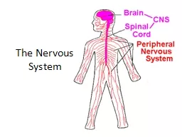
glial cells Pages 227234 Astro cytes Braceanchor neurons provide chemical barrier Most abundant glial cells Microglia Destroy threatening particlescells phagocytes Ependymal cells ID: 692608

myelin cells nervous neuron cells myelin neuron nervous neurons sheath cell tissue glial figure axon body pearson education 2015

Share:

Link:

Embed:

Download Presentation from below link

Download Presentation The PPT/PDF document "Nervous Tissue: Neurons and Support Cell..." is the property of its rightful owner. Permission is granted to download and print the materials on this web site for personal, non-commercial use only, and to display it on your personal computer provided you do not modify the materials and that you retain all copyright notices contained in the materials. By downloading content from our website, you accept the terms of this agreement.


Presentation Transcript

Slide1

Nervous Tissue:Neurons and Support Cells (glial cells)

Pages 227-234Slide2

AstrocytesBrace/anchor neurons, provide chemical barrier

Most abundant

glial cellsMicrogliaDestroy threatening particles/cells; phagocytesEpendymal cellsLine ventricles and central canal; cilia circulate CSFOligodendrocytesProduce myelin sheath around axons (in the CNS)

© 2015 Pearson Education, Inc.

Support Cells:

NeurogliaSlide3

Figure

7.3a Supporting (glial) cells of nervous tissue.

Capillary

Neuron

Astrocyte

(a

) Astrocytes are

the most abundant

and versatile neuroglia.

ASTROCYTESSlide4

Figure

7.3b Supporting (glial) cells of nervous tissue.

Neuron

Microglial

cell

(b) Microglial cells are phagocytes that

defend CNS cells.

MICROGLIASlide5

Figure

7.3c Supporting (glial) cells of nervous tissue.

Ependymal

cells

Brain or

spinal cord

tissue

Fluid-filled cavity

(c) Ependymal cells line cerebrospinal

fluid-filled cavities.

EPENDYMAL CELLSSlide6

Figure

7.3d Supporting (glial) cells of nervous tissue.

Myelin sheath

Process of

oligodendrocyte

Nerve

fibers

(d)

Oligodendrocytes

have processes that form

myelin sheaths around CNS nerve fibers.

OLIGODENDROCYTESSlide7

Satellite cellsProtect neuron cell bodiesSchwann cells

Form myelin sheath

around axons (in the PNS)© 2015 Pearson Education, Inc.Support Cells: PNS glial cellsSlide8

Figure

7.3e Supporting (glial) cells of nervous tissue.

Satellite

cells

Cell body of neuron

Schwann cells

(forming myelin sheath)

Nerve fiber

(e) Satellite cells and Schwann cells (which

form myelin) surround neurons in the PNS.Slide9

specialized to transmit messagesMajor regions of neuronsCell

body

—contains the nucleusmetabolic center of the cellProcesses—fibers that extend from the cell body Dendrites : pick up sensory stimuliAxons : receive impulse from cell bodyImpulses are UNIDIRECTIONAL along the axon

© 2015 Pearson Education, Inc.

Neurons (nerve cells)Slide10

Figure

7.4a Structure of a typical motor neuron.

Mitochondrion

Nucleus

Dendrite

Cell body

Axon

One Schwann cell

Node of Ranvier

Schwann cells,

forming the myelin

sheath on axon

Axon

terminal

(a)Slide11

Figure

7.4b Structure of a typical motor neuron.

Neuron

cell body

Dendrite

(b)Slide12

End in axon terminalsTerminals contain vesicles filled with neurotransmitters

Neurons are separated from other cells by

a gap called the synaptic cleft:Synapse—junction between nervesNeurons NEVER touch each other© 2015 Pearson Education, Inc.

Axons and the Synaptic CleftSlide13

The Myelin Sheath

whitish

, fatty material covering axonsNodes of Ranviergaps in myelin sheath along the axonimpulse jumps from one node to the nextThe current cannot flow across the axon where there is myelin so it jumps between the myelin bundles

The presence of the myelin sheath speeds the nerve impulse

transmission

© 2015 Pearson Education, Inc.Slide14

Myelin sheath becomes destroyedReplaced with hardened sclerosesSclerosis: hardening of body tissue

Incomplete/staggered transmission of impulse

Autoimmune disease- potientially caused by:GenesSmokingViral infectionsVitamin D deficiencyMultiple SclerosisSlide15

Sensory (afferent) neuronsCarry impulses from

sensory

receptors to the CNSCutaneous (skin) sense organsPain receptorsTemperature receptorsTouch/pressure receptorsProprioceptors—detect stretch or tension

Muscle spindle in musclesGolgi tendon organ in tendonw

© 2015 Pearson Education, Inc.

Classification

of NeuronsSlide16

Motor (efferent) neuronsCarry impulses from the central nervous system to viscera, muscles, or glands

Interneurons

(association neurons)Located in the spinal cord (CNS)Connect sensory and motor neuronsPlay role in reflex arc – allow quicker response without involving the brain© 2015 Pearson Education, Inc.

Functional Classification of NeuronsSlide17

Figure

7.6 Neurons classified by function.

Dendrites

Peripheral

process (axon)

Ganglion

Cell

body

Sensory

neuron

Central process (axon)

Spinal cord

(central nervous system)

Interneuron

(association

neuron)

Afferent

transmission

Peripheral

nervous system

Receptors

Efferent transmission

Motor neuron

To effectors

(muscles and glands)