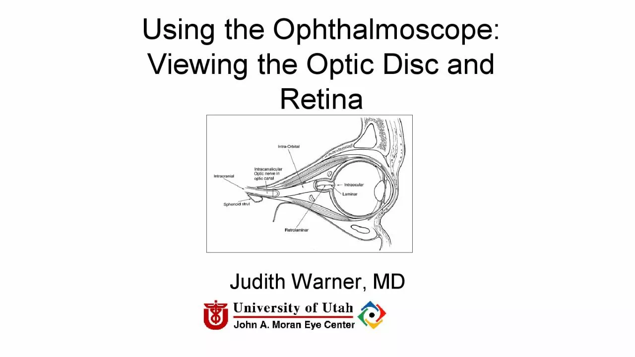
/
Using the Ophthalmoscope Viewing the Optic Disc and RetinaJudith Warn Using the Ophthalmoscope Viewing the Optic Disc and RetinaJudith Warn

Using the Ophthalmoscope Viewing the Optic Disc and RetinaJudith Warn - PDF document

elysha
elysha . @elysha
Follow
348 views
Uploaded On 2022-09-05

Using the Ophthalmoscope Viewing the Optic Disc and RetinaJudith Warn - PPT Presentation

THE OPHTHALMOSCOPE DIRECT OPHTHALMOSCOPYJan Purkinje 1823Hermann von Helmholtz 1851Hand held ophthalmoscopeDirect upright image Dials of the Ophthalmoscope REDFREE FILTER GREEN LIGHT 450 nm monochro ID: 949778

cup disc optic ophthalmoscope disc cup ophthalmoscope optic direct ratio ciliary supply artery retinal posterior avi wmv venous eye

Share:

Link:

Embed:

Download Presentation from below link

Download Pdf The PPT/PDF document "Using the Ophthalmoscope Viewing the Opt..." is the property of its rightful owner. Permission is granted to download and print the materials on this web site for personal, non-commercial use only, and to display it on your personal computer provided you do not modify the materials and that you retain all copyright notices contained in the materials. By downloading content from our website, you accept the terms of this agreement.


Presentation Transcript

Using the Ophthalmoscope: Viewing the Optic Disc and RetinaJudith Warn

er, MDUniversity of Utah THE OPHTHALMOSCOPE DIRECT OPHTHALMOSCOPYJan P

urkinje 1823Hermann von Helmholtz 1851Hand held ophthalmoscopeDirect u

pright image Dials of the Ophthalmoscope REDFREE FILTER (GREEN LIGHT)

450 nm monochromatic lightnerve fiber layeroptic nerve drusen OTHER DI

ALSUsed for measuring lesion sizeLooking for the center of fixation OT

HER DIALS: SLIT BEAM The wheel has lenses of power Panopticophthalmosc

ope Direct typeWider field of viewDistance from pt greaterSimilar aper

turesNot as easy to carrySlightly dimmer light sourceNot as magnified

view ofDiscClean the rubber cup between patientsPhotographs: http://pa

noptic.welchallyn.com/faq.html WHEN EVER POSSIBLE: DILATE THE PATI

ENT Steps to Direct OphthalmoscopyDimly lit roomDilating dropsPatient

fixates distant targetAlign yourselfRed reflexDial in HOW TO USE THE D

IRECT Ophthalmoscope.avi ophthalmoscope.wmv THE RED REFLEX The layers

you will go through to see the optic disc THE OPTIC NERVE WHAT YOU SHO

ULD OBSERVE IN EVERYONE RIGHT EYE AND LEFT EYE THE NORMAL DISCThe disc

is 1.62 mm or 1 million fibersCentral retinal artery and veinLamina C

ribrosaThe optic cup The Normal Disc Appearance The lamina cribrosa is

an important disc structureMeans SieveAnatomically present in all dis

csVisible in about 1/3Shallow in myopia Look at the Cupdisc ratio: WHA

T IS THE CUPDISC RATIO? NO CUP 0.3 CUP 0.1 CUP 0.7 CUP0.9 CUP What is

the cup to disc ratio? 0.60.3 The color of the retinais determined by

the choroid and RPEand the amount of melanin individuals haveTHE COLO

ROF THE RETINA VIEW THE MACULA VASCULAR SUPPLY TO THE GLOBE AND DISC T

WO MAJOR SOURCES byway of the ophthalmic artery+Posterior Ciliary arte

riesCentral retinal artery BLOOD SUPPLY TO THE DISC is from the poste

rior ciliary arteriesThe posterior ciliary arteries supply the disc, t

he choroid and the retinal pigment epithelium CILIORETINAL ARTERYPres

ent in about 32% of peopleEnters separate from the CRAIsolated occlusi

on = Temporal arteritisSpared in CRAO A TOUR OF THE FUNDUS AND VENOUS

PULSATIONS Spontaneous_Venous_Pulse.wmv Spontaneous_Venous_Pulse.avi