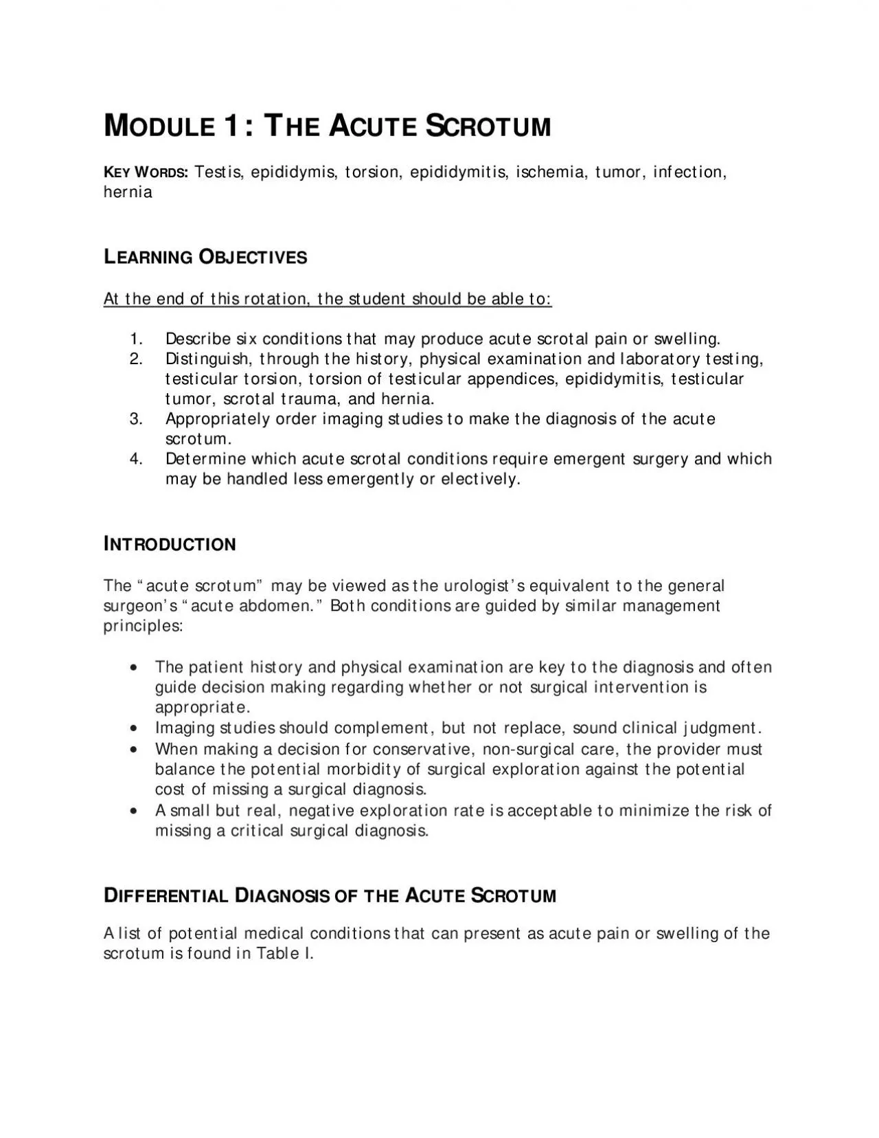
/
itis ischemia tumor infection BJECTIVESAt the end of this rotation itis ischemia tumor infection BJECTIVESAt the end of this rotation

itis ischemia tumor infection BJECTIVESAt the end of this rotation - PDF document

garcia
garcia . @garcia
Follow
342 views
Uploaded On 2022-08-16

itis ischemia tumor infection BJECTIVESAt the end of this rotation - PPT Presentation

uce acute scrotal pain or swelling Distinguish through the history physictesticular torsion torsion of testicultumor scrotal trauma and hernia Appropriately order imaging studies toDetermine wh ID: 936296

testicular testis torsion scrotal testis testicular scrotal torsion acute scrotum surgical pain exploration conditions diagnosis ultrasound tunica swelling epididymitis

Share:

Link:

Embed:

Download Presentation from below link

Download Pdf The PPT/PDF document "itis ischemia tumor infection BJECTIVESA..." is the property of its rightful owner. Permission is granted to download and print the materials on this web site for personal, non-commercial use only, and to display it on your personal computer provided you do not modify the materials and that you retain all copyright notices contained in the materials. By downloading content from our website, you accept the terms of this agreement.


Presentation Transcript

itis, ischemia, tumor, infection, BJECTIVESAt the end of this rotation, the student should be able to: uce acute scrotal pain or swelling. Distinguish, through the history, physictesticular torsion, torsion of testicultumor, scrotal trauma, and hernia. Appropriately order imaging studies toDetermine which acute scrotal conditions require emergent surgery and which may be handled less emergently or electively. NTRODUCTIONThe “acute scrotum” may be viewed as the urologist’s equivalent to the general surgeon’s “acute abdomen.” Both conditions are guided by similar management ion are key to the diagnosis and often guide decision making regarding whetheappropriate. t replace, sound clinical judgment. When making a decision for conservativebalance the potential morbidity of surgical exploration against the potential cost of missing a surgical diagnosis. A small but real, negative exploration rate is acceptable to minimize the risk of missing a critical surgical diagnosis. itions that can present as acute pain or swelling of the scrotum is found in Table I. Table 1: Causes of Acute Scrotal Pain and Swelling Torsion of the testis (synonymous with torsion of the spermatic cord) (prenatal or neonatal) Appendiceal torsion, testis, or epididymis Testicular infarction due to other vascular insult (cord injury, thrombosis, Testicular rupture Intratesticular hematoma, testicular contusion Hematocele Infectious conditions: Acute epididymitis Acute epididymoorchitis Abscess (intratesticular, intravaginal, scrotal cutaneous cysts) Gangrenous infections (Fournier’s gangrene) Henoch-Schonlein purpura (HSP) vasculitis of scrotal wall Fat necrosis, scrotal wallIncarcerated, strangulated inguinal hernia, with or without associated testicular ischemia Acute on chronic events: Spermatocele, rupture

or hemorrhage Hydrocele, rupture, hemorrhage, or infection Testicular tumor with rupture, heWhile the differential diagnosis is broad, ancan frequently precisely define the conditiocan complement clinical judgment and expedite therapeutic decisions. A discussion of at cause acute scrotal pain or swelling ORSIONTesticular torsion The testicle is typically covered by the tunica vaginalis, creating a potential space around the testis. Normally, the tunica vaginalis attaches to the posterior surface of the testicle and allows for very little mobility of the testicle within the scrotum. that the testicle can rotate freely on the spermatic cord within the tunica vaginalis 1). This congenital anomaly, called the “bell clapper deformity,” consists of a transveraffected testis; it can be unilaThis congenital abnormality is present in approximately 12% of human males. During testis torsion, the testicle twists spontaneously on the spermatic cord, causing venous occlusion and engorgement, with suthrough the testicular artery and result in ischemia. In neonates, the testicle ded into the scrotum, after which it becomes attached within the tunica vaginalis. This increased mobility of the testicle predisposes it to Testis torsion is the most common cause ofmales ately 1:4000. Torsion more often involves the left testicle. Among neonatal testicular torsion age rate approaches 100% in patients who undergo detorsion within 6 hours of the start of pain% y;êrs;&#x old;&#x is ; ppr;&#xoxim;&#x-5.5;if detorsion occurs 12 hours; and virtuallyhours (Figure 2). Figure 2. Testis histology during early (A) hemorrhagic phase and chronic late (B) decreased seminiferous tubule diameter and loss of germ cells in late relative to early phases. Figure 1: Bell clapper deformity. Normal testis lie is on

the left and the classic “bell clapper” lie is in the middle. The right side shows a bell clapper variation. Testicular torsion presents with the rapid onset of severe testicular pain and swelling. The onset of pain may be preceded trauma, physical activity, or by no activity (e.g. during sleep). It most often occurs in children or adolescents, but this diagnosis should be considered in evaluating men with scrotal pain of any age, as it may occasionally occur in men 40-50 years old. In this age group, the diagnosis is often delayed or missed due to a low suspicion because of age. Torsion should be in the differential for any sudden acute scrotal pain or swelling. The classic physical examination findings with testis torsion are an exquisitely tender testicle with a high, horizontal lie. Normally the testicle has a vertical lie within the tunica vaginalis of the scrotum – that is, the longitudinal axis of the testis is oriented vertically. With torsion and twisting of the spermatic cord, the testis may assume an altered lie based on the degree of twisting. After venous outflow is occluded, there is swelling and occlusion of arterial flow. Early on, one may be able to palpate the torsed cord and the testis below it; later in the course, however, progressive edema and inflammation ensues, such that after 12-24 hours, the entire hemiscrotum appears as a confluent mass without identifiable landmarks. At this stage, the e from that seen with epididymoorchitis. Importantly, with torsion, signs of infection are usually absent: patients are usually afebrile, free of irritative voiding symptomsse to the inflammation). With a high degree of suspicion, one may reasonably recowithout delay. If scrotal ultrasonography is readily available, and especially if the ngle most useful adjunct to the hist

ory and physical examination in the diagnosis of torsion. The ultra-sonographer should use Doppler flow to assess arterial flow within the affected testis; if arterial flow is absent, torsion is highly likely. It is helpful to compare the flow patterns between both testes to help make this diagnosis. Ultrasonography may also exclude significant testicular trauma, show a hernia extending into the scrotum, and can distinguish epididymitis from torsion by demonstrating increased flow to the epididymis and adnexal structures along with preserved testicular perfusion. Beware of the ultra-ss” exists above the testis that might represent an inflamed epididymis; the torsed cord with edema and inflammation is testicular is the key to the ultrasound diagnosis of torsion. Tests such as nuclear testicular scans, CT or MRI, have essentiaof the acute scrotum. When torsion is diagnosed, urgent surgical exploration and detorsion is mandated, as testicular torsion is a true vascular emergency. Testicular preservation is excellent when corrected within 4-6 hours of onset. Beyond 12 hours, the risk of subsequent testis atrophy is significant with detorsion. Testis salvage is often still appropriate if the testicular appearance at exploration improves with observation following detorsion. The alternative to detorsion is scrotal orchiectomy for pain relief in affected patients. After sharply entering the scrotum, the tunica vaginalis is opened. then undergoes orchidopexy to prevent torsion on that side. The affected testis is (“pinking up”) (Figure 3). If the testis appears viable, or the timeframe suggests that salvage is reasonable then orchiopexy is performed by anchoring the tunica albuginea of the testis to the overlying parietal tunica vaginalis and scrotal dartos muscle. In general, scrotal expl

oration is a procedurseldom results in long-term complications. When weighing conservative treatment exploration. In cases of “late torsion” or “established torsion” exploration generally reveals a hemorrhagic, frankly necrotic testis for which orchiectomy should be “Intermittent” testicular torsion is a well-ren and ultrasound findings are normal. In such cases, it is reasonable to offer an elective bilateral scrotal orchiopexy for the becoming full-fTorsion of testicular or epididymal appendages Small polypoid appendages are often found attached to the testis or epididymis and ts (Figure 4). Similar to testis torsion, torsion of the appendix testis or appendix epididymis can also present with the acute onset of scrotal pain and mass. In most cases, however, the testis is palpable and has the torsed appendage is ecchymotic, it can presents the "blue-dot sign." Doppler ed testis, often with hypervascularity in the area of the appendage. This process is often self-limited, with the infarcted th time. If exploration is pursued, the appendage is Figure 3: Exploration of torsed testis. Note dark, cyanotic color of testis following 30 minutes of detorsion suggesting nonviability. ed. Later in its course, it can be more difficult to distinguish this entity from testicular torsion or epididymitis, as global enlargement and edema of the scrotal compartment may occur. Ultrasound is al blood flow to the testis. Testicular rupture results when there is laceration of the tunica albuginea of the testis, such that testicular parenchyma may extrude. It may occur from either blunt or penetrating trauma. As a general principle, penetrating injuries to the scrotum should be surgically explored. The risk of teinjuries. Even penetrating injuries with a tangential trajectory have a high likelihood of

injuring the testis. In cases of blunt trauma, however, the incidence of testicular rupture varies widely, and depends on the forces exerted, the mechanism of injury, and testis mobility. Following blunt injury, the physical examination findings may include swelling, tenderness or ecchymosis. If one can clearly palpate the testis and it e is significant scrotal wall thickening from edema or hematoma, testicular palpation may be difficult or impossible, and scrotal ultrasonography can determine the degree of testis injury. In addition to demonstrating a break in the continuity of the tunica albuginea or evidence of extruded parenchyma, ultrasound evidence of a marked loss of internal homogeneity of the testis is highly predictive of testicular rupture and warrants surgical exploration. Blunt injury may result in testicular rupture, intratesticular these, only testicular rupture requires surgical repair. t from drainage. For intratesticular hematoma (intact tunica albuginea, localizeservation, rest, cold packs and analgesics Surgical exploration for trauma is performed through incisions that anticipate the structures at risk. For peneinto the groin to expose the spermatic cordover the injured scrotal compartment is effe Figure 4: Illustration of the common appendices of the testis and epididymis. The appendix testis is most commonly affected by torsion tunica vaginalis space, any extruded testicular parenchyma is inspected, irrigated and resected or retained and tunical lacerations repaired. The testicular compartment may be drained, generally with a small Penrose drain. With trauma, most testicular injuries are amenable to repair. Orchiectomy is indicated when there is major injury to the spermatic cord with organ devitalization, and destruction of parenchyma is so extensive that no

significant tissue can be salvaged. NFECTIONSEpididymitis and epididymoorchitis Although they may be difficult to distinguish on physical examination from scrotal trauma or testis torsion, it is important to accurately diagnosis epididymitis and orchitis, as their management is entirely nonsurgical. Epididymitis is usually caused by exposure, recent sexual activity, epididymgonococcal infection, and is amendable to standard antibiotic treatment. In older men and those with problems such as significant benign prostatic hypertrophy (BPH), a history of UTI’s, or urethral stricture disease, enteric, gram negative bacteria initial broad spectrum antibiotics are used with therapy furtheculture results. There are also noninfectious or inflammatory forms of epididymitis. medications, urinary reflux within the ejaculatory ducts, and sperm and fluid extravasation after vasectomy. When epididymitis extends into the testis and causes testicular tenderness and enlargement, it is termed epididymoorchitis. There are several features in the patient history that may indicate epididymitis, such as a history of previous STI, recent sexual activity, irritative voiding symptoms, BPH/incomplete emptying of the bladder, or UTI. The very sudden onset of pain and swelling is more typical of torsion, whil(often greater than 24 hours) suggests epididymitis. On physical examination, tenderness posterior and lateral to the testis (the usual location of the epididymis). Scrotal ultrasoepididymis with normal or increased blood flow to the testis, which will distinguish ess formation within the epididymis or in the peri- epididymal tissues, can also be detected by ultrasound. The diagnostic ididymoorchitis from late torsion. In both entities, there is typically a confluent mass in the scrotum with edema and fixation of

the overlying scrotal wall that obliterate normal anatomic landmarks. Furthermore, advanced epididymoorchitis can result in testicular ischemia and infarction due to compression of the testicular vasculature from epididymal inflammation. On ultrasound, this may prestorsion. In either case, the lack of testis blood flow on Doppler ultrasound requires surgical exploration which allows these conditions to be differentiated. When diagnosed, epididymitis and orchitis are managed conservatively with rest and scrotal elevation. If abscess formation occurs, surgical drainage and/or orchiectomy may be necessary. Infectious conditions within the scrotal wall are also classified under the acute scrotum and include cellulitis and fasciitisabscess formation are distinguishable testicular conditions on physical examination, as the testis is usually palpably normal and nontender, if it can be palpated without rotal wall infections may result from infected sebaceous cysts, folliculitis, or other dermatologic conditions. Incision and drainage with gauze packing and broad-spectrum antibiotics are prescribed for these superficial conditions. Fasciitis of scrotum and groin, termed Fournier’s gangrene, involves a rapidly progressive, life threatening infection of the genital soft tissues. It is associated with predisposing issues inmunocompromised or diabetic patient. On e may be necrotic black or ecchymotic The most diagnostic is the finding of crepitus, a spongy, cracking feeling within the skin that indicates gas-producing microorganisms underneath that can be felt in the scrotum or perineum. When left untreated, genital gangrene will progress over hours and result in overwhelming bacterial sepsis with an associated high mortality rate. Therefore, broad spectrum antibiotics that cover aerobicand u

rgent and repeated surgical drainagethe infection. At the time of surgical treatment, cystoscopy and proctoscopy may be al and rectal abnormalities. NFLAMMATIONHenoch-Schonlein purpura (HSP) is a vasculitisand erythema in the absence of infection. Idiopathic scrotal edema and filarial Figure 5: Fournier’s gangrene of the scrotum. Note necrotic, black patch of scrotal skin with Fournier's gangrene Nat Clin Pract Urol 3: 54–57) infections (rare in the US) can also cause chlymphadenopathy are also rare but significant conditions that may occur under the aegis of the acute scrotum. In most of these conditions, the history of a slowly progressive disease process helps differentiate them from more classically acute conditions. Treatment of the underlying, non-scrotal cause is most effective to relieve the scrotal symptoms. also present as an acute scrotum. In this case, pain and swelling involve both the scrotal contents and the groin area. Although important to differentiate, it may be difficult to distinguish an incarcerated inguinal hernia from as hydrocele, scrotal trauma, or scrotal abscess. An incarcerated inguinal hernia involves bowel that is obstructed and is a true surgical emergency. In selected, less acute cases, groin and scrotal ultrasound or pelvic CT scans can clarify the diagnosis ben may be associated with vas deferens obstruction and infertility later on. Other scrotal conditions that are chronic symptoms and include testicular neoplasmcase of testis tumors, patients may only become aware of the mass after it has been present for many months, after it affects the appearance of the scrotum. However, y if they undergo hemorrhage or necrosis, and produce swelling, pain and soreness. In rotal ultrasound demonstrates a solid lihood of being a germ cell tumor. The suspicion

of tumor is important for the approach to exploratory surgery in the acute to testis cancer is through an inguinal incision and not transcrotally. In addition, out intact, to minimize tumor spillage duridone in the inguinal region to further contain the spread of cancer. Other chronic scrotal lesions which can present acutely include hydroceles (increased fluid within the tunical vaginalis space) and ducts that lead from the rete testis to trauma, or become infected. In additicharacterized by dilated pampiniform plexus veins and that occurs in 15% of men at puberty, can be present for years but become acutely symptomatic. These dilated veins surround the spermatic cord. If the varicocele has acute onset, is only right-sided, or persists in the supine position, then inferior vena caval (IVC) obstruction must be excluded (i.e., IVC thrombus, abdominal mass, etc.). A careful history, physical examination and ultrasound examination is usually sufficient to diagnose these usually benign acute on chronic events. Urgent surgical intervention is rarely needed for drainage of a loculated infectioA full range of scrotal pathology must be considered in acute scrotum cases. e scrotum require surgical exploration, making this a very time sensitive condition. A high value is place on the history, physical examination and ultrasound imaging for acute scrotum diagnoses. Meacham RB: Potential for vasal occlusion amJoyner B & Walsh T: Evaluation of the Pediatric Patient with a Non-Traumatic Acute Kim SH et al.: Significant predictors for determination of testicular rupture on Lin EP et al.: Testicular torsion: twists and turns. Semin Ultrasound CT MR. Tracy CR et al.: Diagnosis and management of epididymitis. Urol Clin North Amer ong JH, Breyer BN, Broghammer JA, et al. Urotrauma: AUA guideline. Jo