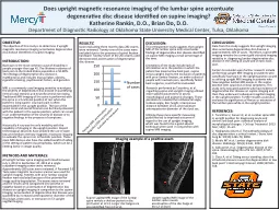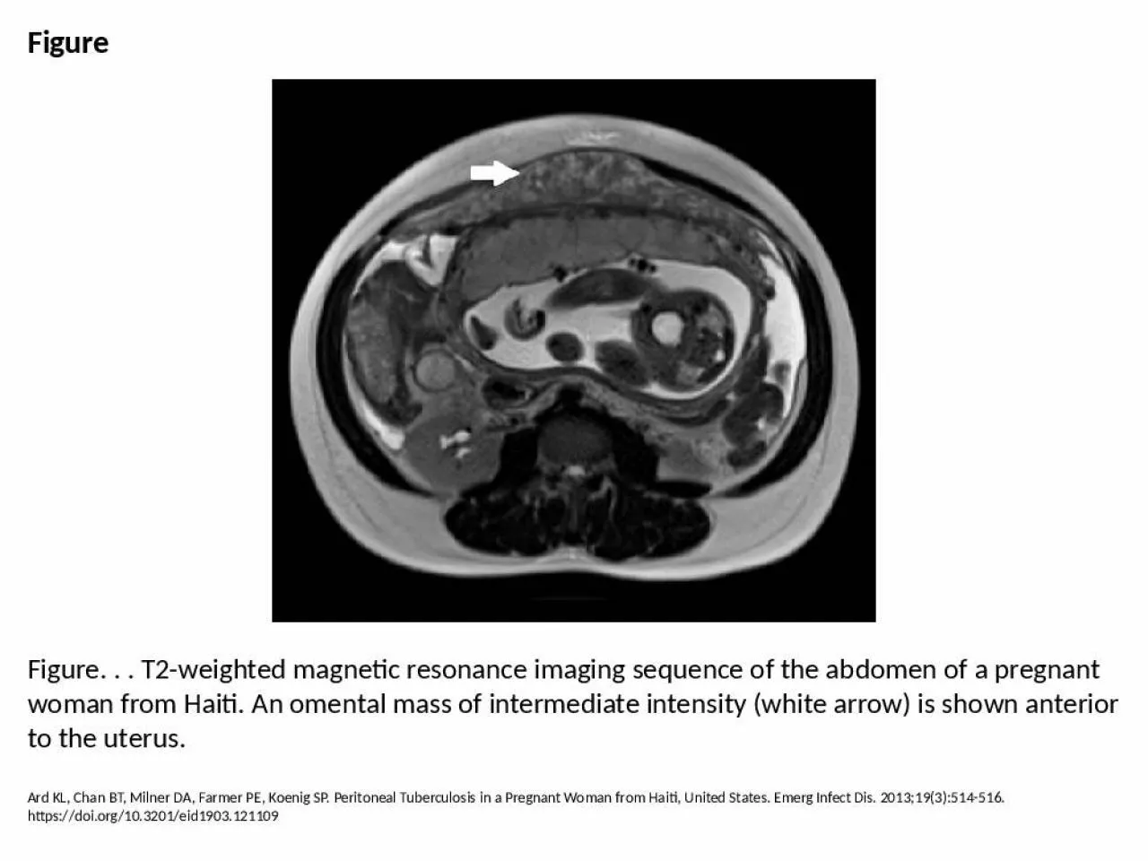
PPT-Figure Figure. . . T2-weighted magnetic resonance imaging sequence of the abdomen of a
Author : isabella | Published Date : 2024-09-06
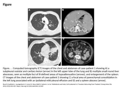
Ard KL Chan BT Milner DA Farmer PE Koenig SP Peritoneal Tuberculosis in a Pregnant Woman from Haiti United States Emerg Infect Dis 2013193514516 httpsdoiorg103201eid1903121109
Presentation Embed Code
Download Presentation
Download Presentation The PPT/PDF document "Figure Figure. . . T2-weighted magnetic ..." is the property of its rightful owner. Permission is granted to download and print the materials on this website for personal, non-commercial use only, and to display it on your personal computer provided you do not modify the materials and that you retain all copyright notices contained in the materials. By downloading content from our website, you accept the terms of this agreement.
Figure Figure. . . T2-weighted magnetic resonance imaging sequence of the abdomen of a: Transcript
Download Rules Of Document
"Figure Figure. . . T2-weighted magnetic resonance imaging sequence of the abdomen of a"The content belongs to its owner. You may download and print it for personal use, without modification, and keep all copyright notices. By downloading, you agree to these terms.
Related Documents