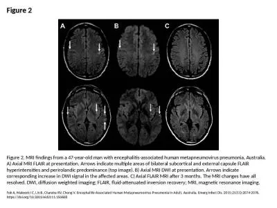
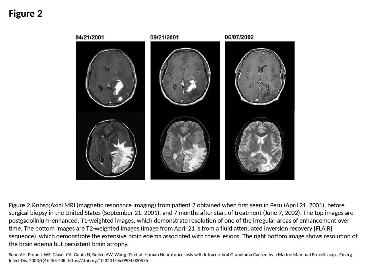
PPT-Figure 2 Figure 2. Axial MRI (magnetic resonance imaging) from patient 2 obtained

Author : cora | Published Date : 2024-01-13

Sohn AH Probert WS Glaser CA Gupta N Bollen AW Wong JD et al Human Neurobrucellosis with Intracerebral Granuloma Caused by a Marine Mammal Brucella spp Emerg Infect

Presentation Embed Code

Download Presentation

Download Presentation The PPT/PDF document "Figure 2 Figure 2. Axial MRI (m..." is the property of its rightful owner. Permission is granted to download and print the materials on this website for personal, non-commercial use only, and to display it on your personal computer provided you do not modify the materials and that you retain all copyright notices contained in the materials. By downloading content from our website, you accept the terms of this agreement.

Figure 2 Figure 2. Axial MRI (magnetic resonance imaging) from patient 2 obtained: Transcript


Sohn AH Probert WS Glaser CA Gupta N Bollen AW Wong JD et al Human Neurobrucellosis with Intracerebral Granuloma Caused by a Marine Mammal Brucella spp Emerg Infect Dis 200394485488 httpsdoiorg103201eid0904020576. Principle first observed in 1946. Used for spectroscopy and imaging . Imaging techniques are a form of tomography, where slices are ’cut’ and depict. MRI utilizes signals from the body . MRI is non-ionizing, operating in radiofrequency range, unlike CT, PET, SPECT. TEHRAN FIR (OIIX) BAGHDAD FIR (ORBB) (OSTT) ANKARA FIR (LTAA) BAKU FIR (UBBB) YEREVAN FIR/UIR (UDDD) TBILISI FIR (UGGG) ROSTOV-NA- DONU FIR (URRV) SIMFEROPOL' FIR (UKFV) ASTRAKHAN FIR (URWA) ODESA FIR Principles of MRI. Some terms:. Nuclear Magnetic Resonance (NMR). quantum property of protons. energy absorbed when precession frequency matches radio frequency. Magnetic Resonance Imaging (MRI). uses spatial differences in resonance frequencies to form an image. . Kulkarni. 1000722132. LIST OF ACRONYMS. CAT - Computed Axial Tomography. CT - Computed Tomography. DICOM - Digital Imaging and Communications in Medicine. EEG - Electroencephalography. EKG - Electrocardiography. Part 1. Magnetic Resonance Imaging. A great multidisciplinary application of physics and other sciences. And example of a “pure science” discovery given a practical application. Based on NMR – Nuclear Magnetic Resonance – discovered in the 1940s. ). B: Sagittal T2-weighted . TSE C. : Sagittal T2-weighted SE image . . Axial T2-weighted turbo spin-echo (TSE) (4902/132) magnetic resonance image of the uterus in the axial (A) and coronal (B) planes . —a measure of X-ray absorption at several positions around the head, maps tissue density. Magnetic resonance imaging (MRI). gives higher resolution images in three steps:. Strong magnets cause protons in brain tissue to line up in parallel. A pulse of radio waves knocks protons over. Protons reconfigure themselves, emitting radio waves that differ by tissue density.. 1. Neju Mathew Philip. Inspire Fellow. Optoelectronic Materials & Devices Lab. NITT. Introduction. Theory. Instrumentation &Working. Applications. Review. 2. OVERVIEW. INTRODUCTION. Invented by Raymond V . test that helps doctors look for and treat many different medical problems. MRI scanners use a strong magnetic field and computers to create detailed pictures. For an MRI arthrogram , a small a – Site of Service Page 1 of 8 UnitedHealthcare Commercial Utilization Review Guideline Effective 04/01/2022 Proprietary Information of UnitedHealthcare. Copyright 202 2 United HealthCare Ser Shirwadkar CG, Samant R, Sankhe M, Deshpande R, Yagi S, Schuster FL, et al. Acanthamoeba Encephalitis in Patient with Systemic Lupus, India. Emerg Infect Dis. 2006;12(6):984-986. https://doi.org/10.3201/eid1206.060087. ot. the sample. What is the Hamiltonian for the system? The magnetic fields are as below.. The Hamiltonian is given by,. . where,. The spin matrices are:. Magnetic Resonance. The Hamiltonian is:. Our growing brain. British Science Week 2022. Michael S Stringer. Medical Physicist, University of Edinburgh. I. Indeed the first evidence we have of surgery is on the brain (known as trepanation). Introduction – Where to start?. Fok A, Mateevici C, Lin B, Chandra RV, Chong V. Encephalitis-Associated Human Metapneumovirus Pneumonia in Adult, Australia. Emerg Infect Dis. 2015;21(11):2074-2076. https://doi.org/10.3201/eid2111.150608.

Download Document

Here is the link to download the presentation.
"Figure 2 Figure 2. Axial MRI (magnetic resonance imaging) from patient 2 obtained"The content belongs to its owner. You may download and print it for personal use, without modification, and keep all copyright notices. By downloading, you agree to these terms.

Related Documents