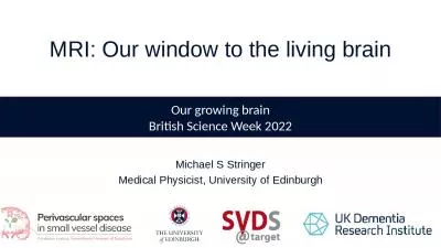
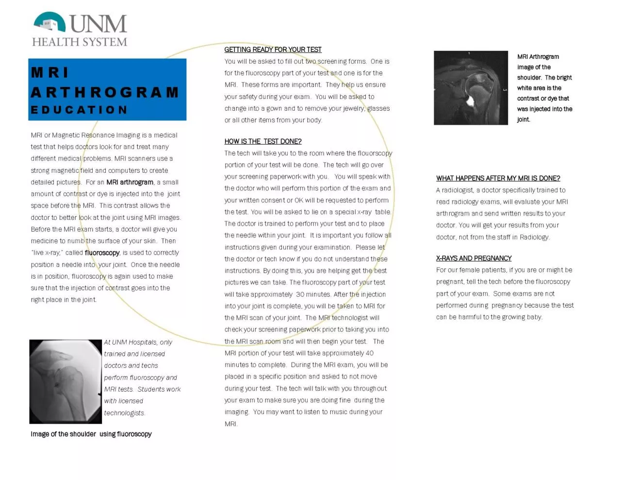
/
MRI or Magnetic Resonance Imaging is a medical MRI or Magnetic Resonance Imaging is a medical

MRI or Magnetic Resonance Imaging is a medical - PDF document

jacey
jacey . @jacey
Follow
342 views
Uploaded On 2022-08-16

MRI or Magnetic Resonance Imaging is a medical - PPT Presentation

test that helps doctors look for and treat many different medical problems MRI scanners use a strong magnetic field and computers to create detailed pictures For an MRI arthrogram a small a ID: 936553

test mri joint doctor mri test doctor joint fluoroscopy exam tech perform asked contrast arthrogram needle trained screening part

Share:

Link:

Embed:

Download Presentation from below link

Download Pdf The PPT/PDF document "MRI or Magnetic Resonance Imaging is a m..." is the property of its rightful owner. Permission is granted to download and print the materials on this web site for personal, non-commercial use only, and to display it on your personal computer provided you do not modify the materials and that you retain all copyright notices contained in the materials. By downloading content from our website, you accept the terms of this agreement.


Presentation Transcript

MRI or Magnetic Resonance Imaging is a medical test that helps doctors look for and treat many different medical problems. MRI scanners use a strong magnetic field and computers to create detailed pictures. For an MRI arthrogram , a small amount of contrast or dye is injected into the joint space before the MRI. This contrast allows the doctor to better look at the joint using MRI images. Before the MRI exam starts, a doctor will give you medicine to numb the surface of your skin. Then “live x - ray,” called fluoroscopy , is used to correctly position a needle into your joint. Once the needle is in position, fluoroscopy is again used to make sure that the injection of contrast goes into the right place in the joint. At UNM Hospitals, only trained and licensed doctors and techs perform fluoroscopy and MRI tests. Students work with licensed technologists. Image of the shoulder using fluoroscopy GETTING READY FOR YOUR TEST You will be asked to fill out two screening forms. One is for the fluoroscopy part of your test and one is for the MRI. These forms are important. They help us ensure your safety during your exam. You will be asked to change into a gown and to remove your jewelry, glasses or all other items from your body. HOW IS THE TEST DONE? The tech will take you to the room where the flouorscopy portion of your test will be done. The tech will go over your screening paperwork with you. You will speak with the doctor who will perform this portion of the exam and your written consent or OK will be requested to perform the test. You will be asked to lie on a special x - ray table. The doctor is trained to perform your test and to place the needle within your joint. It is important you follow all instructions given during your examination. Please let the doctor or tech know if you do not understand these instructions. By doing this, you are helping get the best pictures we can take. The fluoroscopy part of your test will take approximately 30 minutes. After the injection into your joint is complete, you will be taken to MRI for the MRI scan of your joint. The MRI technologist will check your screening paperwork prior to taking you into the MRI scan room and will then begin your test. The MRI portion of your test will take approximately 40 minutes to complete. During the MRI exam, you will be placed in a specific position and asked to not move during your test. The tech will talk with you throughout your exam to make sure you are doing fine during the imaging. You may want to listen to music during your MRI. WHAT HAPPENS AFTER MY MRI IS DONE? A radiologist, a doctor specifically trained to read radiology exams, will evaluate your MRI arthrogram and send written results to your doctor. You will get your results from your doctor, not from the staff in Radiology. X - RAYS AND PREGNANCY For our female patients, if you are or might be pregnant, tell the tech before the fluoroscopy part of your exam. Some exams are not performed during pregnancy because the test can be harmful to the growing baby. MRI Arthrogram image of the shoulder. The bright white area is the contrast or dye that was injected into the joint. MRI ARTHROGRAM EDUCATION