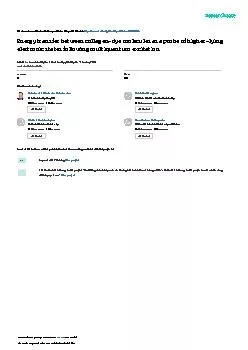
PPT-Combination of fluorescence FRET microscopy and optical tweezers for mechanical studies
Author : isabella2 | Published Date : 2023-09-21
Camille Dubois Biophotonics group Laboratoire Charles Fabry Institut dOptique Under the supervision of Nathalie Westbrook SFP 7 juillet 2023 Ludivine Houel
Presentation Embed Code
Download Presentation
Download Presentation The PPT/PDF document "Combination of fluorescence FRET microsc..." is the property of its rightful owner. Permission is granted to download and print the materials on this website for personal, non-commercial use only, and to display it on your personal computer provided you do not modify the materials and that you retain all copyright notices contained in the materials. By downloading content from our website, you accept the terms of this agreement.
Combination of fluorescence FRET microscopy and optical tweezers for mechanical studies: Transcript
Camille Dubois Biophotonics group Laboratoire Charles Fabry Institut dOptique Under the supervision of Nathalie Westbrook SFP 7 juillet 2023 Ludivine Houel Renault Marie . Chelsea Aitken. Peter Aspinall. Advantages Over Light Microscopy. Resolution of light microscopy limited by Rayleigh Criterion. If two objects cannot be seen as distinct structures, then they may be considered coincident in space. Energy Transfer). The problem. The use of fluorescent probes (ex. GFP) permits direct observation of the dynamic properties of specific proteins in live cells .. But, It is difficult to observe functional information like protein-protein interaction using fluorescent probes.. Lecture 13:. Super-resolution microscopy: Part I. Lecture 13: . Fluorescent labeling, multi-. sprectral. imaging and FRET. Review of previous lecture. FRET. FLIM. Super resolution . microscopy. NSOM. Ozkans. ’. laboratory in UC-Riverside to perform defect analysis and surface metrology of large-area CVD-graphene sheets. This method utilizes the quenching of fluorescence from dye molecules by graphene via resonant energy transfer to increase the visibility of graphene on a glass substrate. A large-area fluorescence montage image of the dyed graphene sample is collected and processed to identify the graphene and indicate the graphene layer thickness throughout the entire graphene sheet. Furthermore, chemically functionalized (doped with fluorine) parts of graphene film is visualized using the same technique. The emission of the dye is quenched to a different extent by fluorinated and pristine graphene, which provides the fluorescence-imaging contrast essential for this method. The regions with pristine graphene appear darker on the fluorescence images than the regions with fluorinated graphene, enabling large-scale mapping of the functionalized regions in CVD grown graphene sheets. Complex circular patterned regions of doped and pristine graphene regions are resolved with great accuracy. This method is posed for widespread adoption by graphene manufacturers as a basis for facile and high throughput metrology of large scale graphene sheets. Finally, this work featured as cover articles in . Mentor: Prof. Dr Igor Poberaj. University of Ljubljana. Faculty of Mathematics and Physics. Microrheology with optical tweezers. Ljubljana, December 4th, 2012. Introduction. Microrheology. Optical tweezers. lecture. 16. November 2011. Fourier. plane. Point . object. Image. f. f. f. f. a. a. ’. Magnification. : m=1. Angles. : sin(. a. ’)=sin(. a) . . a. ’=. a. Magnification and resolution: the Abbe limit. ( Fluorescence lifetime imaging). —. Molecular interraction (FRET). —. intracellular pH. etc. etc etc. . Pulsed IR-laser. ( Multiphoton exitation). —. Intracellular Tracking. —. Uncaging & Photostimulation . Lecture 16:. Super-resolution microscopy: Part . 2. Lecture 16: Super-resolution microscopy and TIRFM. Single molecule imaging. Total internal reflection fluorescence microscopy (TIRFM). Super-resolution techniques . and Kavantzas N., "Computer vision algorithms in DNA ploidy image analysis", Imaging, Manipulation and Analysis of Biomolecules, Cells and Tissues IV, Proc. SPIE, 6088:60880O (2006) and Loukas S., "A KAARTHIK J. INTRODUCTION. A fluorescence microscope is a optical microscope that uses fluorescence and phosphorescence instead of , or I addition to reflection and absorption to study properties of organic or inorganic substances.. are held together at their centromeres binds specific proteins, which in turn make up a disk-like structure called the kinetochore. The kinetochore is an attachment site for spindle fibers, which pla COLLEGE BOTANY: SEM - VI, PAPER - DSE4T: ANALYTICAL TECHNIQUES IN PLANT SCIENCES, UNIT - 1: FLUORESCENCE MICROSCOPY What is fluorescence microscopy? A fluorescence microscope is an optical microscope Reif. Outline. Fluorescent resonance energy transfer (FRET). Fluorescent Labels. Fluorophores. , quantum dots. Single FRET. Molecular Beacon. DNA Tweezers. Multiple FRET. Homo-FRET. Hetero-FRET. Optically-induced molecules. (a guide to designing and . analysing. FRET experiments). Joe. Jan 2018. Fluorescence resonance energy transfer (FRET). . Conceptually FRET is a very simple method to understand. D. A. Donor and Acceptor.
Download Document
Here is the link to download the presentation.
"Combination of fluorescence FRET microscopy and optical tweezers for mechanical studies"The content belongs to its owner. You may download and print it for personal use, without modification, and keep all copyright notices. By downloading, you agree to these terms.
Related Documents