/
A Seven novel TaySachs mutations detected by chemical mismatch cleav A Seven novel TaySachs mutations detected by chemical mismatch cleav

A Seven novel TaySachs mutations detected by chemical mismatch cleav - PDF document

joanne
joanne . @joanne
Follow
343 views
Uploaded On 2022-10-14

A Seven novel TaySachs mutations detected by chemical mismatch cleav - PPT Presentation

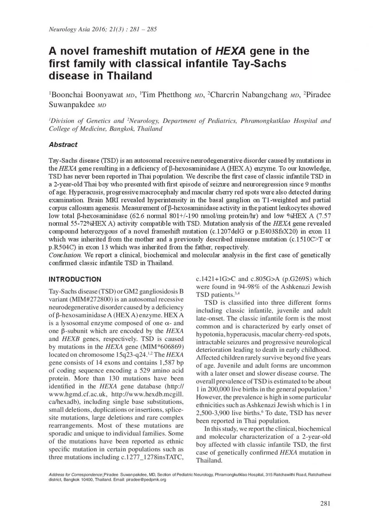
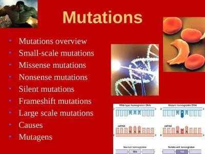
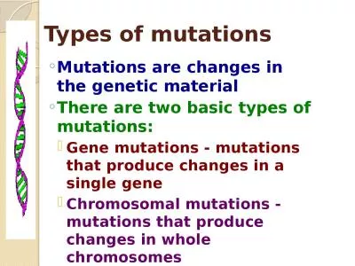
starts and is usually fatal by age 4 or 5 yearsOur patient presented with early infantileonset of intractable seizures and progressive neurological deterioration leading to death at the age of 4 yea ID: 960158

tsd mutation patient mutations mutation tsd mutations patient gene hex disease infantile hexosaminidase sachs tay activity heterozygous dna exon

Share:

Link:

Embed:

Download Presentation from below link

Download Pdf The PPT/PDF document "A Seven novel TaySachs mutations detecte..." is the property of its rightful owner. Permission is granted to download and print the materials on this web site for personal, non-commercial use only, and to display it on your personal computer provided you do not modify the materials and that you retain all copyright notices contained in the materials. By downloading content from our website, you accept the terms of this agreement.


Presentation Transcript

A. Seven novel Tay-Sachs mutations detected by chemical mismatch cleavage of PCR-amplied cDNA 1991; 11:124-34. Pastores GM,Gianutsos Zaroff CM,Kolodny EH. Late-onsetTay-Sachs disease: phenotypic characterization and genotypic correlations in 21 affected patients. Genet Med2005; 7:119-23. Paw BH,Moskowitz SM,Wright Neufeld EF. Juvenile GM2 gangliosidosis caused by substitution of histidine for arginine at position 499 or 504 of the alpha-subunit of beta-hexosaminidase. 1990; 265:9452-7. Neufeld EF. A frameshift mutation in a patient with Tay-Sachs disease causes premature termination and defective intracellular transport of the alpha-subunit of beta-hexosaminidase. J Biol 1989; 264:21376-80. starts and is usually fatal by age 4 or 5 years.Our patient presented with early infantile-onset of intractable seizures and progressive neurological deterioration leading to death at the age of 4 years which is compatible with classical acute infantile form of TSD. Even though the %HEX A activity in the patient leukocytes was slightly higher (7%) than in the individuals with acute infantile form (0-5%), mutation analysis revealed compound heterozygous of two null alleles which were predicted to result in no or extremely low enzymatic activity. Therefore, the residual enzymatic activity alone may not be the good indicator for the severity of the disease. Once our index case has been identied, familial investigation must be required according to the possibility of autosomal recessive transmission. Mutation analysis of both parents was performed. �The missense mutation c.1510CT (p.R504C) was identied in the paternal DNA while the novel frameshift mutation c.1207delG (p.E403SfsX20) was detected in the maternal DNA, indicating that our proband’s parents are both carriers for TSD. Since there is currently no effective treatment for subsequent pregnancies should be recommended to this family due to high recurrence risk. In conclusion, we report a compound heterozygous of a novel frameshift and a previous described missense mutations in the rst Thai family with classic infantile TSD which has never been reported in Thailand. The authors would like to thank Professor W-L Hwu from Department of Pediatrics and Medical Genetics, National Taiwan University Hospital for the support of biochemical diagnosis -hexosaminidase activity) in our patient. We also thank Laura Yeh, medical technologist, for the technical assistance. 1.Myerowitz R,Piekarz R,Neufeld EF,Shows Suzuki K. Human beta-hexosaminidase alpha chain: coding sequence and homology with the beta Proc Natl Acad Sci USA 1985; 82:7830-4. 2.Triggs-Raine BL,Akerman BR,Clarke JT,Gravel RA. Sequence of DNA anking the exons of the gene, and identication of mutations in Tay-Sachs 1991; 49:1041-54. Myerowitz RCostigan FC. The major defect in Ashkenazi Jews withTay-Sachs diseaseis hexosaminidase. 1988; 263:18587-9. Myerowitz RHogikyan ND. Different AshkenaziJewishnon-Jewish French Canadians with Tay-Sachs disease. 1986; 232:1646-8. Gravel

RA, Kabak MM, Proia RL, Sandhoff K, Suzuki K, Suzuki Kuni. The GM2 gangliosidoses. In: Scriver CR, Beaudet AL, Valle D, eds: The metabolic and molecular basis of inherited disease. ed. New York: McGraw-Hill; 2001: 3827-76.Desnick Kornreich R. Experience with carrier screening and prenatal diagnosis for 16 Ashkenazi Jewish 2010; 31:1240-50. Vavougios G. Novel Tay-Sachs 1992; Tamhankar PM. Identication of novel mutations in gene in children affected with Tay Sachs disease from2012; 7:e39122. Tanaka A,Hoang LT,Nishi Y,Maniwa S,Oka Yamano T. Different attenuated phenotypes of GM2 gangliosidosis variant B in Japanese patients mutations at codon 499, and ve novel mutations responsible for infantile acute form. J Hum 2003; 48:571-4. . Newly observed thalamic involvement and mutations of the HEXA gene in a Korean patient with juvenile GM2 gangliosidosis. Metab Brain Dis 2008; 23:235-42. Chan LYJamalia Alagaratnam J. Tay-Sach disease with “cherry-red spot” rst reported case in Malaysia. Taeranawich PNitiapinyasakul . Identification of Sandhoff family: clinical and biochemical characterization. J Med Assoc Thai 2010; 93:1088-92. Macías-Vidal JSpanish GM2 Working Group. GM2 gangliosidoses in Spain: genes in 34 Tay-Sachs and 14 Sandhoff patients. 2012; 506:25-30. Montalvo A,. Molecular in Argentinean patients affected with Tay-Sachs disease: possible common origin of the prev�alent c.459+5AG mutation. 2012; 499:262-5. MontalvoVlahovicek K, gene in Italian patients with infantile and late onset Tay-Sachs disease: detection of fourteen novel alleles. Hum 2005; 26:282. PawWood LC,Neufeld EF. A third mutation at the CpG dinucleotide of codon 504 and a silent Am J Hum 1991; 48:1139-46. 17.MatsuzawaF,Aikawa S,basis of the GM2 gangliosidosis B variant. 2003; 48:582-9. Chelly J,Kahn Neurology Asia Fig. 1: gene in the patient DNA revealed a heterozygous c.1207delG mutation in exon 11 (�) and a heterozygous c.1510CT mutation in exon ) which is inherited from the father ( leading to the clinical characteristic of TSD as demonstrated in our patient. According to the unavailability of skin broblast culture in our HEXA gene was used to conrm the diagnosis of TSD in our patient. After molecular analysis of the gene was performed in our patient DNA, compound heterozygous of one previously described and one novel mutation have been identied. The �rst mutation is missense mutation c.1510CT (p.R504C) in exon 13.This mutation was rst described in German siblings affected with chronic GM2 gangliosidosis. The substitution of R504 to C causes a defective in -subunit dimerization which results in no enzymatic activity. The high frequency of this mutation is probably caused by the presence of CpG dinucleotides which are known as mutagenic Other reported mutations in codon 504 were R504H, R504L and deletion of cytosine in this codon. The second mutation is a novel heterozygous frameshift mutation c.1207delG (p.E403SfsX20) in exon 11. This mutation is predicted

to result in a premature termination at the codon 422 causing a truncation subunit of HEX A enzyme. To date, more than 130 different mutations have been identied including various types of mutation. Most of the mutations are either nonsense or frameshift which are located throughout the gene, whereas most of the missense mutations are clustered in highly conserved amino acids in ligand-like binding domain. As in our patient who mutation in Thailand, the missense mutation is located in exon 13 which is located in the extra- Compound heterozygous of these two null alleles explained the clinical severity and infantile-onset of TSD in our patient.Molecular genetic testing is clinically important not only for diagnostic conrmation but it can sometimes be used to prognostic predictions and can contribute to the management of the patient and family. Classic infantile TSD is the most common form. Affected infants typically appear normal until the age of 3 to 6 months when progressive loss of neurological function CASE REPORTT.D. is a Thai boy who is the only child of non-consanguineous parents. He was born at term with uneventful prenatal and perinatal history. He was healthy and normal milestone until 9 months of age. At time, he experienced his rst episode of seizure then deteriorated in term of seizures and developmental milestone. His neurological deterioration was rapidly progressive, followed by loss of motor ability and nally bed-ridden by the age of 1 year. He was initially diagnosed as myoclonic seizures with encephalopathy and spastic cerebral palsy. He was admitted to the hospital several times according to gastro-esophageal reux disease and recurrent pneumonia. His parents noticed that he also had hyperacusis to noise and tactile stimuli. He was evaluated by pediatric neurologist and geneticist at the age of 2 years when progressive macrocephaly was detected. There were no other dysmorphic features. On neurological examination, he had axial hypotonia and increased deep tendon reexes all extremities. Opthalmological examination revealed bilateral macular cherry red spots. Other examinations were unremarkable. Routine hematological and biochemical investigations were normal. Investigation for inborn error of metabolisms (IEMs) revealed normal serum ammonia, serum lactate, plasma amino acids and urine organic acids. A brain magnetic resonance imaging (MRI) exhibited hyperintensity in the basal ganglion on T1-weighted images and partial agenesis of the corpus callosum. The diagnosis was established by measurement of leukocyte -hexosaminidase activity including total -hexosaminidase and the percentage of HEX A (%HEX A) activity. Total -hexosaminidase activity (62.60 normal 801+/-190 nmol/mg protein/hr) was markedly below the normal range and a low percentage of HEX A activity (7.57% normal 55-72%HEX A) was found, compatible with the diagnosis of TSD. gene After informed consent was obtained, peripheral blood EDTA samples from the patient and the parents were collected. Geno

mic DNA was extracted from peripheral blood lymphocytes commercial available kitsaccording to manufacturer’s protocol. Mutation analysis was characterized by direct DNA sequencing of all 14 coding exons and exon-intron boundaries of gene according to protocols described previously.The nucleotides and amino acids were numbered according to the RefSeq NM_000520.4 and NP_000511.2, respectively. Putative mutations were conrmed in two separate PCR from the patient DNA. Mutation analysis by direct DNA sequencing of all 14 coding exons and exon-intron junction of gene revealed compound heterozygous of one frameshift and one missense mutation in the patient DNA. The heterozygous frameshift mutation was deletion of G at nucleotide 1207 (c.1207delG) which is located in exon 11 (Figure 1A). This frameshift mutation resulted in a premature termination codon at the codon 422 (p.E403SfsX20) and was predicted to encode a -subunit of HEX A.The heterozygous missense mutation was C to T change at nucleotide REPORT1510 (c.1510CT), resulted in the conversion of amino acid from arginine to cysteine at codon 504 (p.R504C) in exon 13 (Figure 1B). Molecular analysis of both parental DNA revealed that the heterozygous frameshift mutation (c.1207delG) was inherited from the mother, whereas the REPORTheterozygous missense mutation (c.1510CT) was inherited from the father (Figure 1C and 1D). TSD is an autosomal recessive lysosomal storage diseases caused by deciency of HEX A enzyme. HEX A is a heterodimeric protein composed -subunits. Mutations in the HEXA -subunit cause TSD, gene which -subunit are causative of Sandhoff disease (MIM#268800). In Asia, TSD has been occasionally reported in central and south Asian populations including Chinese, Indian, Japanese, South Korean and Malaysian. To date, only Sandhoff disease has been reported in Our patient described in this study is the rst case of classic infantile TSD in Thai population. The clinical and neuroimaging ndings of the patient are almost similar to other reported infantile-onset TSD patients from different ethnic groups. The diagnosis of TSD was conrmed by determination of leukocyte -hexosaminidase activity. Both total -hexosaminidase and the percentage of HEX A (%HEX A) activity were low in our patient leukocytes. The enzyme deciency results in the neuronal accumulation of GM2 ganglioside A novel frameshift mutation of HEXA gene in the �rst family with classical infantile Tay-Sachs disease in ThailandBoonchai Boonyawat Tim Phetthong Charcrin Nabangchang Piradee Suwanpakdee Neurology, Department of Pediatrics, Phramongkutklao Hospital and College of Medicine, Bangkok, ThailandAbstract Tay-Sachs disease (TSD) is an autosomal recessive neurodegenerative disorder caused by mutations in gene resulting in a de�ciency of β-hexosaminidase A (HEX A) enzyme. To our knowledge, TSD has never been reported in Thai population. We describe the �rst case of classic infantile TSD in a 2-year-old Thai boy who presente

d with �rst episode of seizure and neuroregression since 9 months of age. Hyperacusis, progressive macrocephaly and macular cherry red spots were also detected during examination. Brain MRI revealed hyperintensity in the basal ganglion on T1-weighted and partial corpus callosum agenesis. Measurement of β-hexosaminidase activityin the patient leukocytes showed low total β-hexosaminidase (62.6 normal 801+/-190 nmol/mg protein/hr) and low %HEX A (7.57 normal 55-72%HEX A) activity compatible with TSD. Mutation analysis of the HEXA gene revealed compound heterozygous of a novel frameshift mutation (c.1207delG or p.E403SfsX20) in exon 11 which was inherited from the mother and a previously described missense mutation (c.1510C>T or p.R504C) in exon 13 which was inherited from the father, respectively. . We report a clinical, biochemical and molecular analysis in the �rst case of genetically con�rmed classic infantile TSD in Thailand. Neurology Asia 2016; 21(3) : 281 – 285Tay-Sachs disease (TSD) or GM2 gangliosidosis B variant (MIM#272800) is an autosomal recessive neurodegenerative disorder caused by a deciency -hexosaminidase A (HEX A) enzyme. HEX A - and -subunit which are encoded by the genes, respectively. TSD is caused gene (MIM*606869) gene consists of 14 exons and contains 1,587 bp of coding sequence encoding a 529 amino acid protein. More than 130 mutations have been www.hgmd.cf.ac.ukhttp://www.hexdb.mcgill.ca/hexadb), including single base substitutions, site mutations, large deletions and rare complex rearrangements. Most of these mutations are sporadic and unique to individual families. Some of the mutations have been reported as ethnic specic mutation in certain populations such as three mutations including c.1277_1278insTATC, c.1421+1GC and c.805GA (p.G269S) which were found in 94-98% of the Ashkenazi Jewish TSD is classied into three different forms including classic infantile, juvenile and adult late-onset. The classic infantile form is the most common and is characterized by early onset of hypotonia, hyperacusis, macular cherry-red spots, intractable seizures and progressive neurological deterioration leading to death in early childhood. Affected children rarely survive beyond ve years of age. Juvenile and adult forms are uncommon with a later onset and slower disease course. The overall prevalence of TSD is estimated to be about 1 in 200,000 live births in the general population.However, the prevalence is high in some particular ethnicities such as Ashkenazi Jewish which is 1 in 2,500-3,900 live births.To date, TSD has never been reported in Thai population. In this study, we report the clinical, biochemical and molecular characterization of a 2-year-old boy affected with classic infantile TSD, the rst mutation in Thailand. Address for Correspondence:Piradee Suwanpakdee, MD, Section of Pediatric Neurology, Phramongkutklao Hospital, 315 Ratchawithi Road, Ratchathewi district, Bangkok 10400, Thailand. Email: piradee@pedpmk.org