/
Central Nervous System The central nervous system (CNS) appears at the beginning of the Central Nervous System The central nervous system (CNS) appears at the beginning of the

Central Nervous System The central nervous system (CNS) appears at the beginning of the - PowerPoint Presentation

joedanone
joedanone . @joedanone
Follow
346 views
Uploaded On 2020-06-17

Central Nervous System The central nervous system (CNS) appears at the beginning of the - PPT Presentation

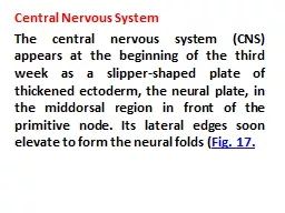
middorsal region in front of the primitive node Its lateral edges soon elevate to form the neural folds Fig 17 With further development the neural folds continue to elevate approach each other in the midline and finally fuse forming the neural tube ID: 779593

group fig spinal cord fig group cord spinal motor efferent visceral plates plate nerves nerve alar neural layer metencephalon

Share:

Link:

Embed:

Download Presentation from below link

Download The PPT/PDF document "Central Nervous System The central nervo..." is the property of its rightful owner. Permission is granted to download and print the materials on this web site for personal, non-commercial use only, and to display it on your personal computer provided you do not modify the materials and that you retain all copyright notices contained in the materials. By downloading content from our website, you accept the terms of this agreement.


Presentation Transcript

Slide1

Central Nervous System

The central nervous system (CNS) appears at the beginning of the third week as a slipper-shaped plate of thickened ectoderm, the neural plate, in the

middorsal

region in front of the primitive node. Its lateral edges soon elevate to form the neural folds (

Fig. 17.

Slide2

With further development, the neural folds continue to elevate, approach each other in the midline, and finally fuse, forming the neural tube (

Figs. 17.2

and

17.3

). Fusion begins in the cervical region and proceeds in cephalic and caudal directions (

Fig. 17.3A

). Once fusion is initiated, the open ends of the neural tube form the cranial and caudal

neuropores

that communicate with the overlying amniotic cavity

Slide3

Closure of the cranial neuropore proceeds cranially from the initial closure site in the cervical region (

Fig. 17.3A

) and from a site in the forebrain that forms later. This latter site proceeds cranially, to close the rostral-most region of the neural tube, and caudally to meet advancing closure from the cervical site (

Fig. 17.3B

). Final closure of the cranial neuropore occurs at the 18- to 20-somite stage (25th day); closure of the caudal neuropore occurs approximately 3 days later.

Slide4

Slide5

Slide6

Slide7

Slide8

Slide9

The cephalic end of the neural tube shows three dilations, the primary brain vesicles:

(a) the

prosencephalon

, or forebrain;

(b) the mesencephalon, or midbrain; and (c) the rhombencephalon, or hindbrain (Fig. 17.4). Simultaneously, it forms two flexures: (a) the

cervical flexure

at the junction of the hindbrain and the spinal cord and

(

b) the

cephalic flexure

in the midbrain region (

Fig. 17.4

).

Slide10

When the embryo is 5 weeks old, the

prosencephalon

consists of two parts:

(a) the

telencephalon, formed by a midportion and two lateral outpocketings, the primitive cerebral hemispheres; and (b) the

diencephalon,

characterized by outgrowth of the optic vesicles (

Fig. 17.5

). A deep furrow, the

rhombencephalic

isthmus, separates the mesencephalon from the

rhombencephalon

.

Slide11

The

rhombencephalon

also consists of two parts:

(a) the

metencephalon, which later forms the pons and cerebellum and (b) the myelencephalon. The boundary between these two portions is marked by the pontine flexure .It form

medulla oblongata .

(

Fig. 17.5

Slide12

The lumen of the spinal cord, the

central canal

, is continuous with that of the brain vesicles. The cavity of the

rhombencephalon

is the fourth ventricle, that of the diencephalon is the third ventricle, and those of the cerebral hemispheres are the

lateral ventricles

(

Fig. 17.5

). The lumen of the mesencephalon connects the third and fourth ventricles. This lumen becomes very narrow and is then known as the

aqueduct of

Sylvius

. The lateral ventricles communicate with the third ventricle through the

interventricular

foramina of

Monro

(

Fig. 17.5

).

Slide13

Slide14

Slide15

Slide16

SPINAL CORD

Neuroepithelial

, Mantle, and Marginal Layers

The wall of a recently closed neural tube consists of

neuroepithelial cells. These cells extend over the entire thickness of the wall and form a P.295

thick

pseudostratified

epithelium

(

Fig. 17.6

). Junctional complexes at the lumen connect them. During the neural groove stage and immediately after closure of the tube, they divide rapidly, producing more and more

neuroepithelial

cells. Collectively, they constitute the

neuroepithelial

layer or

neuroepithelium

.

Slide17

Once the neural tube closes,

neuroepithelial

cells begin to give rise to another cell type characterized by a large round nucleus with pale nucleoplasm and a dark-staining nucleolus. These are the

primitive nerve cells, or

neuroblasts (

Fig. 17.7

). They form the

mantle layer

, a zone around the

neuroepithelial

layer (

Fig. 17.8

). The mantle layer later forms the

gray matter of the spinal cord

.

The outermost layer of the spinal cord,

the marginal layer,

contains nerve fibers emerging from

neuroblasts

in the mantle layer. As a result of myelination of nerve fibers, this layer takes on a white appearance and therefore is called

the white matter of the spinal cord

(

Fig. 17.8

).

Slide18

Basal, Alar, Roof, and Floor Plates

As a result of continuous addition of

neuroblasts

to the mantle layer, each side of the neural tube shows a

ventral and a dorsal thickening. The ventral thickenings, the basal plates, which contain ventral motor horn cells, form the motor areas of the spinal cord; the dorsal thickenings,

the alar plates

, form the sensory areas (

Fig. 17.8A

). A longitudinal groove, the sulcus

limitans

, marks the boundary between the two. The dorsal and ventral midline portions of the neural tube, known as

the roof and floor plates

, respectively, do not contain

neuroblasts

; they serve primarily as pathways for nerve fibers crossing from one side to the other.

Slide19

Slide20

In addition to the ventral motor horn and the dorsal sensory horn, a group of neurons accumulates between the two areas and forms a small

intermediate horn

(

Fig. 17.8B

). This horn, containing neurons of the sympathetic portion of the autonomic nervous system (ANS), is present only at thoracic (T1-T12) and upper lumbar levels (L2 or L3) of the spinal cord.

Slide21

Positional Changes of the Cord

In the third month of development, the spinal cord extends the entire length of the embryo, and spinal nerves pass through the intervertebral foramina at their level of origin (

Fig. 17.13A

). With increasing age, however, the vertebral column and

dura lengthen more rapidly than the neural tube, and the terminal end of the spinal cord gradually shifts to a higher level. At birth, this end is at the level of the third lumbar vertebra (

Fig. 17.13C

). As a result of this disproportionate growth, spinal nerves run obliquely from their segment of origin in the spinal cord to the corresponding level of the vertebral column. The

dura

remains attached to the vertebral column at the coccygeal level

Slide22

In the adult, the spinal cord terminates at the level of

L2 to L3

, whereas the

dural

sac and subarachnoid space extend to S2. At the end of the cord, a thread-like extension of pia mater passes caudally, goes through the dura

, which provides a covering layer at S2 and extends to the first coccygeal vertebra. This structure is called the

filum

terminale

, and it marks the tract of regression of the spinal cord as well as providing support for the cord (the part covered by

dura

and extending from S2, to the coccyx is also called the

coccygeal ligament

). Nerve fibers below the terminal end of the cord collectively constitute the

cauda

equina

. When cerebrospinal fluid is tapped during a lumbar puncture, the needle is inserted at the lower lumbar level (L4-L5), avoiding the lower end of the cord.

Slide23

CRANIAL NERVES

By the fourth week of development, nuclei for all 12 cranial nerves are present. All except the olfactory (I) and optic (II) nerves arise from the brain

stem { pons , MO and midbrain},

and of these, only the

oculomotor (III) arises outside the region of the hindbrain. In the hindbrain, proliferation centers in the neuroepithelium establish eight distinct segments, the rhombomeres

. These

rhombomeres

give rise to motor nuclei of cranial nerves IV, V, VI, VII, IX, X, XI, and XII (

Figs. 17.17

and

17.40

).

Slide24

Three cranial nerves (I, II, and VIII

)

128

are entirely sensory; four (IV, VI, XI, and XII) {4 ,6,11,12} are entirely motor;

three

(VII, IX, and X

)

{ 7,9,10}

have motor, sensory, and parasympathetic fibers;

and

one (

III

) has only motor

and parasympathetic

components. In contrast, each spinal nerve has motor and sensory fibers

.

V

is motor and sensory

Slide25

BRAIN

Distinct basal and alar plates, representing motor and sensory areas, respectively, are found on each side of the midline in the

rhombencephalon

and mesencephalon. In the

prosencephalon, however, the alar plates are accentuated and the basal plates regress.Rhombencephalon

:

Hindbrain

The

rhombencephalon

consists of the

myelencephalon

, the most caudal of the brain vesicles, and the

metencephalon

, which extends from the

pontine

flexure to the

rhombencephalic

isthmus (

Figs. 17.5

and

17.17

).

Slide26

Myelencephalon

The

myelencephalon

is a brain vesicle that gives rise to the mo. It differs from the spinal cord in that its lateral walls are everted (

Fig. 17.18

). Alar and basal plates separated by the sulcus

limitans

can be clearly distinguished. The basal plate, similar to that of the spinal cord, contains motor nuclei. These nuclei are divided into three groups:

(a) a medial somatic efferent group,

(b) an intermediate special visceral efferent group, and

(c) a lateral general visceral efferent group

Slide27

Slide28

The first group contains motor neurons, which form the cephalic continuation of the anterior horn cells. Since this somatic efferent group continues

rostrally

into the mesencephalon, it is called the

somatic efferent motor column

. In the myelencephalon, it includes neurons of the {12}hypoglossal nerve that supply the tongue musculature. In the

metencephalon

and the mesencephalon

, the column contains neurons of the

abducens

(

Fig. 17.19

), trochlear, and

oculomotor

nerves{3,4,6}

(

Fig. 17.23

), respectively. These nerves supply the eye musculature

Slide29

Slide30

The

special visceral efferent

group extends into the

metencephalon

, forming the special visceral efferent motor column. Its motor neurons supply striated muscles of the pharyngeal arches. In the myelencephalon, the column is represented by neurons of the accessory,

vagus

, and

glossopharyngeal {9,10,11}

nerves.

The

general visceral efferent

group contains motor neurons that supply involuntary musculature of the respiratory tract, intestinal tract, and heart.

Slide31

Slide32

The

alar plate

contains three groups of sensory relay nuclei (

Fig. 17.18C

). The most lateral of these, the somatic afferent (sensory) group, receives impulses from the ear and surface of the head by way of the vestibulocochlear and trigeminal

nerves ( 5,8) .

The

intermediate, or special visceral afferent,

group receives impulses from taste buds of the tongue and from the palate, oropharynx, and

epiglottis

( 7 )

.

The medial, or general visceral

afferent, group receives

interoceptive

information from the gastrointestinal tract and heart.

Slide33

The

roof plate

of the

myelencephalon

consists of a single layer of ependymal cells covered by vascular mesenchyme, the pia mater (Fig. 17.18C). The two combined are known as the tela

choroidea

. Because of active proliferation of the vascular mesenchyme, a number of sac-like invaginations project into the underlying ventricular cavity (

Figs. 17.18C

). These tuft-like invaginations form the

choroid plexus

, which produces cerebrospinal fluid

Slide34

Metencephalon

The

metencephalon

, similar to the myelencephalon, is characterized by basal and alar plates (Fig. 17.19). Two new components form (

a) the cerebellum, a coordination center for posture and movement (

Fig. 17.20

), and

(b

) the pons, the pathway for nerve fibers between the spinal cord and the cerebral and cerebellar cortices

Slide35

Each basal plate of the

metencephalon

(

Fig. 17.19

) contains three groups of motor neurons: (a) the medial somatic efferent group, which gives rise to the nucleus of the abducens (6) nerve; (b) the

special visceral efferent

group, containing nuclei of the

trigeminal and facial

nerves (5,7)

,

which innervate the musculature of the first and second pharyngeal arches; and (c) the

general visceral efferent

group, with axons that supply the

submandibular and sublingual glands.

Slide36

The alar plates of the

metencephalon

contain three groups of sensory nuclei:

(

a) a lateral somatic afferent group, which contains neurons of the trigeminal (5) nerve and a small portion of the vestibulocochlear(8)

complex;

(

b) the special visceral afferent group;

and

(c) the general visceral afferent group (

Fig. 17.19

Slide37

Each basal plate of the

metencephalon

(

Fig. 17.19

) contains three groups of motor neurons: (a) the medial somatic efferent group, which gives rise to the nucleus of the abducens nerve (6)

;

(

b) the

special visceral efferent group

, containing nuclei of the

trigeminal and facial

nerves(5.7)

,

which innervate the musculature of the first and second pharyngeal arches;

and

(c) the

general visceral efferent group

, with axons that supply the

submandibular and sublingual glands.

Slide38

The marginal layer of the basal plates of the

metencephalon

expands as it makes a bridge for nerve fibers connecting the cerebral cortex and cerebellar cortex with the spinal cord. Hence, this portion of the

metencephalon

is known as the pons (bridge). In addition to nerve fibers, the pons contains the pontine nuclei, which originate in the alar plates of the metencephalon and

myelencephalon

mk

Slide39

The alar plates of the

metencephalon

contain three groups of sensory nuclei:

(

a) a lateral somatic afferent group, which contains neurons of the trigeminal nerve and a small portion of the vestibulocochlear (5, 8)

complex

;

(

b) the special visceral afferent group; and

(

c) the general visceral afferent group (

Fig. 17.19

).

Slide40

Mesencephalon: Midbrain

In the mesencephalon (

Fig. 17.23

), each basal plate contains two groups of motor nuclei:

(a) a medial somatic efferent group, represented by the oculomotor and trochlear nerves, which innervate the eye musculature and

(

b) a

small general visceral efferent group

, represented by the nucleus of

Edinger-Westphal

,

which innervates the sphincter pupillary muscle (

Fig. 17.23B

Slide41

Diencephalon

Roof

Plate

The diencephalon, which develops from the median portion of the

prosencephalon (Figs. 17.5 and 17.17), is thought to consist of a

roof plate and two alar plates but to lack floor and basal plates

(interestingly, sonic hedgehog, a ventral midline marker, is expressed in the floor of the diencephalon, suggesting that a floor plate does exist). The roof plate of the diencephalon consists of a single layer of ependymal cells covered by vascular mesenchyme. Together, these layers give rise to the choroid plexus of the third ventricle

Slide42

Alar Plate, Thalamus, and Hypothalamus

The alar plates form the lateral walls of the diencephalon. A groove, the hypothalamic sulcus, divides the plate into a dorsal and a ventral region, the thalamus and hypothalamus, respectively (

Figs. 17.24

and

17.25).

Slide43