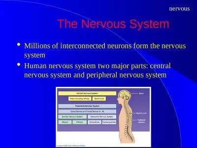
The Somatic System Grants Atlas 12 2009 Lawrence M Witmer PhD Professor of Anatomy Dept of Biomedical Sciences Heritage College of Osteopathic Medicine Ohio University Athens Ohio 45701 ID: 203707
Download Presentation The PPT/PDF document "Peripheral Nervous System 1:" is the property of its rightful owner. Permission is granted to download and print the materials on this web site for personal, non-commercial use only, and to display it on your personal computer provided you do not modify the materials and that you retain all copyright notices contained in the materials. By downloading content from our website, you accept the terms of this agreement.
Slide1
Peripheral Nervous System 1:The Somatic System
Grant’s Atlas
12 2009
Lawrence M. Witmer, PhDProfessor of AnatomyDept. of Biomedical SciencesHeritage College of Osteopathic Medicine, Ohio UniversityAthens, Ohio 45701witmerL@ohio.edu
Taken from and modified……Slide2
Dichotomies
1. Tissues: neurons vs. glia
2. Position: CNS vs. PNS3. Function 1: sensory vs. motor4. Function 2: somatic vs. visceral
Gray’s Anatomy 38 1999
neuron
glial cellSlide3
Neurons-a quick review
cell
body
dendrites
axon with
myelin sheath
synapses
Schwann
cell
Moore’s
COA6
2010
• Dendrites: carry nerve impulses toward cell body
• Axon: carries impulses away from cell body
• Synapses: site of communication between neurons using chemical neurotransmitters
• Myelin & myelin sheath: lipoprotein covering produced by glial cells (e.g., Schwann cells in PNS) that increases axonal conduction velocity
• Demyelinating diseases: e.g., Multiple Sclerosis (MS) in CNS or Guillain- Barré Syndrome in PNSSlide4
CNS vs. PNS
Moore’s
COA6 2010
Central Nervous System
• brain & spinal cord• integration of info passing to & from the periphery
Peripheral Nervous System
• 12 cranial nerves
• 31 pairs of spinal nerves
• Naming convention changes at C7/T1
Collection of nerve
cell bodies:
• CNS: nucleus
• PNS: ganglion Slide5
Sensory (Afferent) vs. Motor (Efferent)
CNS
CNS
e.g., skin
e.g., muscle
Gray’s Anatomy 38
1999
sensory (afferent) nerve
motor (efferent) nerve
(pseudo-) unipolar neurons conducting impulses
from sensory organs to the CNS
multipolar neurons conducting impulses
from the CNS to effector organs (muscles & glands)Slide6
Somatic vs. Visceral
attribute
Somatic SystemVisceral System
embryological origin of tissue
“body wall:” somatic (parietal) mesoderm (dermatome, myotome)
“organs:” splanchnic (visceral) mesoderm, endoderm
examples of adult tissues
dermis of skin, skeletal muscles, connective tissues
glands, cardiac muscle, smooth muscle
perception
conscious, voluntary
unconscious, involuntary
Langman’s Embryo 9
2004Slide7
Sensory/Motor + Somatic/Visceral
Somatic
Visceral
Sensory(Afferent)
somatic sensory
[General Somatic Afferent (GSA)]
visceral sensory
[General Visceral Afferent (GVA)]
Motor
(Efferent)
somatic motor
[General Somatic Efferent (GSE)]
visceral motor
[General Visceral Efferent (GVE)]
Somatic
Nervous
System
Autonomic
Nervous
SystemSlide8
Structure of the Spinal Cord
white matter
(axons)
gray matter (cell bodies)
• dorsal (posterior) horn
• ventral (anterior) horn
meninges
pia •
arachnoid •
dura •
denticulate
ligament
dorsal
rootlets
ventral
rootlets
• dura
• arachnoid
• pia
meninges
dorsal root
(spinal) ganglion
spinal nerve
• dorsal primary ramus
• ventral primary ramus
ventral root
Moore’s
COA6
2010
subarachnoid
space
(CSF)Slide9
Structure of Spinal Nerves: Somatic Pathways
dorsal root
dorsal root
ganglion
ventral root
spinal
nerve
dorsal
ramus
ventral
ramus
gray ramus
communicans
white ramus
communicans
sympathetic
ganglion
dorsal
horn
ventral
horn
somatic
sensory
nerve
(GSA)
somatic
motor
nerve
(GSE)
CNS
inter-
neuron
Mixed Spinal
NerveSlide10
Structure of Spinal Nerves: Somatic Pathways
dorsal root
dorsal root
ganglion
ventral root
spinal
nerve
dorsal
ramus
gray ramus
communicans
white ramus
communicans
sympathetic
ganglion
dorsal
horn
ventral
horn
somatic
sensory
nerve
(GSA)
somatic
motor
nerve
(GSE)
CNS
inter-
neuron
Mixed Spinal
Nerve
ventral
ramus
Somatic sensations
• touch, pain, temperature,
pressure
• proprioception: joints, muscles
Somatic motor activity
: innervate skeletal musclesSlide11
Somatic Nervous systemClearly we can see that this aspect of the peripheral system gathers information from the senses or receptors which are simply organs that detect a change in the internal (bladder full) or external(change in temp/light intensity) and respond in some way.The trigger to a “response” is simply called a stimulus.
The skin responds to many of these stimuliSlide12
Skin/ears/eyesSkin protects us from the outside world as the receptors are constantly alerting us as to the changes we need to respond to, to protect us.The ear groups receptors- sound, and balanceThe eyes clearly enable us to see-msgs
are sent via the optic nerveSee pages 228/229Slide13
DiagramsSlide14
Autonomic Nervous SystemSome activities without you knowing about them-breathing heartbeat sweating etc.-These are controlled by the autonomic nervous system.There are two parts to this system:The Sympathetic-speeds up body functions to increase efficiency
Parasympathetic-slows things down-works when you are at rest-returns the body to normal functioningThese systems are complimentary or opposite to one another in functionSlide15
Function of the sympathetic Nervous System
OrganEffectEyeDilates pupilHeart
Increases rate and force of contractionLungsDilates bronchiolesBlood VesselsConstrictsSweat GlandsActivates sweat secretionDigestive tractInhibits –movement peristalsisKidneyIncreases renin
secretionPg 230 text bookSlide16
Functions of the Parasympathetic Nervous SystemIncreases blood flow to the digestive tractStimulates salivary glands and increases the rate of digestionReduces the diameter of the bronchioles when there is a reduced need for oxygenControls heart beat
Contracts the eye muscles and reduces the diameter of the pupilSlide17
overviewSlide18
Responding to StimuliPage 231- reflex actionsRead and noteDraw basic diagram using text as a guide(teacher to draw on white board)Slide19
References
Agur
, A. M. R. and A. F. Dalley. 2009. Grant’s Atlas of Anatomy, 121th Edition. Lippincott, Williams & Wilkins, New York.Bannister, L. H. et al. 1999. Gray’s Anatomy, 38th Edition. Churchill Livingstone, New York.Moore, K. L. , A. F. Dalley, and A. M. R.
Agur. 2010. Clinically Oriented Anatomy, 6th Edition. Lippincott, Williams & Wilkins, New York.Sadler, T. W. 2004. Langman’s Medical Embryology, 9th Edition. Lippincott, Williams & Wilkins, New York.Stern, J. T., Jr. 1988. Essentials of Gross Anatomy. Davis, Philadelphia.