/
AMERICAN JOURNAL OF FOOD AND NUTRITION AMERICAN JOURNAL OF FOOD AND NUTRITION

AMERICAN JOURNAL OF FOOD AND NUTRITION - PDF document

karlyn-bohler
karlyn-bohler . @karlyn-bohler
Follow
376 views
Uploaded On 2015-10-11

AMERICAN JOURNAL OF FOOD AND NUTRITION - PPT Presentation

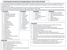
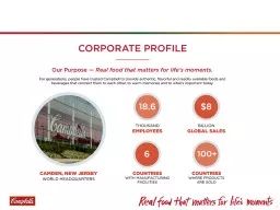
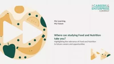
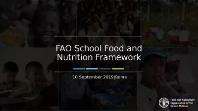
Print ISSN 2157 0167 Online ISSN 2157 1317 doi105251ajfn20133 4 1 82 1 87 ID: 157657

Print: ISSN 2157 - 0167 Online: ISSN

Share:

Link:

Embed:

Download Presentation from below link

Download Pdf The PPT/PDF document "AMERICAN JOURNAL OF FOOD AND NUTRITION" is the property of its rightful owner. Permission is granted to download and print the materials on this web site for personal, non-commercial use only, and to display it on your personal computer provided you do not modify the materials and that you retain all copyright notices contained in the materials. By downloading content from our website, you accept the terms of this agreement.


Presentation Transcript

AMERICAN JOURNAL OF FOOD AND NUTRITION Print: ISSN 2157 - 0167, Online: ISSN 2157 - 1317, doi:10.5251/ajfn.2013.3. 4 .1 82 .1 87 © 2013, ScienceHuβ, http://www.scihub.org/AJFN Isolation and PCR Detection of Cronobacter sakazakii from Powdered Infa nt Formulae retailed in Nigeria Ezeh Abimbola Rashidat a* , Aboaba Olusimbo Olugbo a , Smith Stella Ifeanyi b , Fesobi Wuraola Adetoun b , Omonigbehin Emmanuel Adedayo b , and Bamidele Moses b a. Department of Microbiology, Faculty of Science, University of Lagos, Nigeria b. Nigerian Institute of Medical Research, Edmund Crescent Road, Yaba, Lagos, Nigeria. ABSTRACT The presence of Cronobacter sakazakii in 154 samples of powdered infant formulae (PIF) retailed in Nigeria was analyzed using real time polymerase chain reaction (PCR) and a chromogenic medium, Druggan - Forsythe - Iversen (DFI) agar. Two pairs of species specific primers (SG - F/SG - R and SI - F/SI - R ) targeting the sequences between 16S rDNA and 23S rDNA (internal transcribed spacer, ITS) were used for the amplification. Out of the 154 samples analyzed, C. sakazakii was detected in 3 (1.95%). A particular isolate detected by SI primer was not detected by SG primer. This study shows that Cronobacter sakazakii can be detected using both the chromogenic DFI agar (cultural method) and molecular method. This report is the first for the isolation and molecular detection of Cronobacter sakazakii from powdered infant formulae retailed in Nigeria. Key words: Cronobacter sakazakii, DFI, powdered infant formula, PCR, INTRODUCTION Cronobacter sakazakii (formerly Enterobacter sakazakii ), is a member of the Enterobacteriace ae family. They are motile, non - spore forming, Gram - negative facultative anaerobes. They are catalase positive, oxidase negative and generally positive for α - glucosidase (Jaradat et al ., 2009). Cronobacter sakazakii is an emerging opportunistic, food - borne pathogen that causes severe meningitis, meningoencephalitis, sepsis, and necrotizing enterocolitis in neonates and infants (Caubilla - Barron et al 2007) as well as infections in elderly immuno - compromised individuals (Lai, 2001; Kucerova et al , 2010). In several C. sakazakii related outbreaks and sporadic cases, powdered infant formula was epidemiologically or microbiologically established as the source of the infection (Biering et al ., 1989, Iversen and Forsythe, 200 3). While it appears that the frequency of C. sakazakii infection is low, reported case - mortality meningitis rates vary from 40 to 80% among infected infants, with the majority of those who survive Cronobacter - associated meningitis (94%) developing an irre versible neurological sequela such as hydrocephalus, quadriplegia and retarded neural development (Willis and Robison, 1988; Iversen and Forsythe 2004) within days of birth and in many cases neonates die (Gurtler et al 2005). The International Commission o n Microbiological Specifications for Foods has ranked C. sakazakii as a severe hazard for restricted populations, life threatening or substantial chronic sequelae or long duration (ICMSF, 2002). The bacterium has been isolated from a variety of foods such as spices, cereals, cheese, vegetables, herbs grain and bread. Numerous investigators have isolated C. sakazakii from infant formula (Postupa and Aldova, 1984, Muytjens et al ., 1988, Zink, 2003, Iversen and Forsythe 2004, Jung and Park, 2006, Drudy et al ., 2006). Cronobacter spp. have been isolated at low levels from powdered infant formulae, and the organisms’ high tolerance to desiccation provides a competitive advantage in the dry environments of milk powder factories, increasing the risk of product cont amination. Two main routes by which C. sakazakii can enter infant formula are intrinsic contamination either through contaminated ingredients and/or through external contamination during reconstitution and handling (Mullane et al ., 2006). The bacterium has also been isolated from the household, food production units (Kandhai et al ., 2004) and from insects. The aim of this study was to isolate and detect the presence of C. sakazakii in powdered infant formula retailed in Nigeria using cultural method and po lymerase chain reaction. MATERIALS AND METHODS Sample Collection : A total of 154 samples of commercially produced infant formula were A m . J . Food . N utr , 201 3 , 3 ( 4 ): 182 - 187 183 purchased from local markets and supermarkets in different parts of Nigeria. Isolation of Cronobacter spp (New FDA Method) : Briefly, 25g of powdered infant formula was added to 225ml of buffered peptone water (BPW; pH 7.2 + 0.2). The suspended powdered infant formula was then incubated at 37 0 C for 24h without shaking. Four aliquots of 40ml each were removed from the solution and placed into 50ml centrifuge tubes. The tubes were subjected to centrifugation at 3000 x g for 10 min. The supernatants of each tube were discarded and the resultant pellets were suspended in 200µL of phosphate - buffered saline (PBS; pH 7.2 + 0.2). Templa te DNA for PCR was prepared from 200µL of suspended cells. Isolated DNA was assessed by PCR assay developed by Liu et al ., 2005. Two 100µL samples were plated onto two DFI chromogenic agar plates. The agar plates were then incubated at 37 0 C for 18 to 24 h (Lampel and Chen, 2009). Presumptive C. sakazakii colonies showing blue green color were picked and were confirmed by PCR. DNA Extraction : Genomic DNA was extracted from overnight cultures and re - suspended pellets by the boiling method. Overnight culture s and resuspended pellet (200µl) was added to 1ml of sterile water. The mixture was vortexed and then centrifuged at 10,000 rpm for 5minutes. The supernatant was discarded and the pellet was blotted on paper towel. One ml of sterile distilled water was add ed to the pellet, vortexed and centrifuged at 10,000rpm for 5 minutes. The supernatant was discarded and the pellet blotted and 200µl of sterile distilled water was added, vortexed to homogenize and then boiled in a water bath for 5 minutes at 100 0 C. The m ixture was vortexed and centrifuged at 10,000 rpm for 5 minutes after which the supernatant was transferred into another eppendorf tube by gentle aspiration and used for PCR. PCR detection of C. sakazakii with species specific primers : Species specific amplification for detection of C. sakazakii was optimized in a 50 - μl reaction mixture consisting of 5 μl of the bacterial genome DNA solution, 10 mM Tris - HCl (pH 8.3); 50mM KCl; 3 mM MgCl 2; 200 μM (each) dATP, dCTP, dTTP and dGTP; 2 U of T aq DNA polymerase (Solis BioDyne) and 0.25 μM (each) primers SG - F and SG - R (or SI - F and SI - R) (biomers.net). Thermal cycling parameters were as follows: 94 0 C for 10 min, followed by 30 cycles of 94 0 C for 30 s, 57 0 C for 1 min, 72 0 C for 1 min; and a termina l extension step of 72 0 C for 5 min. The amplification reactions were performed using Eppendorf vapo protect. The products of PCR (10 - μl aliquot) were analyzed by 1% (w/v) agarose gel (Merck, Germany) electrophoresis in 1 X TAE buffer (40 mM Tris - HCl, 1.18 ml acetic acid, 2 mM EDTA, pH 8.0) and a constant voltage of 5 V/cm to confirm the presence of the amplified DNA. The gel was stained for 30 min in 1x TAE buffer containing 0.5 μg/ml ethidium bromides (Amersham bioscience, Sweden) and visualized under UV G el Doc XR (Bio Rad). RESULTS A total of 154 powdered infant formula samples were examined for the presence of Cronobacter sakazakii in this study. Out of the samples examined on the chromogenic DFI agar, only 3 (1.95%) of the samples were observed showing the presumptive blue green colonies of C. sakazakii . The PCR detection of Cronobacter sakazakii using the specific primers SG - F and SG - R and SI - F and SI - R to target the Internal Transcribed Sequence (ITS) of C. sakazakii is shown in Figures 1 and 2 below. A particular isolate (ULD A) was detected by the SI primer but was not detected by the SG primer. Table I: S pecific primers used for the detection of Cronobacter sakazakii (Liu et al ., 2006) Primers and probes Sequence Targeted site SG - F SG - R SI - F SI - R GGGTTGTCTGCGAAAGCGAA GTCTTCGTGCTGCGAGTTTG CAGGAGTTGAAGAGGTTTAACT GTGCTGCGAGTTTGAGAGACTC ITS - G ITS - G &ITS - IA ITS - IA ITS - G & ITS - IA A m . J . Food . N utr , 201 3 , 3 ( 4 ): 182 - 187 184 Fig. 1: Detection of C. sakazakii by PCR with specific primers SI - F and SI - R. The PCR products were analyzed by electrophoresis in 1% agarose gel. A 251 bp DNA fragment was amplified from ITS of C. sakazakii. Key: Lane M - marker, - ve – negative control, Lane 3 - ULD A, Lane 5 - SPC 1, Lane 7 - ULD C, Lane 8 - ULD D. (ULD - University of Lagos Dairy Strain). Fig. 2: Detection of C. sakazakii by PCR with the specific primers SG - F and SG - R. The PCR products were analyzed by electrophore sis in 1% agarose gel. A 282 bp DNA fragment was amplified from ITS of C. sakazakii. KEY: Lane M - marker, Lane 5 - SPC 1, Lane 7 - ULD C, Lane 8 - ULD D. (ULD - University of Lagos Dairy Strain) . A m . J . Food . N utr , 201 3 , 3 ( 4 ): 182 - 187 185 Table 2 : Comparison of the various detection methods Sample Direct Detection Cultural detection on chromogenic agar Primer 1(SG) Primer 2(SI) ULD A - + + ULD B - - + ULD C + + + ULD D + + + SPC 11 + + + SPC 17 - - + DISCUSSION Powdered infant formula (PIF) is not a sterile product and is an excellent medium to support bacterial growth because of its high nutritional content. Bovine milk is an essential ingredient of PIF and a potential source of pathogenic bacteria to humans (Drudy et al ; 2006) . Agostoni et al (2004) reported the isolation of bacteria pathogens such as Citrobacter, Enterobacter, Staphylococcus, Klebsiella, Streptococcus and Yersinia species from PIF. In this study, Cronobacter sakazakii was isolated and detected in 1.95% of the samples analyzed. A definitive link has been reported between the presence of Enterobacter sakazakii in an unopened can of PIF and an outbreak of infection (CDC, 2002). Should C. sakazakii multiply in PIF, it can result in infection especially in neonates and immuno compromised infants. PIF is manufactured using a wet - mix process, a dry mix process, or a combination of these processes (Kim et al ., 2008). The raw materials used may be important source of C. sakaza kii as a contaminant in the finished products. Pasteurization is a process of sterilization meant to eliminate contaminating microorganisms but the addition of dry ingredients such as vitamins and minerals to powdered infant formula further exposes the pro duct to contamination by microorganisms such as C. sakazakii. C. sakazakii can produce trehalose, which is a compatible solute that protects the bacteria from dry conditions by stabilizing phospholipid membranes and proteins (Welsh and Herbert, 1999). The accumulation of trehalose in the cell may play an important role in the high heat tolerance of C. sakazakii (Breeuwer et al ., 2003). Additionally, infant formula has high total solids, fat and sugar contents, which may further protect the microorganism fro m heat (Nazarowec - White and Farber, 1997) . Several media have been used in the isolation of C. sakazakii including OK (Oh and Kang, 2004), TSBA (Guillaume - Gentil et al ., 2005), ESPM (Restaino et al ., 2006) and DFI (Iversen et al ., 2004). The Druggan - Forsyt he - Iversen (DFI) agar which was used in this study contains a chromogenic substrate 5 - bromo - 4 - chloro - 3 - indolyl - α - D - glucopryranoside (X - α - Glc) which is used as an indicator of α - glucosidase activity (Iversen et al ., 2004). The chromogenic substrate is cleav ed by α - glucosidase creating blue - green C. sakazakii colonies. The detection of C. sakazakii by PCR using 2 different primers the sequences between 16S rDNA and 23S rDNA confirmed its presence in the samples used in this study. PCR has been used to detect the presence of C. sakazakii in infant formula in a number of studies. Liu et al (2006) also used primers targeting the sequences between 16S rDNA and 23S rDNA. Seo and Brackett (2005) described a quantitative real - time PCR technique in which primers and a TaqMan probe were developed to target the macromolecular synthesis operon of C. sakazakii. Liu et al (2005) developed 2 real - time PCR assays based on TaqMan and SYBR green technology. Both of these assays used primers that target the 16 - 23S rRNA spacer re gion. The inability of the SG primer to detect the presence of isolate ULD A when the SI primer detected it could be due to the presence of inhibitors (Gieffers et al ., 2000). The findings showed that some of the PIF retailed in Nigeria were contaminated with Cronobacter sakazakii which can pose a health risk to infants. C. sakazakii can proliferate once a feed is reconstituted with water under warm environmental conditions as obtained in Nigeria. Most times the infant formula are reconstituted and kept wa rm for the next meal. Left overs of feeds are usually not discarded but are kept (usually not refrigerated) for the next meal. Under these conditions, C. sakazakii can proliferate sufficiently to cause an infection. CONCLUSION The presence of this organism in infant formula and its potential effects in infants could be a significant public health problem in developing countries such as Nigeria where there is dearth of information about C. sakazakii . A m . J . Food . N utr , 201 3 , 3 ( 4 ): 182 - 187 186 REFERENCES Agostoni, C., Axelsson, I., Goulet, O. Koletzko, B., Michaelsen, K.F., Puntis, J.W. ( 2004 ) . Preparation and handling of powdered infant formula: acommentary by ESPGHAN committee on nutrition. J. Pediatr. Gastroenterol. Nutr. 39(4): 320 - 322. Biering, G., Karlsson , S., Clark, N. C., Jônsdôttir, K. E., Ludvigsson, P., Steingrimsson, O. ( 1989 ) . Three Cases of Neonatal Meningitis Caused by Enterobacter sakazakii in Powdered Milk . J. Clin. Microbiol. 27(9 ): 2054 - 2056. Breeuwer, P., Lardeau, A., Peterz, M., Joosten, H. M., ( 2003 ) . Desiccation and heat tolerance of Enterobacter sakazakii . J . Appl . Microbiol . 95 : 967 – 973 Caubilla_Barron, J., Hurrell, E., Townsend, S., Cheetham, P., Loc - Carrillo, C., Fayet, O. Prere M., Forsythe, S. J. ( 2007 ) . Genotypic and phenotypic analysis of Enterobacter sakazakii strains from an outbreak resulting in a neonatal intensive care unit in France. J. Clin. Microbiol. 45 : 3979 - 3985. Centers for Disease Control and Prevention. ( 2002 ) . Enterobacter sakazakii infections associated with the use of powdered infant formula - Tennessee, ( 2001 ) . MMWR Morb. Mortal. Wkly Rep . 51: 297 - 300. Drudy, D., Mullane, N.R., Quinn, T., Wall, P.G., Fanning, S. ( 2006 ) . Enterobac ter sakazakii : an emerging pathogen in powdered infant formula. Clin . Infect . Dis . 42: 996 - 1002. Gieffers, J ., Reusche, E ., Solbach, W ., Maass, M . ( 2000 ) . Failure to detect Chlamydia pneumoniae in brain sections of Alzheimer's disease patients. J. Clin. Microbiol. 38(2) : 881 - 882. Guil laume - Gentil, O., Sonnard, V., Kandhai, M.C., Marugg, J.D., Joosten, H. ( 2005 ) . A simple and rapid cultural method for detection of Enterobacter sakazakii i n environmental samples. J. Food Protect. 68(1) : 64 - 69. Gurtler J.B. Kornacki J.L., Beuchat L.R. ( 2005 ) . Enterobacter sakazakii : A coliform of increased concern to infant health. Int . J . Food Microbiol . 104 : 1 - 34. ICMSF (International Commission for Microbiological Specifications for Foods). ( 2002 ) . Microorganisms in food 7: Microbiological testing in food safety management . Kluver Academic/Plenum Publishers. Iversen, C., Forsythe, S. J. ( 2003 ) . Risk profile of Enterobacter sakazakii , an emergent pathogen associated with infant milk formula. Trends Food Sci . Tech . 14 : 443 – 454. Iversen , C., Forsythe S. J. ( 2004 ) . Isolation of Enterobacter sakazakii and other Enterobacteriaceae from powdered infant formula milk and related products. Food Microbio l . 21 : 771 – 776. Iversen, C., Druggan, P., Forsythe, S., ( 2004 ) . A selective differential medium for Enterobacter sakazakii , a preliminary study. Int . J . Food Microbio l. 96: 133 – 139. Jaradat, Z.W., Ababneh, Q.O., Saadoun, I.M., Samara, N.A. and Rashdan, M.A. ( 2009 ) . Isolation of Cronobacter spp. (formerly Enterobacter s akazakii ) from infant food, herbs and environmental samples and the subsequent identification and confirmation of the isolates using biochemical, chromogenic assays, PCR and 16S rRNA sequencing. BMC Microbiol . 9: 225. Jung, M - K., Park, J - H. ( 2006 ) . Prevalence and thermal stability of Enterobacter sakazakii from unprocessed ready - to - eat agricultural products and powdered infant formulas. Food Sci . Biotech nol . 15 : 152 - 157. Kandhai, M. C., Reij, M. W., Gorris, L. G. M., Guillaume - Gentil, O., and Van S chothorst, M. ( 2004 ) . Occurrence of Enterobacter sakazakii in food production environments and households. Lancet 39 – 40. Kim, K., Jang, S.S., Kim, S.K., Park, J.H., Heu, S., Ryu, S., ( 2008 ) . Prevalence and genetic diversity of Enterobacter sakazakii in ingredients and of infant foods. Int . J . Food Microbiol. 122: 196 – 203. Kucerova, E., Clifton, S.W., Xia, X., Long, F., Porwollik, S., Fulton, L., Fronick, C., Minx, P., Kyung, K., Warren, W., Fulton, R., Feng, D., Wollam, A., Shah, N., Bhonag iri, V., Nash W. E., Hallsworth - Pepin K., Wilson R.K., McClelland, M., Forsythe, S.J. ( 2010 ) . Genome Sequence of Cronobacter sakazakii BAA - 894 and Comparative Genomic Hybridization Analysis with Other Cronobacter Species. PLoS ONE 5(3): 9556 Lai, K. K. ( 2 001 ) . . Enterobacter sakazakii infections among neonates, infants, children and adults. Case reports and a review of the literature. Medicine 80 : 113 - 22. Lampel, K.A. and Chen,Y. ( 2009 ) . Method for the isolation and detection of Enterobacter sakazakii ( Cronobacter ) from powdered infant formula. Int . J . Food Microbiol. 136: 179 - 184. Liu, Y., Gao, Q. Zhang, X., Hou, Y., Yang, J. and Huang, X. ( 2006 ) . PCR and oligonucleotide array detection of Enterobacter sakazakii in infant formula. Mol . Cell . Probes 20: 11 - 17. Liu, Y ., Cai, X ., Zhang, X ., Gao, Q ., Yang, X ., Zheng, Z ., Luo, M ., Huang, X . ( 2006 ) . Real time PCR using TaqMan and SYBR Green for detection of Enterobacter sakazakii in infant formula. J . Microbiol . Methods. 65(1): 21 - 31. Mullane, N.R. Drudy, D. Whyte, P. O’Mahong M ., Scannell, A.G.M., Wall, P.G., Fanning, S. ( 2006 ) . Enterobacter sakazakii : biological properties and significance in A m . J . Food . N utr , 201 3 , 3 ( 4 ): 182 - 187 187 dried infant milk formula (IMF) powder. Int . J . Dairy Technol . 59 : 102 - 111. Muytjens, H. L., Roelfos, W. H., Jaspar, G. H. J. ( 1988 ) . Qua lity of powdered substitutes for breast milk with regard to members of family Enterobacteriaceae . J. Clin. Microbiol. 26 : 743 – 746. Nazarowec - White, M., Farber J. M. ( 1997 ) . Incidence, survival, and growth of Enterobacter sakazakii in infant formula. J . Food Protect. 60 : 226 – 230. Oh, S. W., Kang, D. H. ( 2004 ) . Fluorogenic selective and differential medium for isolation of Enterobacter sakazakii . Appl . Environ . Microbiol . 70 : 5692 - 5694. Postupa, R., & Aldova, E. ( 1984 ) . Enterobacter sakazakii : a Tween 80 esterase - positive representative of the genus Enterobacter isolated from powdered milk specimens. J. Hyg. Epid. Microb. Im. 28 : 435 – 440. Restaino, L., Frampton, E.W., Lionberg, W.C., Becker, R.J. ( 2006 ) . A chromogenic plating medium for the is olation and identification of Enterobacter sakazakii from foods, food ingredients and environmental sources. J . Food Protect . 69 : 315 - 322. Seo,K.H.,Brackett, R.E. ( 2005 ) . Rapid, specific detection of Enterobacter sakazakii in infant formula using a real time PCR assay. J. Food Protect. 68 : 59 - 63. Welsh, D.T., Herbert, R. A. ( 1999 ) . Osmotically induced intracellular trehalose but not glycine betaine accumulation promotes desiccation tolerance in Escherichia coli. FEMS Microbiol . Lett . 174: 57 - 63. Willis, J., Robinson, J.E. ( 1988 ) . Enterobacter sakazakii meningitis in neonates. Pediatr . Infect . Dis . J . 7: 196 - 199. Zink, D. ( 2003 ) . FDA Field Survey of powdered infant formula manufacturing. U.S. Food and Drug Administration Food Advisory Committee Mtg. Marc h 18 - 19, Washington, DC.