/
Psychiatric Aspects of Spatz Syndrome A Report Psychiatric aspects of Psychiatric Aspects of Spatz Syndrome A Report Psychiatric aspects of

Psychiatric Aspects of Spatz Syndrome A Report Psychiatric aspects of - PDF document

lam
lam . @lam
Follow
342 views
Uploaded On 2022-10-13

Psychiatric Aspects of Spatz Syndrome A Report Psychiatric aspects of - PPT Presentation

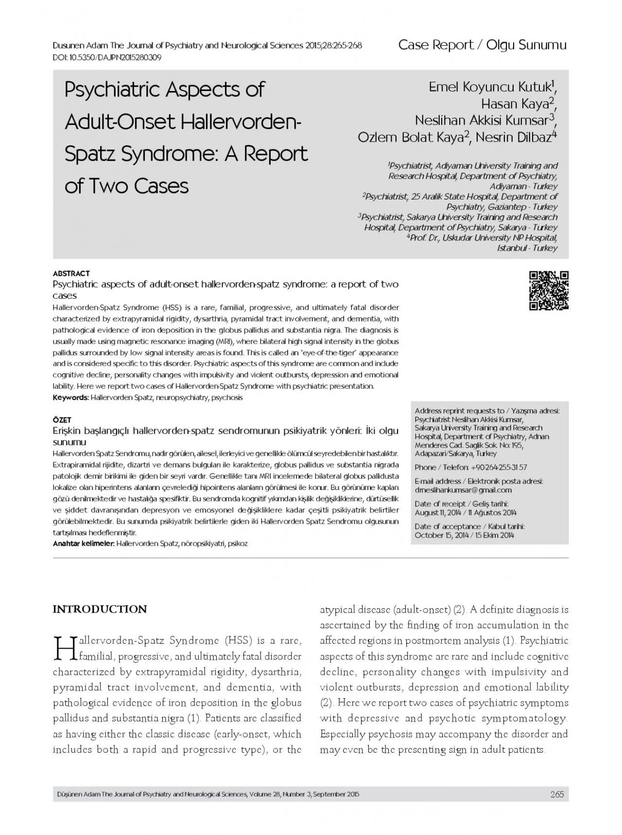
265 allervordenSpatz Syndrome HSS is a rare familial progressive and ultimately fatal disorder characterized by extrapyramidal rigidity dysarthria pyramidal tract involvement and dementia wi ID: 959369

spatz symptoms onset case symptoms spatz case onset disease hallervorden psychiatric syndrome hss examination mri rigidity clinical observed extrapyramidal

Share:

Link:

Embed:

Download Presentation from below link

Download Pdf The PPT/PDF document "Psychiatric Aspects of Spatz Syndrome A ..." is the property of its rightful owner. Permission is granted to download and print the materials on this web site for personal, non-commercial use only, and to display it on your personal computer provided you do not modify the materials and that you retain all copyright notices contained in the materials. By downloading content from our website, you accept the terms of this agreement.


Presentation Transcript

265 Psychiatric Aspects of Spatz Syndrome: A Report Psychiatric aspects of adult-onset hallervorden-spatz syndrome: a report of two Hallervorden-Spatz Syndrome (HSS) is a rare, familial, progressive, and ultimately fatal disorder characterized by extrapyramidal rigidity, dysarthria, pyramidal tract involvement, and dementia, with pathological evidence of iron deposition in the globus pallidus and substantia nigra. The diagnosis is allervorden-Spatz Syndrome (HSS) is a rare, familial, progressive, and ultimately fatal disorder characterized by extrapyramidal rigidity, dysarthria, pyramidal tract involvement, and dementia, with pathological evidence of iron deposition in the globus pallidus and substantia nigra (1). Patients are classified as having either the classic disease (early-onset, which includes both a rapid and progressive type), or the atypical disease (adult-onset) (2). A definite diagnosis is ascertained by the finding of iron accumulation in the affected regions in postmortem analysis (1). Psychiatric aspects of this syndrome are rare and include cognitive decline, personality changes with impulsivity and violent outbursts, depression and emotional lability (2). Here we report two cases of psychiatric symptoms with depressive and psychotic symptomatology. Especially psychosis may accompany the disorder and Düşünen Adam The Journal of Psychiatry and Neurological Sciences, Volume 28, Number 3, September 2015 266 A 32-year-old married housewife was brought to the emergency unit with generalized dystonia, paranoid delusional ideation, depressive mood, irritability, hypomnesia, anxiety, significant speech disturbances, and dysphagia. The neurological examination revealed limited cooperation, bilateral extrapyramidal signs (prominent rigidity and Parkinsonian gait), and pyramidal spasticity with increased tendon reflexes. The onset of her illness had been a year before, with complaints including non-steroid anti-inflammatory drug-resistant headache, vertigo, difficulty in onset psychiatric signs, such as paranoid delusions, mood disturbances, psychomotor agitation, and auditory-visual complex hallucinations, she had been admitted to a psychiatry outpatient unit and diagnosed with schizophreniform disorder three months before our examination. She was treated with zuclopenthixol decanoate (200mg every two weeks), and her psychotic symptoms partially responded to the treatment. Fifteen days after our initial examination, motor signs consisting of extrapyramidal rigidity, mutism and hypersomnia were observed. During physical examination, common rigidity and restriction in her movements were prominent. In the neurological examination, she was conscious, her cooperation and orientation was limited. Motor activity examination was not possible because of common rigidity of all extremities and marked spasticity of the upper extr

emities. Leakage of saliva was present, and she did not communicate verbally. No pathological sign was observed in clinical EEG results and cerebrospinal fluid examination. A brain MRI showed low signal intensity in the globus pallidus and substantia nigra bilaterally, consistent with iron deposition (Figure 1). On the eighth day of hospitalization, she became clinically more unstable, respiratory distress developed, and low arterial O2 pressure was detected. She was transferred to the intensive care unit. In response to the development of visual hallucinations and agitation in the patient, 100mg/day of oral quetiapine was added to the treatment of baclofen and L-dopa. The patient was discharged since she did not show any improvement in the psychotic symptoms, except for the decrease in psychomotor agitation and partial improvement in spasticity and rigidity. Due to gradually increased difficulty in swallowing, her oral intake became limited and in the second month of follow-up, she was brought to the emergency unit. After being treated in intensive care for 2 days, she became unstable, respiratory year history of hypomnesia, difficulties in concentrating and sleeping, dysarthria and frequent lumbago and intermittent headache. Approximately one year later, his complaints of dysarthria and pain increased gradually, and paranoid ideation, violent behavior, cognitive deterioration, hand tremor complicated the clinical presentation. Complaints including unhappiness, hopelessness, lack of desire, crying during daytime, feeling weakness, balance disorder, and falling occurred in addition to the previous complaints which had also increased. Initial examination of the patient revealed dysarthria, substantia nigra bilaterally in MRI 267 extrapyramidal rigidity, bradykinesia, bilaterally hyperactive deep tendon reflexes, tremor in the upper extremities, mild impairment in remote memory and type preoccupations secondary to depressive thought content, depressive mood, and anxious affect. His complaints of hypomnesia and loss of attention showed a gradual increase. Deep tendon reflexes were increased. Resting tremor was observed in his hands, and the cogwheel sign, which is an abnormal rigor in muscle tissue characterized by jerky movements when the muscle is passively stretched, was observed in his right upper extremity with an additional Myerson’s sign, which is a sign of frontal lobe involvement in the basal ganglia disease, repeated blinking of the eyes on tapping the forehead. His speech was dysarthric. Mild impairment was detected in attention tasks and in remote memory using the Wechsler Memory Scale – Revised form. His baseline score on the 17-item Hamilton Depression Rating Scale was 34, indicating severe depressive symptoms. No pathological sign was observed in clinical EEG results and cerebrospinal fluid In the T2A slices

of the cranial MRI, symmetrical hyperintense signal changes and a surrounding hypointense rim (“eye-of-the-tiger” appearance) were For his depressive symptoms, 15mg/day (night) of mirtazapine was administered. After approximately four days, the dosage was increased to 30mg/day. After all investigations for differential diagnoses were completed, the patient was discharged on the 18th day of hospitalization, with an invitation for neurological In this case report involving two patients, we described HSS featuring a case with psychotic symptoms and another case with depressive symptoms. Both were diagnosed with HSS by using imaging methods and progressive examination findings. The clinical phenotype generally falls into one of two categories (2). The classical disease begins with symptoms related to the extrapyramidal system, such as dystonia or dysarthria, and shows a dramatic progression resulting in death. However, atypical disease manifestation, with late onset and symptoms related to extrapyramidal systems, progresses slowly and shows a wide range of clinical heterogeneity. Due to the late age of onset, slow progression of symptoms, presence of speech disorders and psychiatric symptoms, and absence of pigmentary retinopathy in the male case; and the late age of onset, presence of speech disorders and psychiatric symptoms, and absence of pigmentary retinopathy in the female case, both of our In the female case, the onset of symptoms in the third decade of life was contradictory to standard, classical diagnostic criteria. In the literature, however, age of onset for HSS has varied within a wide range, between the ages sign was detected in the MRI findings for the female case, she met the criterion of “presence of a hypointense area in the region of basal ganglia involvement”, a supporting feature for diagnosis developed by Swaiman (9). The corticospinal involvement, the progressive intellectual deterioration, and the MRI findings all supported the Figure 2: “Eye-of-the-tiger” appearance in the T2A slices of the cranial MRI Düşünen Adam The Journal of Psychiatry and Neurological Sciences, Volume 28, Number 3, September 2015 268 There was another remarkable point: Atypical subjects generally show slow progression; however, our female case showed rapid progression after the beginning of the symptoms. If we consider this fact and similar ones, maybe another, third form to be called mixed type can be described for patients where criteria of diagnosis fit the atypical form, while the clinic progression fits the classical type. In view of the fact that general clinical classification remains the best approach at this time, it seems reasonable to attempt a classification until definitive molecular markers are The different MRI findings in the two cases can be explained by genetic factors. The etiopathoge

nesis of the disease has focused on a pantothenate kinase enzyme 2 (PANK2) gene mutation on chromosome 20. Hayflick et al. (3) investigated the association between the classical MRI pattern and PANK2 gene mutation in 123 patients with HSS who were selected from 98 separate families. The mutation was detected in all patients with the classical disease and one third of the-tiger” sign detected in all MRI data, regardless of the disease classification, was strongly associated with a pantothenate kinase enzyme defect (3). There was no For the etiopathogenesis of the psychiatric symptoms, it would be helpful to investigate the features of the basal ganglia that are involved in HSS. The visual and auditory hallucinations and paranoid delusions observed in the female case could have resulted from the aforementioned disorders. Another mechanism that might explain the occurrence of psychiatric symptoms is the intellectual deterioration seen in HSS, which was observed in both cases. It might resemble the clinical presentation of subcortical dementia, in which psychotic and mood symptoms In the literature related to psychiatric symptoms in HSS, there are three case reports presenting psychotic symptoms published in 2003, 2011, and 2013, Although a significant improvement in understanding etiopathogenesis has been achieved since HSS was first diagnosed, we have observed that comorbidity of psychiatric symptomatology is often neglected. Thus, clinicians should be careful when 1.Hallervorden J, Spatz H. Eigenartige Erkrankung im extrapyramidalen System mit besonderer Beteiligung des Globus Globus 2.Hayflick SJ. Unraveling the Hallervorden-Spatz syndrome: 3.   Hayflick SJ, Westaway SK, Levinson B, Zhou B, Johnson MA, Ching KH, Gitschier J. Genetic, clinical, and radiographic 4.   Oner O, Oner P, Deda G, Icagasioglu D. Psychotic disorder in a case with Hallervorden-Spatz disease. Acta Psychiatr Scand 2003; 108:394-397. [CrossRef]5.   Pawar Y, Kalra G, Sonavane S, Shah N. A case of Hallervorden-Spatz disease presenting as catatonic schizophrenia. Indian J Psychiatry 2013; 55:386-389. [CrossRef]6.   del Valle-López P, Pérez-García R, Sanguino-Andrés R, González-magnetic resonance imaging in Hallervorden-Spatz disease. Clin Clin 8.   Grimes DA, Lang AE, Bergeron C. Late adult onset chorea with typical pathology of Hallervorden-Spatz syndrome. J Neurol Neurosurg Psychiatry 2000; 69:392-395. [CrossRef]9.Swaiman KF. Hallervorden-Spatz syndrome and brain iron 10.   Cooper GE, Rizzo M, Jones RD. Adult-onset Hallervorden-Spatz syndrome presenting as cortical dementia. Alzheimer Dis Assoc Disord 2000; 14:120-126. [CrossRef]11.Swaiman KF. Hallervorden-Spatz syndrome. Pediatr Neurol Düşünen Adam The Journal of Psychiatry and Neurological Sciences, Volume 28, Number 3, September 2015