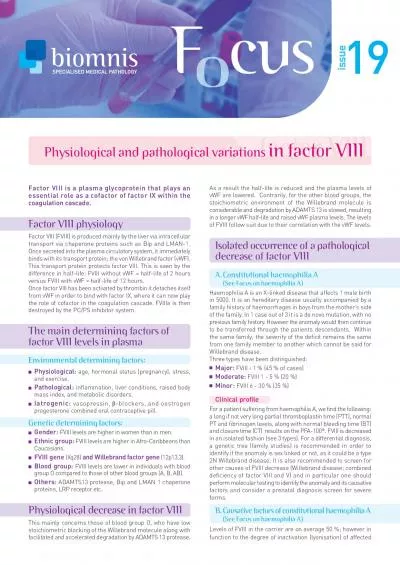
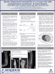
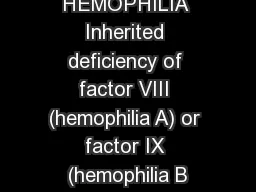
/
Hemophilia HEMOPHILIA Inherited deficiency of factor VIII (hemophilia A) or factor IX Hemophilia HEMOPHILIA Inherited deficiency of factor VIII (hemophilia A) or factor IX

Hemophilia HEMOPHILIA Inherited deficiency of factor VIII (hemophilia A) or factor IX - PowerPoint Presentation

liane-varnes
liane-varnes . @liane-varnes
Follow
354 views
Uploaded On 2019-03-15

Hemophilia HEMOPHILIA Inherited deficiency of factor VIII (hemophilia A) or factor IX - PPT Presentation

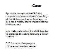
Sexlinked inheritance almost all patients male Female carriers may have mild symptoms Most bleeding into joints muscles mucosal and CNS bleeding uncommon Severity inversely proportional to factor level ID: 756367

bleeding factor platelet vwf factor bleeding vwf platelet deficiency hemophilia viii mutations type vwd activity clinical mutation inherited syndrome

Share:

Link:

Embed:

Download Presentation from below link

Download Presentation The PPT/PDF document "Hemophilia HEMOPHILIA Inherited deficien..." is the property of its rightful owner. Permission is granted to download and print the materials on this web site for personal, non-commercial use only, and to display it on your personal computer provided you do not modify the materials and that you retain all copyright notices contained in the materials. By downloading content from our website, you accept the terms of this agreement.


Presentation Transcript

Slide1

HemophiliaSlide2

HEMOPHILIA

Inherited deficiency of factor VIII (hemophilia A) or factor IX (hemophilia B)

Sex-linked inheritance; almost all patients male

Female carriers may have mild symptoms

Most bleeding into joints, muscles; mucosal and CNS bleeding uncommon

Severity inversely proportional to factor level

< 1%: severe, bleeding after minimal injury

1-5%: moderate, bleeding after mild injury

> 5%: mild, bleeding after significant trauma or surgerySlide3

GENETICS OF HEMOPHILIA A

About half of cases of hemophilia A due to an inversion mutation in intron 1 (5%) or 22 (45%)

Remainder genetically heterogeneous

Nonsense/stop mutations prevent factor production

Missense mutations may affect factor production, activity or half-life

15-20% of cases due to new mutations

Over 600 missense mutations identifiedSlide4

The factor VIII gene

Nested gene (“F8A”) of uncertain function in intron 22; 2 additional copies of this gene near the tip of the X chromosomeSlide5

The “flip tip” inversion in the factor VIII gene

Crossover between internal F8A and one of the two external copiesSlide6

GENETICS OF HEMOPHILIA B

Most cases associated with point mutations

Deletions in about 3% of cases

Promoter mutations in about 2%

In these cases an androgen response element near transcription start site may allow factor level to rise after puberty (“hemophilia B Leyden”)

Severe disease (<1% factor) less common than in hemophilia ASlide7

Deficiency of factor VIII or IX affects the propagation phase of coagulation

Most likely to cause bleeding in situations where tissue factor exposure is relatively lowSlide8

ACUTE COMPLICATIONS OF HEMOPHILIA

Muscle hematoma (pseudotumor)

Hemarthrosis (joint bleeding)Slide9

LONG-TERM COMPLICATIONS OF HEMOPHILIA

Joint destruction

Nerve damageSlide10

Hemophilic arthropathy

“Target joint” = irreversibly damaged joint with vicious cycle of injury and repeated bleeding Slide11

Management of hemophilic arthropathy

Physical therapy

Weight control

COX-2 inhibitors (eg, celecoxib) safe and effective

Judicious use of opioids

Surgical or radionuclide synovectomy

Joint replacementSlide12
Slide13

Bleeding rates decreased dramatically from 1999 to 2010

This change occurred in parallel with increased use of prophylaxis

Prophylaxis started before age 4 preserved joint function

Blood 2017;129:2368-2374Slide14

OTHER COMPLICATIONS OF HEMOPHILIA

Pseudotumor: gradually enlarging cyst in soft tissue or bone (requires surgery)

Retroperitoneal hemorrhage

Bowel wall hematoma

Hematuria

→ renal colic (rule out structural lesion)

Intracranial or intraspinal bleeding (rare but deadly) – usually after traumaSlide15

HEMOPHILIA

Treatment of bleeding episodes

Unexplained pain in a hemophilia should be considered due to bleeding unless proven otherwise

External signs of bleeding may be absent

Treatment: factor replacement, pain control, rest or immobilize joint

Test for inhibitor if unexpectedly low response to factor replacementSlide16

Dosing clotting factor concentrate

1 U/kg of factor VIII should increase plasma level by about 2% (vs 1% for factor IX)

Half-life of factor VIII 8-12 hours, factor IX 18-24 hours

Volume of distribution of factor IX about twice as high as for factor VIII

Steady state dosing about the same for both factors – initial dose of factor IX should be higherSlide17

Give factor q 12 hours for 2-3 days after major surgery, continue with daily infusions for 7-10 days

Trough

factor levels with q 12 h dosing after major surgery should be at least 50%

Most joint and muscle bleeds can be treated with “minor” (50%) doses for 1-3 days without monitoringSlide18

FACTOR VIII CONCENTRATE

Recombinant

Virus-free, most expensive replacement

Treatment of choice for younger/newly diagnosed hemophiliacs

Somewhat lower plasma recovery than with plasma-derived concentrate

Highly purified

Solvent/detergent treated, no reports of HIV or hepatitis transmissionIntermediate purity (Humate-P

™)Contains both factor VIII and von Willebrand factorSolvent/detergent treated, no reports of HIV or hepatitis transmissionMainly used to treat von Willebrand diseaseSlide19

FACTOR IX CONCENTRATE

Recombinant (slightly lower plasma recovery)

Highly purified (solvent/detergent treated, no reports of virus transmission)

Prothrombin complex concentrate

Mixture of IX, X, II, VII

Low risk of virus transmission

Some risk of thrombosis Mostly used to reverse warfarin effect Slide20

DDAVP

Releases vWF/fVIII from endothelial cells

Factor VIII levels typically rise 2-4 fold after 30-60 min (IV form) or 60-90 min (intranasal)

Enhanced platelet adhesion due to

↑ vWF

Useful for mild hemophilia (VIII activity > 5%) prior to dental work, minor surgery etc

Trial dose needed to ensure adequate responseCardiovascular complications possible in older patientsSlide21

Inhibitor formation in hemophilia

More common in hemophilia A

< 1% of hemophilia B patients develop inhibitors

7-10 x more common in severe hemophilia

About 30% of patients with intron 22 inversion develop inhibitors

More common with use of recombinant factor

Other genetic factors also involvedSlide22

When to test for an inhibitor?

If factor

replacment

less effective than usual

Prior to major surgery

Routine screening?

Current pediatric recommendations recommend frequent screeningScreening every 3-6 mo reasonable in high risk patientsSlide23

TREATMENT OF HEMOPHILIACS WITH INHIBITORS

Recombinant factor

VIIa

Enhances TF-driven thrombin formation

FEIBA (

F

actor Eight Inhibitor B

ypassing Activity)Mixture of partially activated vitamin K-dependent clotting proteases including VIIaPorcine factor VIII (if available)Some inhibitors active against porcine VIII (need to test)High dose factor VIII (if low titer inhibitor)Induction of tolerance with daily factor VIII infusionsOptimal dose not establishedRole for concomitant immunosuppression?Slide24

Liver disease in hemophilia

Hepatitis C still a problem, though incidence falling with safer factor concentrates

Liver transplantation done occasionally (cures hemophilia)

All newly diagnosed hemophiliacs should be vaccinated against hepatitis A and BSlide25
Slide26

Hemophilia: carrier testing

Factor level alone should not be used

VIII:VWF ratio may be helpful

DNA testing should be done if possible

Identification of causative mutation in an affected relative helpful, particularly for families with missense mutationsSlide27

von Willebrand diseaseSlide28

VON WILLEBRAND DISEASE

Common (most common?) inherited bleeding disorder

Partial lack of VWF causes mild or moderate bleeding tendency

Menorrhagia, bleeding after surgery, bruising

Typically autosomal dominant with variable penetrance

Laboratory:

Defective platelet adherence (PFA-100) or long bleeding time

Subnormal levels of von Willebrand antigen and factor VIII in plasmaLow Ristocetin cofactor activity or VWF activitySlide29

VON WILLEBRAND FACTOR

Single very large molecules visualized by electron microscopy

Electrophoresis showing range of multimer sizesSlide30

Biosynthesis of VWF

NEJM 2016;375:2067Slide31

VWF multimer formationSlide32

Weibel-Palade body

(arrows) in the cytoplasm of endothelial cell. N - nucleus. Scale = 100 nm. (Human, skin.)

Endothelial cellSlide33

Metcalf D J et al. J Cell Sci 2008;121:19-27

Tubular VWF arrays within Weibel-Pallade bodiesSlide34

VWF Mediates Platelet Adhesion and Facilitates Platelet Activation

NEJM 2016;375:2067Slide35

VON WILLEBRAND DISEASE

Type 1: VWF antigen and activity reduced proportionately

VWF levels range from < 20% to

~30%

Levels of >30-50% (“Low VWF”) fairly common, may cause mild bleeding

Complex genetics – only 65% of cases associated with VWF gene mutations

Autosomal dominant inheritance (dominant negative or null alleles)

Variable penetrance (affected by blood type, other factors)Defects in VWF processing, storage or secretion may account for cases lacking VWF gene mutationSome variants cause accelerated VWF clearanceSlide36

VON WILLEBRAND DISEASE

Genetics

Frequency of VWF gene mutations in type I VWD according to degree of deficiency:

Mutations identified in 53% of the Type 1 VWD cohort

Of the Type 1 VWD individuals with VWF levels <40, 74% had VWF gene mutations. 

87% with VWF:Ag of 2-10

93% with VWF:Ag 11-20

71% with VWF:Ag of 21-30 67% with VWF:Ag of 31-40 52% with VWF:Ag of >40Montgomery et al, 2013 ASH abstractSlide37

VON WILLEBRAND DISEASE

Type 2 – qualitative defect (missense mutation)

Four

different types

Usually a disproportionate decrease in vWF activity vs antigen

Type 3 – severe deficiency

Antigen, activity and factor VIII levels < 10%Homozygosity for null allelesHemophilia-like phenotype

Recessively inheritedSlide38

Type 2 vWD

2A: Deficiency of intermediate & large multimers

Defective assembly (mutation in either of two domains involved in multimer formation),

or

Increased susceptibility to proteolysis (mutation in domain cleaved by ADAMTS-13)

2B: Largest multimers missing

Gain of function mutation in platelet Gp Ib binding domainLargest multimers bind spontaneously to platelets and cleared from blood

Rarely, a mutation in Gp Ib may have the same effect (“platelet-type” vWD)2M: Normal multimer patternLoss of function mutation in GP Ib binding domain2N: Decreased binding of factor VIII to vWF (recessive)Slide39

Subtypes of VWD

NEJM 2016;375:2067Slide40
Slide41

Genetics of VWD

Most type 1 VWD due to missense mutations (dominant negative – interference with intracellular transport of dimeric pro-VWF)

Some forms with incomplete penetrance require co-inheritance of blood type O for expression (causes increased VWD proteolysis)

Most type 3 VWD due to null allelesSlide42

Laboratory testing in VWD

Von Willebrand factor activity

Measures binding of patient VWF to latex beads coated with monoclonal Ab to GPIb binding site; sensitive to multimer size and platelet-binding ability

Platelet function screen (PFA)

Measures time necessary for platelet plug to form in collagen coated tube under high shear conditions in the presence of ADP or epinephrineSlide43
Slide44

A Diagnostic Algorithm for VWD

NEJM 2016;375:2067

First level tests: VWF Ag and activity, FVIII activity

Second level: Multimer analysis,

propeptide

level,

ristocetin

-induced platelet aggregation, factor VIII binding assaySlide45

Bleeding in VWD

NEJM 2016;375:2067Slide46

Desmopressin (DDAVP) in vWD

DDAVP releases vWF from endothelial cells

Can be given IV or intranasally

0.3 mcg/kg IV, or 150 mcg per nostril

Typically causes 2-4 fold increase in blood levels of vWF (in type 1 vWD), with half-life of 8+ hours

Response to DDAVP varies considerably

Administration of a trial dose necessary to ensure a given patient responds adequately

Peak responseDuration of responseSlide47

Treatment of VWF

NEJM 2016;375:2067Slide48
Slide49
Slide50

Indications for clotting factor concentrate administration in vWD

Type 2 or 3

vWD

Active bleeding

Surgery or other invasive procedure

Type 1

vWD with inadequate response to DDAVPVery low baseline VWF activityVariants with rapid clearanceSlide51
Slide52

Inherited platelet disordersSlide53

Defects in platelet surface molecules

J

Thromb

Haemost

2011; 9(

suppl

1):77Slide54

Defects in platelet organelles or cytosolic proteins

J Thromb Haemost 2011; 9(suppl 1):77Slide55

Bernard-Soulier syndrome

Pathophysiology:

Deficiency of platelet membrane glycoprotein

Ib

-IX (VWF “receptor”)

Defective platelet adhesion

Clinical: Moderate to severe bleedingInheritance: autosomal recessive

Morphology: Giant plateletsThrombocytopenia (20-100K) (Often confused with ITP)Diagnosis: No agglutination with ristocetin, decr thrombin response, responses to other agonists intactMorphologyDecreased GP Ib expressionSlide56

Bernard-Soulier syndromeSlide57

Glanzmann thrombasthenia

Pathophysiology:

Deficiency of platelet membrane GPIIb-IIIa

Absent platelet aggregation with all agonists; agglutination by ristocetin intact

Clinical: Moderate to severe bleeding

Inheritance: autosomal recessive

Morphology: normalDiagnosis:

Defective platelet aggregationDecreased GP IIb-IIIa expressionSlide58

Gray platelet syndrome

Pathophysiology: Empty platelet alpha granules

Clinical: Mild bleeding

Inheritance: Autosomal dominant or recessive

Morphology:

Hypogranular platelets

Giant plateletsThrombocytopenia (30-100K)

Myelofibrosis in some patientsDiagnosisVariably abnormal platelet aggregation (can be normal)Abnormal platelet appearance on blood smearElectron microscopy showing absent alpha granulesSlide59

Gray platelet syndromeSlide60

Giant platelet syndromes associated with MYH9 mutations

May-

Hegglin

anomaly

Fechtner

syndrome

Sebastian syndromeEpstein syndrome

All associated with mutations in the non-muscle myosin heavy chain gene MYH9Thrombocytopenia with giant platelets, but mild bleedingAutosomal dominant inheritanceNo consistent defects of platelet function detectable in the clinical laboratoryDiagnosis usually based on clinical picture, family history, examination of blood smear for neutrophil inclusionsSlide61

Giant platelet syndromes associated with MYH9 mutations

Syndrome

Neutrophil inclusions

Hereditary nephritis

Deafness

May-

Hegglin

Yes

No

No

Fechtner

Yes

Yes

Yes

Sebastian

Yes*

No

No

Epstein

No

Yes

Yes

*

Neutrophil inclusions have different structure from those in May-HegglinSlide62

Neutrophil inclusions in May-Hegglin anomalySlide63

Wiskott-Aldrich syndrome

Pathophysiology

Mutation in WASP signaling protein

Decreased secretion and aggregation with multiple agonists; defective T-cell function

Clinical:

Mild to severe bleeding

Eczema, immunodeficiencyInheritance: X-linked

Morphology:Thrombocytopenia (20-100K)Small platelets with few granulesDiagnosis: Family hx, clinical picture, genetic testingSlide64

Wiskott-Aldrich syndromeSlide65

Hermansky Pudlak syndrome

Chédiak-Higashi syndrome

Pathophysiology:

Platelet dense granule deficiency: decreased aggregation & secretion with multiple agonists

Defective pigmentation

Defective lysosomal function in other cells

Clinical:

Mild to moderate bleedingOculocutaneous albinism (HPS)Lysosomal storage disorder with ceroid deposition, lung & GI disease (HPS)Immunodeficiency, lymphomas (CHS)Inheritance: autosomal recessiveMorphologyReduced dense granulesAbnormal neutrophil granules (CHS)Diagnosis: clinical picture, neutrophil inclusions (CHS), genetic testingSlide66

Chédiak-Higashi, showing neutrophil inclusions

HPS, with oculocutaneous

albinismSlide67

Platelet type von Willebrand disease

Pathophysiology: Gain of function mutation in GP Ib, with enhanced binding to VWF and clearance of largest multimers from blood

Clinical: Mild to moderate bleeding

Inheritance: Autosomal dominant

Morphology: Normal, but platelet count often low

Diagnosis: Variably low VWF antigen, disproportionately low ristocetin cofactor activity, loss of largest VWF multimers on electrophoresis, enhanced platelet agglutination by low dose ristocetin (indistinguishable from type 2B VWD)

Can distinguish from 2B VWD by mixing studies with normal/pt platelets and plasma and low dose ristocetin, or by genetic testingSlide68

Von Willebrand multimer analysisSlide69

Rare clotting factor deficienciesSlide70

Afibrinogenemia

Prevalence approx 1:1,000,000

Recessive inheritance

Most reported cases from consanguineous parents

Parents typically have asymptomatic hypofibrinogenemia

Genetically heterogeneous (>30 mutations)

May be due to failure of synthesis, intracellular transport or secretion of fibrinogen

Moderate to severe bleeding (typically less than in severe hemophilia)Death from intracranial bleeding in childhood may occurGI and other mucosal hemorrhageMenorrhagiaPlacental abruptionTreat with purified fibrinogen concentrate or cryoprecipitate for bleeding, during pregnancySlide71

Inherited dysfibrinogenemia

Prevalance uncertain (most cases asymptomatic)

Usually exhibits dominant inheritance

Most cases due to missense mutations

Mutations may affect fibrin polymerization, fibrinopeptide cleavage, or fibrin stabilization by FXIIIa

Variable clinical manifestations (mutation-dependent):

Over 50% asymptomatic

Approx 25% with bleeding tendency (mild to severe)20% have a thrombotic tendency (arterial, venous, or both)Decreased thrombin-binding (antithrombin effect) of fibrin?Altered fibrin clot structure?Slide72

Diagnosis of dysfibrinogenemia

Prolonged thrombin & reptilase times

PT, aPTT may be prolonged

Disparity (>30%) between fibrinogen activity and antigen

Family testing

Evaluate for liver diseaseSlide73

Recessively inherited clotting factor deficiencies

Rare

Exceptions: XI, XII deficiency

Homozygotes (often consanguineous parents) or compound heterozygotes

Heterozygous parents usually asymptomatic

Quantitative (“type 1”) deficiency: parallel reduction in antigen and activity

Qualitative (“type 2”) deficiency: reduced activity with near-normal antigen

Genetically heterogeneousComplete deficiency of II, X not described (lethal?)Mutation usually in gene encoding clotting factorExceptions: Combined V, VIII deficiency Combined deficiency of vitamin K-dependent factorsSlide74

Combined deficiency of factors V and VIII

Levels of affected factors 5-20% of normal

Associated with mutations of LMAN-1 (ERGIC-53) or MCFD2, both of which regulate intracellular trafficking of V and VIIISlide75

Deficiency of multiple vitamin-K dependent clotting factors

Levels of II, VII, IX, X, proteins C and S range from <1% to 30% of normal

Bleeding symptoms proportional to degree of deficiency

Usually associated with missense mutations in vitamin K epoxide reductase subunit 1 (VKORC1)Slide76

Relative frequencies of recessively inherited factor deficiencies

Blood 2004; 104:1243Slide77

Clinical features of recessively inherited factor deficiencies

Blood 2004; 104:1243Slide78

Patterns of bleeding in recessively inherited factor deficiency vs hemophilia

Blood 2004; 104:1243Slide79

Severity of bleeding in rare inherited bleeding disorders

J Thromb Haemost 2012;10:615

Number of patients with each condition

Frequency of bleeding episodesSlide80

Factor concentration

vs

bleeding severity in rare coagulation factor deficiencies

Deficiency

Asymptomatic

Grade I bleeding

Grade II

bleedingGrade III bleedingFibrinogen113 mg/dL73 mg/dL

33 mg/

dL

0 mg/

dL

Factor V

12%

6%

0.01%

0%

FV

+ F VIII

43%

34%24%

15%Factor VII25%19%13%8%Factor X56%40%25%10%Factor XI

26%

26%

25%

25%

Factor XIII

31%

17%

3%

0%

European Network of Rare Bleeding Disorders: J

Thromb

Haemost

2012;10:615

Grade 1: Bleeding after trauma or anticoagulant/antiplatelet drug ingestion

Grade 2: Spontaneous minor bleeding

Grade 3: Spontaneous major bleedingSlide81

Treatment of rare clotting factor deficiencies

FFP

Prothrombin complex concentrate (II, VII, IX, X) or specific factor concentrate (XIII – others available in Europe) when appropriate

Goal is to maintain “minimal hemostatic levels”

Antifibrinolytic drugs may be helpful in patients with mucosal hemorrhage

Routine prophylaxis appropriate for F XIII deficiency (long half-life, low levels adequate for hemostasis)

Otherwise treatment appropriate for active bleeding or pre-procedureSlide82

Factor XISlide83

Factor XI deficiency

Recessively inherited

Most common in individuals of Ashkenazi Jewish descent

2 common mutations (one nonsense, one missense)

Allele frequency as high as 10%, 0.1-0.3% homozygous

Most affected patients compound heterozygotes with low but measurable levels of XI activity

Long aPTT, normal PTXI activity < 10% in most patients with bleeding tendencySlide84

Factor XI deficiency

Clinical features & treatment

Variable, generally mild bleeding tendency

Bleeding after trauma & surgery

Spontaneous bleeding uncommon

Bleeding risk does not correlate well with XI level

Treatment: FFP

15 ml/kg loading, 3-6 ml/kg q 12-24hHalf life of factor >48 hoursAmicar useful after dental extraction, surgeryrVIIa is effective but expensive; thrombotic complications reportedSlide85

Factor XIII

Transglutaminase: forms amide bonds between lysine and glutamic acid

residues on different protein molecules

Heterotetramer

(A

2

B2) in plasmaA chains made by megakaryocytes and monocyte/macrophage precursors

Platelet XIII (50% of total XIII) has only A chainsB chains (non-catalytic) made in liverProenzyme activated by thrombinCrosslinks and stabilizes fibrin clotCan crosslink other proteins (e.g., antiplasmin) into clotSlide86

Factor XIII (transglutaminase) mechanism

Enzyme links glutamine side chain on protein A with lysine side chain on protein B

A

B

XIIISlide87

Inherited factor XIII deficiency

Autosomal recessive, rare (consanguineous parents)

Heterozygous woman may have higher incidence of spontaneous abortion

Most have absent or defective A

subunitSlide88

Inherited factor XIII deficiency

Clinical features & treatment

Bleeding begins in infancy (umbilical cord)

Poor wound healing

Intracranial hemorrhage

Oligospermia, infertility

Diagnosis:

Urea solubility testQuantitative measurement of XIII activityRule out acquired deficiency due to autoantibodyTreatment: F XIII concentrate or recombinant factor XIIIlong half life, give every 4-6 weeks as prophylaxisSlide89

Vascular disordersSlide90

Hereditary Hemorrhagic Telangiectasia

Autosomal dominant inheritance

Mutation in endoglin gene that controls vascular remodeling

Molecular diagnosis possible

Multiple small AVMs in skin, mouth, GI tract, lungsSlide91

Hereditary hemorrhagic telangiectasia

J Thromb Haemost 2010;8:1447Slide92

Hereditary Hemorrhagic Telangiectasia

Clinical features

Epistaxis, GI bleeding – may be severe

Severe iron deficiency common

Pulmonary or CNS bleeding often fatal

Gradual increase in bleeding risk with age

AVMs enlarge during pregnancy

Risk of brain abscessHypoxemia from pulmonary HTN and R→L shunting in lungSlide93

Hereditary Hemorrhagic Telangiectasia

Treatment

No consistently effective method for preventing bleeding

Aggressive iron replacement

Antibiotic prophylaxis for dental work etc

Screen for CNS lesions

→ consider surgical interventionSlide94

Ehlers-Danlos syndrome

Defective collagen structure

Mutations in genes for various types of collagen

9 variants

Type IV (mutation in type III collagen gene) most likely to cause bleeding

Bleeding due to weakening of vessel wall

→ vessel ruptureConventional tests of hemostatic integrity normalSlide95

Ehlers-Danlos syndrome

Thin, weak skin with poor healing

“Cigarette paper” scars

Bruising

Hypermobile joints

Spontaneous joint dislocation

Median survival 48 years in type IV EDSDeath from rupture of large vessels or colon perforation