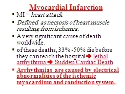
Mostafa Hekmat Cardiologist Electrophysiologist VENTRICULAR TACHYCARDIA VT Arises Distal To The Bifurcation Of The HB In The Specialized Conduction System In The Ventricular Muscle Or In Combination Of Both Tissues ID: 775331
Download Presentation The PPT/PDF document " Ventricular Arrhythmia Dr" is the property of its rightful owner. Permission is granted to download and print the materials on this web site for personal, non-commercial use only, and to display it on your personal computer provided you do not modify the materials and that you retain all copyright notices contained in the materials. By downloading content from our website, you accept the terms of this agreement.
Slide1
Ventricular Arrhythmia
Dr
Mostafa Hekmat
Cardiologist
Electrophysiologist
Slide2VENTRICULAR TACHYCARDIA
VT Arises Distal To The Bifurcation Of The HB In The Specialized Conduction SystemIn The Ventricular MuscleOr In Combination Of Both Tissues
Dr Mostafa Hekmat
2
Slide3Note
Any wide QRS complex tachycardia should be treated as ventricular tachycardia until definitive evidence is found to establish another diagnosis
Dr Mostafa Hekmat
3
Slide4Types of Ventricular Arrhythmia
Premature Ventricular ComplexesVentricular TachycardiaMonomorphicPolymorphicTorsade de pointNormal QTVentricular FlutterVentricular Fibrillation
Dr Mostafa Hekmat
4
Slide5Ventricular ArrhythmiasDefinitions
Premature Ventricular beatsSingle beatsVentricular Bigeminy, the appearance of one PVC after each sinus beatCouplets, two consecutive premature beatsTriplets, three consecutive premature beatsSalvos, runs of 3-10 premature beats
Dr Mostafa Hekmat
5
Slide6Ventricular ArrhythmiasDefinitions
Accelerated Idioventricular Rhythm (Slow VT), rate 60-100 bpm Ventricular Tachycardia (VT), rate over 100 bpm Ventricular Flutter, regular large oscillations at a rate of 150-300 bpmVentricular Fibrillation (VF), irregular undulations of varying contour and amplitude
Dr Mostafa Hekmat
6
Slide7PVC
Dr Mostafa Hekmat
7
60 bpm
Rate?
Regularity?
Occasionally irreg.
None for 7
th QRS
0.08 s (7th wide)
P waves?
PR interval?
0.14 s
QRS duration?
Interpretation?
Sinus Rhythm with 1 PVC
Slide8VT
Dr Mostafa Hekmat
8
160 bpm
Rate?
Regularity?
Regular
None
Wide (> 0.12 sec)
P waves?
PR interval?
None
QRS duration?
Interpretation?
Ventricular Tachycardia
Slide9VF
Dr Mostafa Hekmat
9
None
Rate?
Regularity?
Irregularly irreg.
None
Wide, if recognizable
P waves?
PR interval?
None
QRS duration?
Interpretation?
Ventricular Fibrillation
Slide10Premature Ventricular Complexes
Premature occurrence of a QRS complex that is abnormal in shapeDuration usually exceeding the dominant QRS complex Generally longer than 120 millisecondsThe T wave is usually large and opposite in direction to the major deflection of the QRSA fully compensatory pause usually follows a PVCThe PVC may not produce any pause and may therefore be interpolated
Dr Mostafa Hekmat
10
Slide11Dr Mostafa Hekmat
11
Slide12Ventricular fusion beat
Dr Mostafa Hekmat
12
Slide13PVC and ventricular echo
Dr Mostafa Hekmat
13
Slide14An interpolated PVC
Dr Mostafa Hekmat
14
Slide15Ventricular Premature Complexes
Dr Mostafa Hekmat
15
Compensatory Pause
Interpolated VPC
Slide16Premature Ventricular Complexes
Bigeminy Pairs of complexes and indicates a normal and premature complexTrigeminy Premature complex that follows two normal beatsQuadrigeminyPremature complex that follows three normal beats is called
Dr Mostafa Hekmat
16
Slide17PVC
Pair or couplet Two successive PVCsTriplet Three successive PVCsVentricular tachycardia Three or more successive PVCsPVCs can have different contoursMultifocal Multiform PolymorphicPleomorphic
Dr Mostafa Hekmat
17
Slide18Multiform
Dr Mostafa Hekmat
18
Slide19Salvos
Dr Mostafa Hekmat
19
Slide20PVC
Fixed couplingReentry Triggered activityVariable couplingParasystole Changing conduction in a reentrant circuitChanging discharge rates of triggered activity
Dr Mostafa Hekmat
20
Slide21CLINICAL FEATURES
The prevalence of premature complexes increases with ageMale genderHypokalemiaPVCs are more frequent in the morning in patients after MIThis circadian variation is absent in patients with severe left ventricular (LV) dysfunction.
Dr Mostafa Hekmat
21
Slide22CLINICAL FEATURES
Activity that increases the heart rate can decreaseThe patient’s awareness of the premature systolesReduce their number.SleepUsually associated with a decrease in the frequency of ventricular arrhythmiasBut some patients can experience an increase.
Dr Mostafa Hekmat
22
Slide23The importance of PVCs
Depends on the clinical settingIn the absence of underlying heart disease, the presence of PVCs usually has no impact on longevity or limitation of activityAntiarrhythmic drugs are not indicatedPatients should be reassured if they are symptomatic
Dr Mostafa Hekmat
23
Slide24PVC and MI
Those occurring close to the preceding T waveMore than five or six per minuteBigeminal or multiform complexesThose occurring in salvoes of two or three or moreDo not occur in about 50% of patients in whom VF developsAnd VF does not develop in about 50% of patients who have these PVCsThus, these PVCs are not particularly helpful prognostically
Dr Mostafa Hekmat
24
Slide25Idioventricular Rhythm
Dr Mostafa Hekmat
25
Slide26Accelerated idioventricular rhythm
Dr Mostafa Hekmat
26
Slide27Dr Mostafa Hekmat
27
Slide28ECG Distinction of VT from SVT with Aberrancy
Favors VT Favors SVT with Aberrancy
Dr Mostafa Hekmat
28
Morphology Precordial concordance
If LBBB: V
1
duration > 30
ms
S wave > 70
ms
S wave notched or slurred
V
6
:
qR
or QR R wave monophasic
If RBBB: V
1
: monophasic R wave
qR
If
triphasic
, R > R
1
R < R
1
V
6
: R < S
Slide29VENTRICULAR FLUTTER & VENTRICULAR FIBRILLATION
Dr Mostafa Hekmat
29
Slide30A-V Dissociation, Fusion, and
Capture Beats in VT
Dr Mostafa Hekmat
30
ECTOPY
FUSION
CAPTURE
V1
E
F
C
Slide31Accelerated Idioventricular Rhythm
The arrhythmia occurs as a rule in patients who have heart disease, such as those with acute myocardial infarction or with digitalis toxicity.Reperfusion of a previously occluded coronary artery During resuscitationIt is transient and intermittentEpisodes lasting a few seconds to a minuteDoes not appear to seriously affect the patient’s clinical course or the prognosis
Dr Mostafa Hekmat
31
Slide32Therapy when
Considered when AV dissociation results in loss of sequential AV contractionAn accelerated idioventricular rhythm occurs together with a more rapid VTAn accelerated idioventricular rhythm begins with a PVC discharging in the vulnerable period of the preceding T waveThe ventricular rate is too rapid and produces symptomsVF develops as a result of the accelerated idioventricular rhythm.
Dr Mostafa Hekmat
32
Slide33Therapy
Increasing the sinus rate with Atropine or atrial pacing suppresses the accelerated idioventricular rhythm
Dr Mostafa Hekmat
33
Slide34Dr Mostafa Hekmat
34
Slide35Clinical Impact of VT/VF
PVCs and even runs of nonsustained VT may be frequently seen in people with normal and abnormal heartsSustained VT and VF usually develop in patients with advanced structural heart disease
Dr Mostafa Hekmat
35
Slide36Clinical Impact of VT/VF
Frequent PVCs in the recovery phase after an exercise test are stronger predictors of mortality than ventricular extrasystoles during exercisePredicts an increased mortality in systolic heart failure patientsPost exercise severe ventricular ectopyTripletsNonsustained VTSustained VTSustained VF
Dr Mostafa Hekmat
36
Slide37Clinical Impact of VT/VF
CAD is the most frequent cause of SCD and clinically documented VT and VF 80%There is no reason to neglect the device even if the stable VT has been successfully ablated
Dr Mostafa Hekmat
37
Slide38VT + RBBB
(1) the QRS complex is monophasic or biphasic in V1, with an initial deflection different from that of the sinus-initiated QRS complex(2) the amplitude of the R wave in V1 exceeds the R′(3) a small R and large S wave or a QS pattern in V6 may be present.
Dr Mostafa Hekmat
38
Slide39VT + LBBB
(1) the axis can be rightward, with negative deflections deeper in V1 than in V6,(2) a broad prolonged (more than 40 milliseconds) R wave in V1(3) a small Q–large R wave or QS pattern in V6 can exist
Dr Mostafa Hekmat
39
Slide40VT
QRS duration exceeding 140 millisecondsIn precordial leads with an RS pattern, the duration of the onset of the R to the nadir of the S exceeding 100Fusion beatCapture beatAV dissociation has long been considered a hallmark of VTRetrograde VA conduction to the atria from ventricular beats occurs in at least 25% of patients
Dr Mostafa Hekmat
40
Slide41Supraventricular arrhythmiawith aberrancy
(1) consistent onset of the tachycardia with a premature P wave(2) very short RP interval (0.1 sec) (3) QRS configuration the same as that occurring from known supraventricular conduction at similar rates(4) P wave and QRS rate and rhythm linked to suggest that ventricular activation depends on atrial discharge (an AV Wenckebach block) (5) slowing or termination of the tachycardia by vagal maneuvers
Dr Mostafa Hekmat
41
Slide42Dr Mostafa Hekmat
42
Slide43A QRS complex in V1 - V6, either all negative or all positive favors a VT The presence of a 2 : 1 VA block VTPositive QRS complex in V1 - V6 can also occur from conduction over a left-sided accessory pathway.Supraventricular beats with aberrationTriphasic pattern in V1An initial vector of the abnormal complex similar to that of the normally conducted beatsWide QRS complex with long-short cycle sequence
Dr Mostafa Hekmat
43
Slide44Sustained Monomorphic VT
Dr Mostafa Hekmat
44
Slide45Dr Mostafa Hekmat
45
Slide46Increased risk in VT
Reduced LV functionSpontaneous ventricular arrhythmiasLate potentials on signal-averaged ECGQT interval dispersionT wave alternansProlonged QRS durationHeart rate turbulenceInducible sustained VTs
Dr Mostafa Hekmat
46
Slide47Treatment
Dr Mostafa Hekmat
47
Slide48Treatment
Frequent PVCs, even in the setting of an acute MI, need not be treated unless they directly contribute to hemodynamic compromise
Dr Mostafa Hekmat
48
Slide49Treatment
Beta blockers are often the first line of therapy.If they are ineffective, class IC drugs seem particularly successful in suppressing PVCsFlecainide and Moricizine have been shown to increase mortality in patients treated after MI Should be reserved for patients without coronary artery disease or LV dysfunctionAmiodaroneShould be reserved for highly symptomatic patients and those with structural heart disease.
Dr Mostafa Hekmat
49
Slide50Treatment
VT that precipitatesHypotension Shock Angina Congestive heart failureSymptoms of cerebral hypoperfusionShould be treated promptly with DC cardioversionVery low energies can terminate VT a synchronized shock of 10 to 50 J.Digitalis-induced VT is best treated pharmacologically.
Dr Mostafa Hekmat
50
Slide51Thump Version
Can terminate VT by mechanically inducing a PVC that presumably interrupts the reentrant pathway necessary to support it.Chest stimulation at the time of the vulnerable periodCan accelerate the VT or Possibly provoke VF.Intermittent VT best treated pharmacologically
Dr Mostafa Hekmat
51
Slide52Treatment
In patients in whom procainamideIneffectiveProcainamide may be problematicSevere heart failureRenal failureIntravenous Amiodarone is often effectiveLoading dose of 15 mg/min is given during a 10-minute periodInfusion of 1 mg/min for 6 hoursMaintenance dose of 0.5 mg/min for the remaining 18 hours and for the next several days
Dr Mostafa Hekmat
52
Slide53Reversible conditions
VT related to ischemia antianginal Hypotension vasopressorsHypokalemia potassiumCorrection of HF reduce the frequency of ventricular arrhythmiasSinus bradycardia or AV block PVCs and ventricular tachyarrhythmias, which can be corrected by administrationAtropine Temporary isoproterenol administrationPacing
Dr Mostafa Hekmat
53
Slide54Long-Term Therapy
Asymptomatic nonsustained ventricular arrhythmias in low-risk populations (preserved LV function) often need not be treated.In patients with symptomatic nonsustained tachycardia, beta blockers are frequently effective in preventing recurrences.In patients refractory to beta blockers, class IC agents, Sotalol, or Amiodarone can be effective
Dr Mostafa Hekmat
54
Slide55CLASSIFICATION OF IDIOPATHIC MONOMORPHIC VT
Dr Mostafa Hekmat
55
Slide56RMVT
Dr Mostafa Hekmat
56
Slide57Both Forms Are Characterized By Adenosine SensitivityAnd Are Thought To Be Caused By Cyclic Amp Mediated Triggered Activity
Dr Mostafa Hekmat
57
Slide58VT Can Be Terminated With Adenosine, Verapamil, The Valsalva Maneuver or CSP
Dr Mostafa Hekmat
58
Slide59VERAPAMIL-SENSITIVE VT
Dr Mostafa Hekmat
59
Slide60VERAPAMIL-SENSITIVE VT
90-95% HAS RBBB AND LEFT SUPERIOR-AXIS MORPHOLOGYTHE REMAINDER OF PATIENTS HAVE VT WITH RBBB AND RIGHT INFERIOR-AXIS MORPHOLOGYRS INTERVAL IS USUALLY 60-80 ms (in VTs associated with structural heart disease RS is longer than 100 ms )
Dr Mostafa Hekmat
60
Slide61CPVT
Inherited VTChildren and adolescentsWithout any overt structural heart disease.Patients typically present with syncope or aborted sudden deathHighly reproducible, stress-induced VTBidirectional Mutations of the ryanodine receptor gene result in an autosomal dominantMutations in the calsequestrin gene result in an autosomal recessive
Dr Mostafa Hekmat
61
Slide62CPVT
The treatment of choice is beta blockers and an ICDSympathectomyAvoid vigorous exercise
Dr Mostafa Hekmat
62
Slide63Dr Mostafa Hekmat
63
Slide64Dr Mostafa Hekmat
64
Slide65Long QT Syndrome
Slide66Dr Mostafa Hekmat
66
Slide67Congenital LQTS
The congenital LQTS is a rare disorder (incidence 1:10,000 to 1:15,000) characterized byProlongation of the Q–T interval on the surface ECGRecurrent syncopeSudden death
Dr Mostafa Hekmat
67
Slide68Congenital LQTS
Romano-Ward syndromeAutosomal dominant inheritanceOnly Cardiac ArrhythmiasJervell and Lange-Nielson syndromeAutosomal recessive inheritance Cardiac ArrhythmiasCongenital deafness.Andersen-Tawil syndromeVentricular ArrhythmiasPeriodic paralysis & Facial/Skeletal dysmorphism
68
Slide69The acquired form of long-QT
QuinidineProcainamideN-acetylprocainamideSotalolAmiodaroneDisopyramidePhenothiazinesTricyclic antidepressantsErythromycin Pentamidine
Cisapride Probucol Hypokalemia Hypomagnesemia liquid protein dietStarvation central nervous system lesionsBradyarrhythmias cardiac ganglionitismitral valve prolapse
Dr Mostafa Hekmat
69
Slide70LQTS
Torsades de pointes commonly developsIn patients with the acquired form during periods of bradycardia or after a long pause in the R-R intervalWhereas those with the idiopathic form can have a sinus tachycardia preceding the ventricular arrhythmia.
Dr Mostafa Hekmat
70
Slide71Dr Mostafa Hekmat
71
Slide72Dr Mostafa Hekmat
72
Slide73K or Na
The principal abnormality in LQTS is prolongation of action potential duration caused by a reduction in outward potassium current (LQT1 and LQT2)or, less commonly, a persistent inward sodium current during the plateau phase (LQT3).
Dr Mostafa Hekmat
73
Slide74Action potential prolongation development of early after depolarizations triggered action potential premature beat can initiate a polymorphic VT known as torsades de pointes,Which underlies the clinical symptoms of palpitations, syncope, or sudden death due to VF.
Dr Mostafa Hekmat
74
Slide75Dr Mostafa Hekmat
75
Slide76Dr Mostafa Hekmat
76
Slide77Dr Mostafa Hekmat
77
Slide78Triggers of cardiac event 1
The classic description of events in LQTS involves exercise- or emotion-induced clinical events.Symptoms often begin in adolescence, though they may begin earlier in LQT1.
Dr Mostafa Hekmat
78
Slide79Triggers of cardiac event 2
Adrenergic Stimuli in LQTS with Loss of function of K (iKs-LQTS1 , iKr-LQTS2) TDPAdrenergic Stimuli Transmural disturbances Β Blocker are useful in LQTS1 & LQTS2
Dr Mostafa Hekmat
79
Slide80Dr Mostafa Hekmat
80
Slide81LQTS1
Rhythm disturbances mainly occur during sports especially swimming
Dr Mostafa Hekmat
81
Slide82LQTS 2
Rhythm disturbances mainly triggered by Auditory stimuli
Dr Mostafa Hekmat
82
Slide83LQTS 3
Malfunction of iNaL , Symptom mostly occur at rest or during nightNo adrenergic triggersB Blockers Contraindicated
Dr Mostafa Hekmat
83
Slide84In
10% of patient SCD is the first & tragic symptom.LQTS1 and LQTS2 are more cardiac eventsBenign cardiac symptoms such as palpitation and syncope degenerate more easily to SCD in LQTS3
Dr Mostafa Hekmat
84
Slide85LQTS
The hallmark of this condition is prolongation of the QTc greater than 460 ms, QTc may be normal in up to 1/3 of genotype-positive patients.
Dr Mostafa Hekmat
85
Slide86LQTS
Causes of considerable temporal variation in QTcRepolarization is affected by factors such asSympathetic outflowElectrolyte balancePharmacologic agents
Dr Mostafa Hekmat
86
Slide87LQTS
The mean QTc does not differ between the LQT1, LQT2, and LQT3 typesBut is significantly longer in Jervell and Lange-Neilson syndrome.Other electrocardiographic abnormalities that may be found in LQTS include ST–T wave changesU waves, T wave alternansIncreased QT dispersionSinus bradycardia
Dr Mostafa Hekmat
87
Slide88FactorMechanismBradycardia↑APD↑APD prolongation withclass III agentsDrugs Mainly IKr IKs blockadeElectrolyte disorders (hypokalemia, hypomagnesemia, hypocalcemia) Hypokalemia ↓IKr and ↑IKrsensitivity to pharmacologicblockersLeft ventricular hypertrophy/failure ↓K+ currents (Ito, IKr, IKs)Changes to ICaL andintracellular Ca2+Miscellaneous (e.g., anorexia,cerebrovascular disease,hypothyroidism, ionic contrastmedia)? Reduced repolarization reserveLong QT syndrome
Dr Mostafa Hekmat
88
Slide89MANAGEMENT
For patients who have idiopathic long-QT syndrome but not SyncopeComplex ventricular arrhythmias, Family history of sudden cardiac deathQTc longer than 500 milliseconds,No therapy or treatment with a beta blocker is generally recommended.
Dr Mostafa Hekmat
89
Slide90MANAGEMENT
In asymptomatic patients with Complex ventricular arrhythmias,Family history of early sudden cardiac deathQTc longer than 500 millisecondsBeta adrenoceptor blockers at maximally tolerated doses are recommended.PPM to prevent the bradycardia or pauses that may predispose to the development of TDP may be indicated
Dr Mostafa Hekmat
90
Slide91MANAGEMENT
In patients with syncope caused by ventricular arrhythmias or aborted sudden death, an ICD is warranted.Concomitant beta blockersOverdrive atrial pacing (via the ICD) to minimize the frequency of ICD discharges
Dr Mostafa Hekmat
91
Slide92MANAGEMENT
Most competitive sports are contraindicated for patients with the congenital long-QT syndromeFor patients with the acquired form and TDP, intravenous Mg and atrial or ventricular pacing are initial choices.
Dr Mostafa Hekmat
92
Slide93Survival is dramatically improved by
Aggressive treatment with β-Blocker drugsCardiac pacingLeft cervical sympathectomyImplantable cardioverter defibrillator (ICD).
Dr Mostafa Hekmat
93
Slide94Short QT
Syndrome
Slide95Definition
QT of less than 350 milliseconds at rates of less than 100 beats/min
Dr Mostafa Hekmat
95
Slide96Short-QT
interval has recently been identified to carry an increased risk of sudden death due to VFOne of the syndromes responsible for “idiopathic VF”
Dr Mostafa Hekmat
96
Slide97Causes
Hyperkalemia Hypercalcemia Hyperthermia Acidosis Digitalis Genetic abnormalities
Dr Mostafa Hekmat
97
Slide98Treatment
ICDs are considered the treatment of choice in symptomatic patients to prevent sudden cardiac death. Quinidine
Dr Mostafa Hekmat
98
Slide99Dr Mostafa Hekmat
99