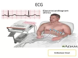
by the SA node causes the P wave P R T Q S SA node AV node With atrial depolarization complete the impulse is delayed at the AV node Ventricular depolarization begins at apex causing the ID: 774591
Download Presentation The PPT/PDF document " Figure 18.17 Atrial depolarizatio..." is the property of its rightful owner. Permission is granted to download and print the materials on this web site for personal, non-commercial use only, and to display it on your personal computer provided you do not modify the materials and that you retain all copyright notices contained in the materials. By downloading content from our website, you accept the terms of this agreement.
Slide1
Slide2Figure 18.17
Atrial depolarization, initiated
by the SA node, causes the
P wave.
P
R
T
Q
S
SA node
AV node
With atrial depolarization
complete, the impulse isdelayed at the AV node.
Ventricular depolarizationbegins at apex, causing theQRS complex. Atrialrepolarization occurs.
P
R
T
Q
S
P
R
T
Q
S
Ventricular depolarizationis complete.
Ventricular repolarizationbegins at apex, causing theT wave.
Ventricular repolarizationis complete.
P
R
T
Q
S
P
R
T
Q
S
P
R
T
Q
S
Depolarization
Repolarization
1
2
3
4
5
6
Slide3Slide4Slide5Figure 18.18
(a) Normal sinus rhythm.
(c) Second-degree heart block.
Some P waves are not conducted through the AV node; hence more P than QRS waves are seen. In this tracing, the ratio of P waves to QRS waves is mostly 2:1.
(d) Ventricular fibrillation. These chaotic, grossly irregular ECG deflections are seen in acute heart attack and electrical shock.
(b) Junctional rhythm.
The SA
node is nonfunctional, P waves
are absent, and heart is paced by
the AV node at 40 - 60 beats/min.
Slide6In catheter ablation, catheters are threaded through the blood vessels to the inner heart, and electrodes at the catheter tips transmit energy to destroy a small spot of heart tissue.
Slide7The answers to the following EKGs is on the
last slide.
Slide81
Slide92
Slide103
Slide114
Slide125
Slide136
Slide147
Slide158
Slide169
Slide1710
Slide1811
Slide1912
Slide2013
Slide2114
Slide2215
Slide2316
Slide241. A flutter
2. PVC3. 1st degree block4. Inverted T wave5. A fib6. A fib with PVC7. Tachycardia8. normal
9. A flutter
10. 1
st
degree block
11. A fib
12. 1
st
degree block
13. A fib
14. PVC
15. A flutter with inverted
16.2
nd
degree