/
Information for women after ultrasound detection of fetal renal pelvic dilatation Information for women after ultrasound detection of fetal renal pelvic dilatation

Information for women after ultrasound detection of fetal renal pelvic dilatation - PDF document

marina-yarberry
marina-yarberry . @marina-yarberry
Follow
379 views
Uploaded On 2017-08-15

Information for women after ultrasound detection of fetal renal pelvic dilatation - PPT Presentation

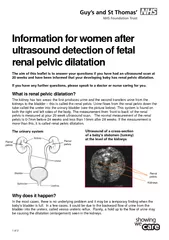
If you have any further questions please speak to a doctor or nurse caring for you What is renal pelvic dila ta tion The kidney has two areas the first produces urine and the second transfers urine from the kidneys to the bladder this is called t h ID: 77041

you have any

Share:

Link:

Embed:

Download Presentation from below link

Download Pdf The PPT/PDF document "Information for women after ultrasound d..." is the property of its rightful owner. Permission is granted to download and print the materials on this web site for personal, non-commercial use only, and to display it on your personal computer provided you do not modify the materials and that you retain all copyright notices contained in the materials. By downloading content from our website, you accept the terms of this agreement.


Presentation Transcript

1 of 2 Information for women after ultrasound detection of fetal renal pelvic dilatation The aim of this leaflet is to answer your questions if you have had an ultrasound scan at 20 weeks and have been informed that your developing baby has renal pelvic dilatati on. If you have any further questions, please speak to a doctor or nurse caring for you. What is renal pelvic dila ta tion ? The kidney has two areas : the first produces urine and the second transfers urine from the kidneys to the bladder – this is called t he renal pelvis. Urine flows from the renal pelvis down the tube called the ureter into the urinary bladder ( s ee the picture below ). This system is found o n both the right and left sides of the body . The measurement from ‘ front to back ’ of the renal pelvis is measured at your 20 week ultrasound scan. The normal measurement of the renal pelvis is 0 - 7mm before 24 weeks and less than 10mm after 2 8 weeks. If the measurement is more than this , it is called renal pelvic dilat at ion. The urinary system Why does it happen ? In the most cases, there is no underlying problem and it may be a temporary finding when the baby’s bladder is full. In a few cases, it could be due to the backward flow of urine from the bladder into the ureters, called vesico ureteric reflux. Rarely, a hold up to the flow of urine may be causing the dilatation (enlargement) seen in the kidneys . Ultrasound of a c ross - section of a baby’s abdomen (tummy) at the level of the kidneys Renal pelvis Kidneys Renal pelvis Renal pelvis 2 of 2 What will happen next ? 1. You will have a follow - up scan in the f etal m edicine u nit at about 28 - 34 weeks of pregnancy , or earlier. The r enal pelvic dilatation may have gone away by the time of this examination. 2. If the renal pelvic dilatation continues , you will be advised to have a repeat ultrasound scan at about 30 - 34 weeks in to your pregnancy . 3. You may also see a paediatric urologist ( a doctor who looks after children with urinary tract problems) who will explain to you what will happen after your baby is born. If the problem continues , what will happen ? During pregnancy: There is no need for any treatment before the baby is born. A fter your baby is born : 1. Your b aby will be started on a small dose of antibiotics to prevent urinary tract infection. 2. Your b aby may need further scans, the timing of which will be decided by the neonatologists (baby doctors). 3. If the scan s are normal, the doctors will write to you or to your GP asking you to stop antibiotics. 4. If the scan s show continuing or increasing dilatation, your baby may need further scans or treatment and an appointment will be made to see the paediatric urologist. Patient Advi ce and Liaison Service (PALS) To make comments or raise concerns about the Trust’s services, please contact PALS. Ask a member of staff to direct you to the PALS office or: e : 020 7188 8801 at St Thomas’ t: 020 7188 8803 at Guy’s e : pals@gstt.n hs.uk Knowledge & Information Centre (KIC) For more information about health conditions, support groups and local services, or to search the internet and send emails, please visit the KIC on the Ground Floor, North Wing, St Thomas’ Hospital. t: 020 7188 3416 Language support services If you need an interpreter or information about your care in a different language or format, please get in touch using the following contact details. t: 020 7188 8815 fax: 020 7188 5953 NHS Choices Provides online infor mation and guidance on all aspects of health and healthcare, to help you make choices about your health. w: www.nhs.uk Contact us If you have any questions or concerns about the scan, please contact the f etal medicine midwives on 020 7188 2321 (Monday to Friday, 9am to 5pm). Leaflet numb er: 3663 /VER 1 Date published: May 2013 Review date: May 2016 © 2013 Guy’s and St Thomas’ NHS Foundation Trust