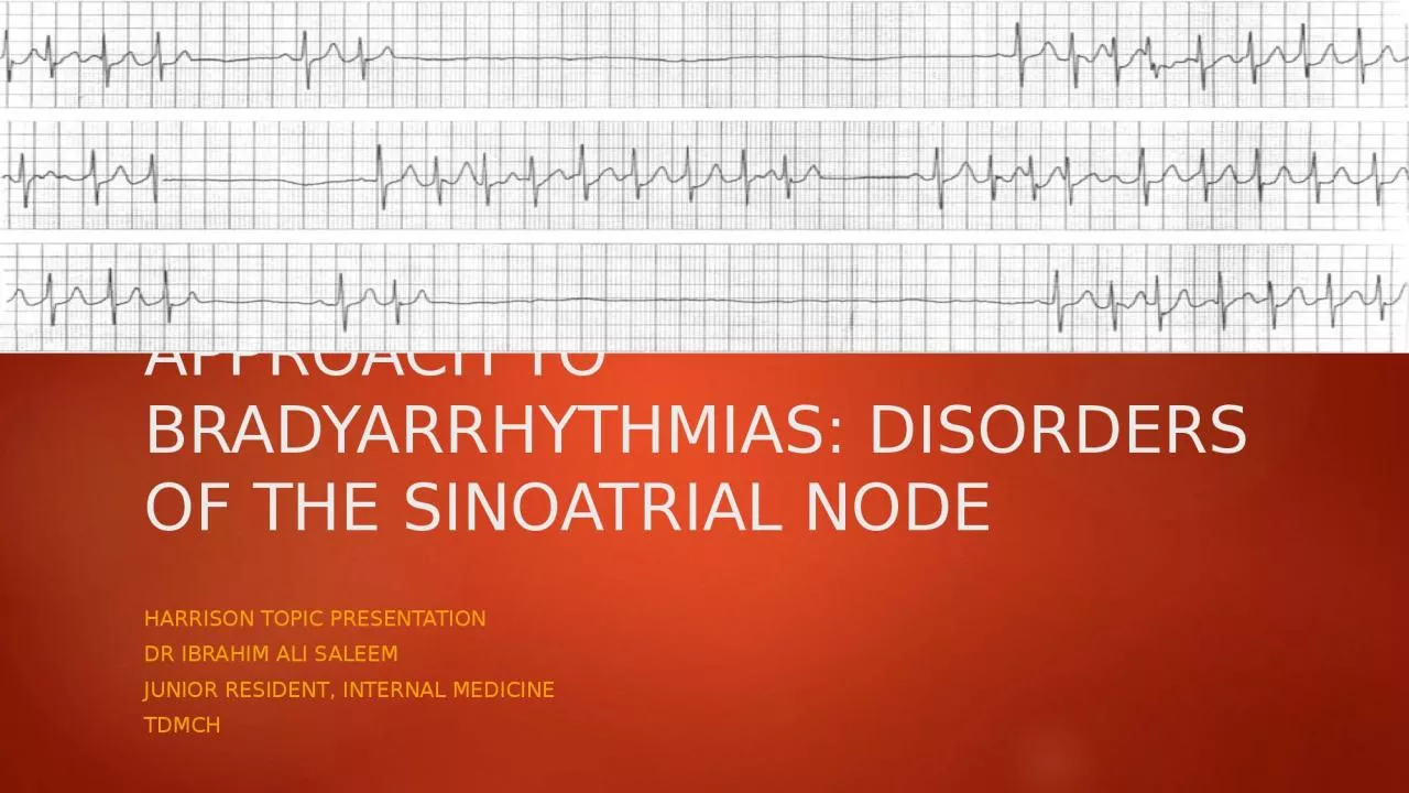
/
APPROACH TO BRADYARRHYTHMIAS: DISORDERS OF THE SINOATRIAL NODE APPROACH TO BRADYARRHYTHMIAS: DISORDERS OF THE SINOATRIAL NODE

APPROACH TO BRADYARRHYTHMIAS: DISORDERS OF THE SINOATRIAL NODE - PowerPoint Presentation

megan
megan . @megan
Follow
28 views
Uploaded On 2024-02-09

APPROACH TO BRADYARRHYTHMIAS: DISORDERS OF THE SINOATRIAL NODE - PPT Presentation

HARRISON TOPIC PRESENTATION DR IBRAHIM ALI SALEEM JUNIOR RESIDENT INTERNAL MEDICINE TDMCH On a Friday night casualty A 62 year old gentleman known case of T2DMSHTN Mild CAD Good LV Function Paroxysmal AF on ID: 1045493

node sinus atrial pacemaker sinus node pacemaker atrial heart rate ecg cells conduction pacing block bradycardia failure absence degree

Share:

Link:

Embed:

Download Presentation from below link

Download Presentation The PPT/PDF document "APPROACH TO BRADYARRHYTHMIAS: DISORDERS ..." is the property of its rightful owner. Permission is granted to download and print the materials on this web site for personal, non-commercial use only, and to display it on your personal computer provided you do not modify the materials and that you retain all copyright notices contained in the materials. By downloading content from our website, you accept the terms of this agreement.


Presentation Transcript

1. APPROACH TO BRADYARRHYTHMIAS: DISORDERS OF THE SINOATRIAL NODEHARRISON TOPIC PRESENTATIONDR IBRAHIM ALI SALEEMJUNIOR RESIDENT, INTERNAL MEDICINETDMCH

2. On a Friday night casualty…A 62 year old gentleman, known case of T2DM,S.HTN, Mild CAD, Good LV Function, Paroxysmal AF on Apixaban, presented with c/o palpitations and presyncope since past 2 months.O/E Pt conscious, orientedHR – 120/’ BP – 110/70 mmHgChest – Clear

3. ECG: AF with FVR.

4. 24 HR HOLTER: TERMINATION PAUSESLong Sinus Pauses after termination of tachycardia?“Termination Pauses”

5. He was treated with Dual Chamber Pacemaker ImplantaionCardioversion T.Sotalol 80 1-0-1He remained in SR.

6. INDEXSTRUCTURE AND PHYSIOLOGY OF THE SA NODEETIOLOGY OF SA NODAL DISEASECLINICAL FEATURES OF SA NODAL DISEASEECG OF SA NODE DISEASEDIAGNOSTIC TESTINGTREATMENTGUIDELINES FOR PACEMAKER IMPLANTATION IN SA NODE DYSFUNCTION

7. STRUCTURE AND PHYSIOLOGY OF THE SA NODESTRUCTURE:CLUSTER OF SMALL FUSIFORM CELLS IN THE SULCUS TERMINALIS ON THE EPICARDIUMAT THE RIGHT ATRIAL-SUPERIOR VENA CAVAL JUNCTIONENVELOPS THE SA NODAL ARTERY Head, body, tail.

8. HISTOLOGY:Weakly coupled heterogenous cellsMesh-like nests of densely packed specialized myocytes = the principal pacemaker cellsNon pacemaker cells Embedded in dense supporting connective matrix

9. P CELLS.Pacemaker cells ( “P cells” as they are relatively pale in appearance in EM) – 3 major classes Elongated spindle-shaped cells – multinucleated, upto 80 micronsSpindle cells – uninucleated, upto 40 micronsSpider cells

10. Transitional zone at the node peripheryMosaic featuresGradation of cellular structural and membrane electrophysiological properties from p cells to atrial myocardiumNodal cells - smaller with fewer myofilamentsPoorly developed sarcomeres and SR, lower cell to cell electrical coupling, and higher density of nuclei

11. PHYSIOLOGY:Sinus Node – predominant pacemakerReduced maximum diastolic membrane potential (-40 to -60mV) and steep phase 4 spontaneous depolarization

12. Depolarized membrane potential - Relative absence of inward rectifier potassium current (IK1) The slow upstroke of phase 0 - absence of available fast sodium current (I Na); mediated by L-type calcium current (ICa-L); Phase 4 depolarization - the aggregate activity of a number of ionic currents. Depolarizing current - Both L- and T-type (ICa-T) calcium currents, the pacemaker current (so-called funny current, or If) – Beta adrenergic stimulation - and the electrogenic sodium-calcium exchanger.

13. Hyperpolarising current - Delayed rectifier (IKr) and acetylcholine-gated (IKACh) potassium currents (-vagal modulation). The slow conduction within the SA node - the absence of INa and poor electrical coupling of cells in the node

14. PACEMAKER HIERARCHY“Pacemaker hierarchy within the SA node” concept.Pacemaker cells at superior compartments (head) of sinus node depolarize at a higher frequency – faster HRSlower firing cells – at inferior compartments (tail) – slower HRA dynamic craniocaudal shift in the “leading pacemaker site produces HR changes.Eg. Sympathetic stimulation shifts leading pacemaker superiorly.

15. BLOOD SUPPLY:SA nodal artery (55-60% from RCA, 40-45% from Left Circumflex A)INNERVATION:Dense innervation with postganglionic adrenergic and cholinergic nerve terminals (esp Right Vagus)3x more innervation than adjacent atrial tissueInfluence the rate of spontaneous depolarization and the point of exit from sinus node complex to atriumThus, subtle changes in p morphology.

16. Response to vagal stimulation – rapid onset and offset Allow beat to beat vagal modulation of HRResponse to sympathetic stimulation – slow

17. ETIOLOGYEXTRINSIC CAUSES – often reversibleDrugs, ANS influences, Hypothyroidism, Sleep Apnea, Critically ill patientsINTRINSIC CAUSES – degenerativeFibrosis of SA node and atrial connectionsSSS,CAD,RHD, Pericarditis, MyocarditisSLE, RA, MCTDSenile Amyloidosis (9th decade of life)Iatrogenic

18. Rare heritable forms:Autosomal dominant SND with SVT - Tachy-brady variant of SSS - HCN4 gene on chr.15, If (pacemaker current) gene – SSS2Autosomal Recessive – cardiac sodium channel gene SCN5A,chr. 3 – SSS1MYH6 gene - SSS3Kearns-Sayre syndromeMyotonic Dystrophy

19.

20. CLINICAL FEATURES OF SA NODE DISEASEMay be completely asymptomaticManifest as sinus bradycardia, sinus arrest and exit blockAlternating SVT, usually AF and bradycardiabradycardia may be associated with hypotension, syncope, presyncope, fatigue, and weaknesstachycardia may be associated with palpitations, angina pectoris, and heart failure,

21. In the setting of SSS (SND), overdrive suppression of the SA node may result in prolonged pauses and syncope upon termination of the tachycardia. In many cases, symptoms associated with SA node dysfunction result from concomitant cardiovascular disease. A significant minority of patients with SSS develop signs and symptoms of heart failure that may be related to slow or fast heart rates. One-third to one-half of patients with SA node dysfunction develop supraventricular tachycardia, usually atrial fibrillation or atrial flutter.

22. Patients with the tachycardia-bradycardia variant of SSS - are at risk for thromboembolism.Those at greatest risk - aged ≥65 years and patients with a prior history of stroke, valvular heart disease, left ventricular dysfunction, or atrial enlargement - anticoagulation. Natural history – varying intensitiy of symptomsOverall longevity not compromised in the absence of other comorbid conditions

23. ECG OF SA NODE DISEASEManifests as sinus bradycardia, sinus pauses, sinus arrest, sinus exit block, tachycardia (in SSS), and chronotropic incompetence. often difficult to distinguish pathologic from physiologic sinus bradycardia A sinus rate of <40 beats/min in the awake state in the absence of physical conditioning generally is considered abnormal.Sinus pauses and sinus arrest result from failure of the SA node to discharge, producing a pause without P waves visible on the ECG . Sinus pauses of up to 3 s are common in awake athletes, and pauses of this duration or longer may be observed in asymptomatic elderly subjects.

24. SINUS BRADYCARDIA<60 BPMAbnormal when persistent,unexplained, inappropriate for physiological circumstances< 40 BPM (not associated with sleep or physical conditioning) – abnormal.

25.

26. SINUS PAUSE / SINUS ARRESTSINUS ARREST AND SINUS PAUSE – often used interchangeablySinus arrest – Total Cessation of impulse formation within SNThe pause produced is random in durationPauses > 3 seconds – rare in normal individualsUsually caused by underlying SND

27.

28. SA NODAL EXIT BLOCKProlongation of conduction from the sinus node will not be apparent on the ECG (FIRST DEGREE)Second-degree SA block - intermittent conduction from the SA node, regularly irregular atrial rhythm. ->Type I - progressive prolongation of SA node conduction with intermittent failure of the impulses originating in the sinus node to conduct to the surrounding atrial tissue. Second-degree SA block appears on the ECG as an intermittent absence of P waves ->Type II second-degree SA block - no change in SA node conduction before the pause.

29. Complete or third-degree SA block results in no P waves on the ECG. Tachycardia-bradycardia syndrome is manifest as alternating sinus bradycardia and atrial tachyarrhythmias. Atrial tachycardia, atrial flutter, and atrial fibrillation (most common tachycardia)Chronotropic incompetence - the inability to increase the heart rate in response to exercise or other stress appropriately.

30. Classification of SA Exit BlocksFIRST DEGREE EXIT BLOCK – Abnormal prolongation of sinoatrial conduction time (SACT)Not seen on surface ECG.Diagnosed only by direct sinus recording or indirect measurement of SACT during EP study

31. SECOND DEGREE EXIT BLOCK: TYPE 1Wenckebach periodicity of P wave on surface ECGProgressive delay in conduction of the sinus-generated impulse through sinus node to atrium (SACT),finally a non conducted sinus impulse, absence o P wave on surface ECG.Sinus discharge – a silent event on surface ecg

32. P-Pintervals become progressively shorter until a P wave fails to occurThe INCREMENT in the delay in impulse conduction through the sinus node tissue is progressively less.The pauses are less than twice the shortest sinus cycle

33. SECOND DEGREE EXIT BLOCK: TYPE 2Abrupt absence of one or more P waves on surface ECGNo previous prolongation of SACTNo shortening of P-P intervalsSinus pause should be an exact multiple of the immediately preceding P-P interval (may be obscured due to sinus arrhythmia)

34. ATRIAL STANDSTILLRare clinical syndromeNo spontaneous atrial activityAtria cannot be electrically stimulatedSurface ECG – Junctional Bradycardia without atrial activityGenerally – fibrotic atria, no functional myocardiumHigh risk of thromboembolism

35.

36. DIAGNOSTIC TESTINGSND – most commonly a clinical or ECG diagnosisSymptoms in the absence of sinus bradyarrhytmias – excludes SND1. Central role in diagnosis : ECGResting ECG, longer-term recording employing Holter or Event monitorsImplantable ECG monitors – long term recording (12-18 months) in patients with infrequent symptoms.

37. “chronotropic incompetence”Failure to increase the heart rate with exercise is referred to as chronotropic incompetence. Alternatively, defined as “failure to reach 85% of predicted maximal heart rate at peak exercise” or “failure to achieve a heart rate >100 beats/min with exercise or a maximal heart rate with exercise less than two standard deviations below that of an age-matched control population.”

38.

39. 2. Exercise testing - useful in discriminating chronotropic incompetence from resting bradycardia

40. 3.ANS TESTINGTo diagnose carotid sinus hypersensitivityPauses > 3 seconds - pathological

41. Intrinsic Heart Rate (IHR) – to distinguish SND from slow HR due to high vagal toneNormal IHR = 117.2 – (0.53 x age) in beats/min , after administering 0.2mg/kg propranolol and 0.04mg/kg atropineLow IHR – SA disease

42. 4. Electrophysiologic TestingIn the assessment of patients with - presumed SA node dysfunction- in the evaluation of syncope (particularly in the setting of structural heart disease). - to rule out more malignant etiologies of syncope, such as ventricular tachyarrhythmias and AV conduction block.

43. SNRT,SACT…Invasive assessment of SA node function: Sinus node recovery time (SNRT) - the longest pause after cessation of overdrive pacing of the right atrium near the SA node Normal: <1500 ms or, corrected for sinus cycle length, <550 ms)Sinoatrial conduction time (SACT), defined as one-half the difference between the intrinsic sinus cycle length and a noncompensatory pause after a premature atrial stimulus (normal <125 ms).

44. The combination of an abnormal SNRT, an abnormal SACT, and a low IHR is a sensitive and specific indicator of intrinsic SA node disease.

45. TREATMENTAIM – Alleviation of SymptomsExclude Extrinsic Causes of SNDCorrelate cardiac rhythm with symptomsPrimary therapeutic intervention in symptomatic SND – Pacemaker Implantation

46. Pharmacological interventions – limited use in long term mxDigitalis – improve SA node functionAtropine, Isoproterenol – increase Sinus rate acutelyTheophylline – problematic.In pts with tachy-brady syndrome – SVT provocationsIn structural heart disease – VT

47. CAD and Sinus bradycardiaNo specific treatment or only temporary rate support requiredIn AMI , inferior or posterior wallPain – vagal activation, Drugs – morphineIschemia of SA nodal ArteryEven with infarction, SND is transient

48. CAROTID SINUS HYPERSENSITIVITY – with recurrent syncope/presyncope associated with cardioinhibitory component Drug-refractory vasovagal syncopeResponds to pacemaker implantation

49. PERMANENT PACEMAKERSNOMENCLATUREFive Letter code – 1st - chamber(s) that is paced (O,none; A,atrium; V,ventricle; D,dual; S,single)2nd - chamber(s) in which sensing occurs (O, none; A, atrium; V, ventricle; D, dual; S, single), 3rd - response to a sensed event (O, none; I, inhibition; T, triggered; D, inhibition + triggered), 4th - the programmability or rate response (R, rate responsive)5th - the existence of antitachycardia functions if present (O, none; P, antitachycardia pacing; S, shock; D, pace + shock)

50. Modern pacemakers – multiprogrammableShow rate responsiveness to rate sensors: activity or motion, minute ventilation, QT interval Most commonly programmed – single chamber –VVIR, dual chamber – DDDRIn adults - permanent pacemakers are most commonly implanted with access to the heart by way of the subclavian– superior vena cava venous system.

51.

52. COMPLICATIONSHighly reliable, but Rare, possible, acute complications of transvenous pacemaker implantation:InfectionHematomaPneumothoraxCardiac perforationDiaphragmatic/ phrenic nerve stimulationLead dislodgment

53. TWIDDLER’S SYNDROME“Rotation of the pacemaker pulse generator in its subcutaneous pocket, either intentionally or inadvertently”Can produce dislodgement with failure to sense or pace heart.Now a rare complication – small and lightweight contemporary pacemakersLeadless pacemakers now developed - single chamber ventricular (RV) pacing

54.

55. PACEMAKER SYNDROMEDue to disturbances in AV synchrony and/or left ventricular mechanical synchrony. When Pacing modes interrupt or fail to restore AV synchrony leads to a constellation of signs and symptoms, collectively referred to as pacemaker syndromeneck pulsation, fatigue, palpitations, cough, confusion, exertional dyspnea, dizziness, syncope, elevation in jugular venous pressure, canon A waves, and stigmata of congestive heart failure, including edema, rales, and a third heart sound

56. Right ventricular apical pacing - can induce dyssynchronous activation of the left ventricleCompromised LV systolic function, MR, CCF.Maintenance of AV synchrony - minimizes the sequelae of pacemaker syndrome. Selection of pacing modes that minimize unnecessary ventricular pacing Implantation of a device capable of biventricular pacing

57. PACEMAKER THERAPY IN SA NODE DYSFUNCTIONIndicated to alleviate symptoms of bradycardiaConsensus guidelines published by the American Heart Association (AHA)/American College of Cardiology/Heart Rhythm Society (ACC/HRS) outline the indications for the use of pacemakers Class I conditions - there is evidence or consensus of opinion that therapy is useful and effective.

58. Class II conditions - there is conflicting evidence or a divergence of opinion about the efficacy of a procedure or treatment Class IIa conditions - the weight of evidence or opinion favors treatmentclass IIb conditions - efficacy is less well established by the evidence or opinion of experts. In class III conditions - the evidence or weight of opinion indicates that the therapy is not efficacious or useful and may be harmful

59.

60. Pacing modes that preserve AV synchrony - reduction in the incidence of atrial fibrillation and improved quality of life. Because of the low but finite incidence of AV conduction disease, patients with SA node dysfunction usually undergo dual-chamber pacemaker implantation.

61. Pacemaker Therapy in Carotid Sinus Hypersensitivity and Vasovagal SyncopeCarotid sinus hypersensitivity, if accompanied by a significant cardioinhibitory component, responds well to pacing. Intermittent and single-chamber ventricular pacing is often sufficient. The mechanism of vasovagal syncope - appears to involve activation of cardiac mechanoreceptors with consequent activation of neural centers that mediate vagal activation and withdrawal of sympathetic nervous system tone.

62. Drug-refractory vasovagal syncope - some studies suggested reduction in the frequency and the time to recurrent syncope in patients who were paced compared with those who were not.A recent follow-up study to one of those initial trials, however, found less convincing results.

63. CASE: 260/MDM,HTNFATIGUE X 3monthsHR – 50, BP -120/80JVP – not elevatedChest – ClearECG – VR @ 50/’SINUS RHYTHM

64. TREADMILL EXERCISE TESTING DONEAt 7 minutes (exercise stage 3), he could just walk.Extremely fatigued, HR 113/’Test stopped due to presyncopal symptomsTarget HR = 220 – age = 220- 60 = 160/’% HR achieved = 113/160 x100 = 71% (<85%)Inadequate submaximal HR= CHRONOTROPIC INCOMPETENCE

65. Since pt had daily symptoms,DDDR pacemaker implanted

66. CASE 327/MNO KNOWN COMORBIDITIESC/O DIZZINESS , PALPITATION X 1 monthO/E : BMI – 30.1PR – 70/’BP -100/70CHEST – CLEARECG – ATRIAL ECTOPICS, in singlets, couplets

67. 12 lead Holter SA NODAL TYPE 2 SECOND DEGREE EXIT BLOCK

68. ON FURTHER EVALUATIONHE HAD H/O SNORING, DAYTIME SOMNOLENCESLEEP STUDY DONE:APNEA:HYPOPNEA INDEX – 54.1 DESATURATION – LOWEST – 76%DIAGNOSED TO HAVE SEVERE SLEEP APNEA

69. TREATED WITH CPAP, 8-10 L/DAYSYMPTOMS IMPROVEDREPEAT HOLTER – FEWER ECTOPICS, NO SA BLOCKSLEEP APNEA IS A REVERSIBLE CAUSE OF SINUS NODE DYSFUNCTION

70. THANK YOU.