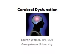
/
BRAIN TUMORS Jeanette Norden, Ph.D. BRAIN TUMORS Jeanette Norden, Ph.D.

BRAIN TUMORS Jeanette Norden, Ph.D. - PowerPoint Presentation

min-jolicoeur
min-jolicoeur . @min-jolicoeur
Follow
376 views
Uploaded On 2018-11-24

BRAIN TUMORS Jeanette Norden, Ph.D. - PPT Presentation

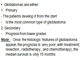
Professor Emerita Vanderbilt University School of Medicine CNS TUMORS CNS tumors neoplasms abnormal masses of cells produced by uncontrolled cellular proliferation can be Primary arising from cells in the CNS ID: 733381

tumors tumor cells brain tumor tumors brain cells clinical case cns cont primary cortex grade iii arise area growing left frontal common

Share:

Link:

Embed:

Download Presentation from below link

Download Presentation The PPT/PDF document "BRAIN TUMORS Jeanette Norden, Ph.D." is the property of its rightful owner. Permission is granted to download and print the materials on this web site for personal, non-commercial use only, and to display it on your personal computer provided you do not modify the materials and that you retain all copyright notices contained in the materials. By downloading content from our website, you accept the terms of this agreement.


Presentation Transcript

Slide1

BRAIN TUMORS

Jeanette Norden, Ph.D.

Professor Emerita

Vanderbilt University School of MedicineSlide2

CNS TUMORS

CNS tumors (neoplasms): abnormal masses of cells produced by

uncontrolled cellular proliferation

; can be

Primary:

arising from cells in the CNS

Secondary:

arising from the spread of cancerous cells from another site in the body

The most common

cancers

to metastasize to brain are lung, breast, and melanoma

CNS involvement can be the 1

st

sign of a cancer in the bodySlide3

CNS TUMORS

In the CNS, many different kinds of cells can give rise to

primary tumors;

more common of these types include:

Meningiomas

:

arise from meningeal

cells; most common primary tumor in CNS

Gliomas:

derived from cells that in development arise from

glioblasts

(immature cells that give rise to “glial” cells in the brain)

Ependymomas

:

arise from ependymal cells that line the ventricles

Oligodendrogliomas

:

arise from oligodendrocytes;

generally

slow-growing and benign

Astrocytomas

:

arise from astrocytes;

the

most common glial cell tumor in adults

There are a number of

genetic risk factors

; the only environmental risk factor identified is exposure to ionizing radiation, particularly when youngSlide4

CNS TUMORS

Brain tumors may be

Benign:

non-cancerous; the vast majority (~85%) of

meningiomas

are benign; slow-growing, non-invasive

Malignant:

cancer; fast-growing and invasive

While brain tumor cells

rarely

metastasize to other parts of the body,

they may metastasize within the brainSlide5

CNS TUMORS

Tumors can also be “graded” by their

malignancy

For example,

Astrocytomas

are “graded” from I- IV (low-grade to high-grade

); low grade are minimally invasive and slow-growing

High-grade (IV): tumors are

rapidly growing, invasive

(the cells invade adjacent normal tissue),

and “undifferentiated”

(cells look immature and in various stages of cell division);

Grade IV

astrocytomas

are also called glioblastoma

multiforme

(GBMs)

High-grade

astrocytomas

can arise de novo (primary) or evolve from low-grade

astrocytomas

(secondary)Slide6

In development,

Glioblasts

give rise to oligodendrocytes, ependymal cells and astrocytes; all of these cell types are capable of mitosis (cell division to create new cells) throughout life

Neuroblasts

give rise to neurons; neurons are post-mitotic cells, meaning that they are not capable of cell division in adults; thus, they do not form tumors in the adult brain

Oligodendrocytes

Non-mitotic

Mitotic

WHY THERE ARE NO “NEURON” TUMORS IN THE ADULT CNSSlide7

CNS TUMORS

CNS TUMORS MAY PRODUCE BOTH GENERALIZED AND LOCALIZED SIGNS/SYMPTOMS

GENERALIZED SYMPTOMS WOULD INCLUDE

HEADACHE, SEIZURES, CONFUSION

LOCALIZED SIGNS/SYMPTOMS DEPEND ON

WHERE

THE TUMOR IS

; ADDITIONAL SIGNS/SYMPTOMS WILL BE SEEN AS THE TUMOR GROWS AND EXPANDS TO INVOLVE NEARBY AREAS

IN GENERAL, ONSET OF SIGNS/SYMPTOMS IS

PROGRESSIVESlide8

In infants and small children, tumors can arise from “immature” cells

Blastoma

:

neoplasms which are composed of immature, undifferentiated cells

Medulloblastoma

:

a highly malignant cancer occurring primarily in infants/young children; named because the tumors are composed of immature cells that look like primitive cells of the medullary area of the developing neural tube

M

ost commonly occur in the vermis (midline area) of the cerebellum – a structure involved in balance, equilibrium, and coordination of learned, skilled motor movementSlide9

ALL BRAIN TUMORS ARE POTENTIALLY LIFE-THREATENING BECAUSE OF THE POSSIBILITY OF BRAIN HERNIATION

Herniation of the brain can cause death; for example, tonsillar herniation produces pressure on cardiovascular and respiratory centers in the medulla, causing deathSlide10

BRAIN TUMORS

While tumors can occur in the spinal cord, we will confine our discussion to brain tumors – reinforcing that understanding the functional anatomy of the brain is the key to understanding how physicians come to an

anatomic diagnosis

Our discussion

of Clinical Cases will

involve 4 types of tumors:

Medulloblastoma

Meningioma

Glioblastoma

multiforme

AstrocytomaSlide11

CLINICAL CASE I

Cindy is a 28 month old girl, brought to the pediatrician by her Mother who was concerned because the little girl was falling frequently and seemed to have very poor balance. The problems had been present since

C

indy had begun to walk (at 13 months), and were getting progressively worse; the child had also become increasingly irritable and withdrawn. The Mother also reported that the child vomited frequently. Upon examination, Cindy was seen to have a broad-based ataxic gait; she required help in sitting, standing, or walkingSlide12

Medulloblastoma

Tumor cells in the midline of the cerebellum; causes

Wide-based gait, ataxia, and truncal instability

Nausea and vomiting probably secondary to increased intracranial pressure, as well as pressure on medulla from expanding tumorSlide13

WHY MEDULLOBLASTOMA IS SO DANGEROUS

It is a malignant cancer

Often not diagnosed early, because infants are “naturally” ataxic (uncoordinated)

Most common site is the midline area of the cerebellum which is close to the 4

th

ventricle (one of the cavities in the brain that contains

Cerebrospinal Fluid [CSF]);

malignant cells get into CSF and “seed”

to

other areas of the brain

4th ventricle can also be blocked by cancer cells – and a hydrocephalus (build-up of CSF in ventricles) can occur which can also cause the brain to herniateSlide14

WHY MEDULLOBLASTOMA IS SO DANGEROUS

4

th

VentricleSlide15

MENINGIOMA

Most common primary brain tumor (~30% of all brain tumors) in adults

Generally, slow-growing benign (~85%) tumors

Meninges (connective tissue sheaths that surround the brain and spinal cord), from outer to inner

DURA

ARACHNOID

PIASlide16

CLINICAL CASE II

A 47

yo

Caucasian woman (Mrs. T. D.) goes to her gynecologist for her yearly examination. She tells the gynecologist that over the last year she has been having headaches and depression. No neurological exam was deemed necessary – on the assumption that her headaches and depression were due to perimenopause.

Two months later, Mrs. D. was found unconscious by her husband and brought into the Emergency Department by ambulance. He reports that she had been having severe headaches and had been very depressed and difficult to get along with over the last few months.Slide17

MENINGIOMA

MENINGIOMA of the left frontal lobe

Depression was the result of compression of the left frontal lobe; headaches and death were due to increased intracranial pressure and eventual herniation of the brainSlide18

GLIOBLASTOMA MULTIFORME

Glioblastoma

multiforme

(GBMs) are highly malignant tumors (Grade IV

astrocytomas

); they are

rapidly growing, invasive, with “undifferentiated” cellsSlide19

CLINICAL CASE III

A 69

yo

African-American male professor (Mr. D. M.) at a major medical school has a seizure during a departmental faculty meeting. He is immediately taken to the Emergency Department where an emergent CT shows a mass in his right hemisphere involving

the frontal/parietal lobe area/underlying axons.

Consultation with neurosurgery indicates that the tumor is inoperable because of how it has invaded surrounding tissue; thus, surgery would require the resection or removal of a large amount of normal tissue. Chemotherapy and radiation were begun in order to shrink the tumor mass and to try and slow the tumor growth.Slide20

CLINICAL CASE III, Cont.

The initial neurological examination revealed

Papilledema

(Due to increased intracranial pressure)

W

eakness on the left

(Involvement of primary motor cortex [Area 4]/underlying axons on the right)

Loss of fine touch, vibration and conscious proprioception on the left

(Involvement of primary somatosensory cortex [Areas 3, 1 & 2]/underlying axons on the right)

When asked if he was experiencing headaches he said “yes” and that they were worse in the morningsSlide21

CLINICAL CASE III, cont.

Normal fundus

Appearance of fundus under conditions of increased intracranial pressure

(

papilladema

)Slide22

CLINICAL CASE III, Cont.

Area 4 – primary motor cortex

Areas 3, 1 & 2 – primary somatosensory cortex

A tumor is

compromising Areas

4 and 3, 1 &

2, and underlying axons

Mr. M. has

headaches

that are worse in the mornings because of the redistribution of CSF (in the presence of a tumor), when going from a lying down to a standing positionSlide23

CLINICAL CASE III, Cont.

After chemotherapy and radiation, Mr. M. had a few good months. He then developed (in addition to previous signs/symptoms)

Contralateral (left-sided) neglect

(Due to growth of the tumor to include posterior parietal cortex/underlying axons on the right)Slide24

CLINICAL CASE III, Cont.

CONTRALATERAL (LEFT) NEGLECT

Tumor has expanded to now include posterior parietal cortex/underlying axons, causing

contralateral (left) neglect of the body & world

; also extends to memory

Posterior Parietal CortexSlide25

CLINICAL CASE III, Cont.

CONTRALATERAL NEGLECTSlide26

CLINICAL CASE III, Cont.

Note how the tumor has invaded normal tissue so that it is difficult to tell the difference between normal tissue and the tumorSlide27

CLINICAL CASE IVAstrocytoma

A 47

yo

Caucasian male (Mr. R. W.) goes to his Primary Care Physician for his yearly “wellness” visit. During the neuro portion of his physical examination, the physician notes that Mr. W. has lost his sense of smell, and that he has a “grasp” reflex.

After being referred to a Neurologist, a CT is done and a large tumor is identified in the anterior cranial fossa.Slide28

CLINICAL CASE IV, Cont.

CT WITH CONTRAST

(Tumors lack a blood-brain barrier which is why this tumor is showing up bright “white” when a contrast agent is given)Slide29

CLINICAL CASE IV, Cont.

The tumor is at the base of the brain, anteriorly

The tumor crosses the midline and has affected both

frontal lobes and olfactory structures

The brain is “upside down” so that the ventral underside of the brain is visible; the blue circle indicates the extent of the tumor

FRONTAL LOBE

OLFACTORY BULBS

FRONTAL

LOBESlide30

CLINICAL CASE IV, Cont.

The loss of smell (anosmia) is because the olfactory bulbs have been affected bilaterally

The “grasp” reflex is a reflex

seen normally

in infants; it is inhibited by the frontal lobe during development; thus, the re-appearance of the reflex in an adult is a pathological sign; it signals compromise of the

prefrontal cortexSlide31

CLINICAL CASE IV, Cont.

Mr. W. had surgery to remove the tumor; he has done well. He does show persistent anosmia (they were unable to save the olfactory bulbs) and difficulty “inhibiting” his behavior. The latter is due to damage to the prefrontal cortex which occurred both because of the tumor and its removal.

Post-surgical MRI; area where the tumor was removed is filled with CSFSlide32

TAKE-HOME MESSAGES

If you or a loved one experience progressive headache, confusion, memory loss, loss of function (motor, sensory,

etc

), or other events (like a seizure), notify your Primary Care Physician!

If a brain tumor is identified, survival depends on many factors – but

early diagnosis

is important if surgery is an option

Also remember: CNS involvement could be the 1

st

sign of a cancer in the body – again early detection is important!