/
The winged helix proteins constitute a subfamily within theof the wing The winged helix proteins constitute a subfamily within theof the wing

The winged helix proteins constitute a subfamily within theof the wing - PDF document

min-jolicoeur
min-jolicoeur . @min-jolicoeur
Follow
447 views
Uploaded On 2015-12-08

The winged helix proteins constitute a subfamily within theof the wing - PPT Presentation

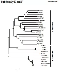
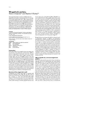
Ketan S Gajiwala SBA115QXD 02172000 1211 Page 110 Winged helix proteins model for LexA interactions with DNA suggests an HNF3glike mode of DNA binding 12 The E2F family of transcription fa ID: 218089

Ketan Gajiwala SBA115.QXD 02/17/2000

Share:

Link:

Embed:

Download Presentation from below link

Download Pdf The PPT/PDF document "The winged helix proteins constitute a s..." is the property of its rightful owner. Permission is granted to download and print the materials on this web site for personal, non-commercial use only, and to display it on your personal computer provided you do not modify the materials and that you retain all copyright notices contained in the materials. By downloading content from our website, you accept the terms of this agreement.


Presentation Transcript

The winged helix proteins constitute a subfamily within theof the winged helix/fork head motif in 1993, a large number ofhave been characterized by X-ray crystallography and solutionNMR spectroscopy. Recently, a winged helix transcriptionfactor (RFX1) was shown to bind DNA using unprecedentedmajor groove. This surprising observation suggests that theclasses with radically different modes of DNA recognition.Addresses Ketan S Gajiwala SBA115.QXD 02/17/2000 12:11 Page 110 Winged helix proteins model for LexA interactions with DNA suggests an HNF-3g-like mode of DNA binding [12]. The E2F family of transcription factors controls genesinvolved in growth and DNA replication [13]. DNA bindingby the E2F proteins is enhanced upon heterodimerizationwith members of the DP family, a distant relative of E2F. The(Figure3) [6Arg-Arg-X-Tyr-Asp motifs found on the H3 recognitionwith one half of a palindromic DNA target (5sion and a shortened helix H3. An arginine sidechain from theof the DNA near the T-rich region of the consensus binding, however, the E2F4–DP2 complex does not containsignificant difference in the behavior of these proteins con-to DNA binding by the E2F4–DP2 heterodimer—they areMoreover, there is an extensive protein–protein interfacebetween helices H1 and H2 of the other. This observation. This observationFokIis a restriction Figure 2 RFX1GH5+Rap30ADAR1DP2HNF-3QQWLLDNYE-------GVSLPRSTLYNHYLLHSQE--LEPVNAASFGKLIRS-FM-GLRTRRLGRGN--KYHYYGEEMIAA-RA-------RGGSSRQSIQKYIKSHYKVGHNADLQIKLSIRR-LAVLKQTKGVTG--SGSFRLAKKQRAFLKLYMITMTEQERLYGLKLLEVLRSEFK-GFK-----NHTEVYRSLHELLDLKQIKVKKE-LQEVVLYQFKDYEAADPNRLRLLSLLARSELCVGDLAQAIGV----------S-AVSHQLRSLRN-----LVSYRKGR------VYYQ-DHH-ALYQEELVKAFKALLKEEKFS-GEIVAALQEQGF-NNQSKVSRMLTKFGGAVRTRMVYCLPAELGNEKTATILITIAKKDFITAAEVREVHPD--------GNAVVNSNIGVLIKK-LVVEKSGDGLIITGEAQDISGEFAVLKALVSHPREPLSRDKLMNLARGR-Y-------MERSIDVQISRLR---IQTVW-LGYVFVMKDNTVPLKLIALLA----EFHSGEQLGETLG-----------AAINKHIQTLRD---VDVFTVPGKGYSLP-----IADKQHVLDMLFSAFKHQYYNLKDLVDITK-------------PVVYLKEILKEI-GVQNVGKGIH----NTWELKPEYRHYARQQEVFDLIRDHI-MPPTRAEIAQRLG-----------RSPNAAEEHLKALA-IQIVSGQ--------SRGIR-----LKEYVRTRRALILEILKAGSLKIEQIQDNLKKLG----------VIWTIENDIKGLI-FIEIKGRFYQLKDHSIY---QEQRILKFLEELGGKATTAHDLSGKLG-------------PKKEINRVLYSLA-LQKEAGTPPLWKIAVKPPYSYIALITMAILQSPQKKLTLSGICEFISNRFP-YREKFPAWQNSIRHNLSLNDCFVKIPREPGNPGKGNYWTLD-QSEDMSRHE-GLLTTKFVSLLQEAKD--LDLKLAADTLA-------------QKRRIYDITNVLE-LIEKKSKNSIGKGLRHFSMKVCEKVQRKGTTSYNEVADELVSEFTN--YDQKNIRRRVYDALNVL-NIISKEKKEIKWIGLPHAKPPYSYISLITMAIQQAPGKMLTLSEIYQWIMDLFPYYRENQQRWQNSIRHSLSFNDCFVKVARSPDKPGKGSYWALHPSSGNMFENGCYLRRQARFKLA|**|*****|***|**|***|*|*||120130140150160170180190200210H1S1H2TH3S2W1S3W2 Current Opinion in Structural Biology respectively. Secondary structure (top, where T denotes turn region),the DBD (indicated by an asterisk) refer to HNF-3(+) indicates that the structure was determined using solution NMRspectroscopy. Residue color coding: red, specific DNA contacts; blue,nonspecific DNA contacts; green, water-mediated DNA contacts;orange, mapped to the protein–DNA interface by solution NMRspectroscopy; yellow, DNA contacts according to LexA dockingcalculation; and cyan, judged to be near DNA using biochemicalassays. Topology of the winged helix fold. H2H3S1S2S3NCW1W2 SBA115.QXD 02/17/2000 12:11 Page 111 Genesis is a 465-residue transcription repressor whoseexpression is restricted to embryonic stem cells and cer-tain tumor cell lines [16]. It contains a winged helix DBDthat has been extensively characterized via solution NMR(Figure3) [17,20]. The major structural differ-(Figure2) [21]. Solution NMR studies of a genesis–DNA2) [21]. Solution NMR studies of a genesis–DNA••]. NMR dynamicalstudies of the free protein have shown that its two wingsundergo collective motions [20]. The motions of wing W2go collective motions [20]. The motions of wing W2••]. Thissituation is reminiscent of the HNF-3g–DNA complex, inwhich a single protein–DNA contact maps to the base ofwing W1.Taken together, these results imply that themarkedly different DNA sequences, when nine out of ten Protein–nucleic acid interactions Winged helix proteins recognizing B-formDNA. RFX1 binds to the X-box as a symmetricdimer. E2F4 and DP2 bind DNA as aheterodimer. For clarity, E2F4 and DP2 areshown separately, with their heteromericpartner in gray. E2F4 and DP2 recognizecontiguous sites, whereas the RFX1within the X-box. Color coding: red,helix;green,strand; orange, random-coil region; yellow,DNA backbone; blue, target DNA sequence.of winged helix DBDs on HNF-3recognition helices are colored red. (b)Yellow,Rap30 (PDB code 1bby); blue, replicationterminator protein (PDB code 1bm9); pink,biotin repressor BirA (PDB code 1bia); cyan,globular domain of histone H5 (GH5; PDBcode 1hst). (c)Gray, I (PDB code 2fok);brown, ADAR1 (PDB code 1qbj); lilac, MotA(PDB code 1bja); blue, OmpR (PDB code1opc). (d)Blue, ArgR (PDB code 1aoy); lilac,LexA (PDB code 1lea); yellow, SmtB (PDB Current Opinion in Structural Biology (b)(c)(d)HNF-3GenesisE2F4RFX1DP2DP2 SBA115.QXD 02/17/2000 12:11 Page 112 tution being serine to asparagine; Figure2). etal.al.20-residue segment adjacent to the N terminus of therecognition helix of HNF-3bwith correspondingsequences from HFH-1 allowed the HNF-3b–HFH-1chimera to recognize the DNA target of HFH-1. Similarderived from HFH-2. This effect is thought to be respon-sible for the observed differences in the DNA-bindingferences in the DNA-bindingpossess an identical pentapeptide (Phe-Pro-Tyr-Tyr-Arg)in the turn between helices H2 and H3 (Figure2); how-ever, the three preceding residues and five residuesfollowing this segment are very different and may beferent and may be•]. The structure of this apo DBD isvery similar to that of HNF-3g. When incubated with anappropriate eukaryotic gene promoter, significantwhich behaves like guanine in pre-mRNAs—giving risegiving risealso binds to the Z conformation of DNA through its N-terminal Zadomain. The co-crystal structure of the Zadomain of human ADAR1 bound to a short Z-DNA frag-ment showed that it is a winged helix protein (Figure3)3)••]. Zabinds to Z-DNA using residues from recogni-tion helix H3 and the C-terminal base of wing W1. Theonly protein–DNA major groove contact is a van der Waalsnot significantly different from its DNA-bound formferent from its DNA-bound form•], effectively ruling out an induced-fit mode ofAtypical DNA recognition by a winged helix-CGTTACCATG-GTAACG-3) (Figure3) (KSGajiwala Gajiwala RFX1–DNA complex revealed that wing W1 of RFX1makes most of the contacts with DNA via its major groove.The so-called recognition helix (H3) overlies the minorgroove, where it makes a single DNA contact using a lysinesidechain. All other well-characterized winged helix DBDsuse their H3 recognition helices to bind the major groove.Like HNF-3g, most RFX1–DNA interactions involvepolar residues making direct or water-mediated interac-tions. Two van der Waals interactions with thymine methylmethylated DNA targets. gets. RFX co-crystal structure, two copies of the protein makea symmetric complex in which the two monomers have nointermolecular interactions (distance of closest approach@10Å). In the absence of direct protein–protein interac-molecule causes the minor groove to widen by over 3Åsite of the X-box (Figure3a).functional classes. Figure4 illustrates the surface elec-4 illustrates the surface elec-gandRFX1. In every case, the most basic face of the proteinis responsible for DNA binding. For HNF-3g, DP2,E2F4 and genesis, this basic surface electrostatic featurecorresponds to the H3 face, which interacts with themajor groove of DNA. In RFX1, the W1 face displays thelargest number of basic residues and is responsible forgest number of basic residues and is responsible for•] and the linker histone proteinGH5 [33] are outliers. These proteins exhibit clusteringof basic residues on their wing W1 surfaces and largelyneutral H3 faces (Figure4), as seen in the RFX1–DNAexample, Lys85 of GH5 is maximally protected fromys85 of GH5 is maximally protected fromequivalent to Arg58 in wing W1 of RFX1. The RFX1-based model of GH5–DNA places Lys85 in closeproximity to DNA. We propose that SmtB and GH5 are Winged helix proteins SBA115.QXD 02/17/2000 12:11 Page 113 thesemolecules.molecules.HNF-3aacts as a transcriptional activator of a genetic pro-gram leading to the orderly differentiation of thecells [37]. Surprisingly, HNF-3tributes to this process, but as a transcriptional repressor,not an activator. Mutations in either of these winged helix. Mutations in either of these winged helixdevelopmental potentiators of endodermal tissue by bind-ing to regulatory sequences in the promoters of targetget)Winged helix proteins also figure prominently in the life. Normally,insulin-like growth factor, more than doubles the life span, more than doubles the life spandepends on the activity of a gene known as daf-16, whichencodes an HFH protein. Together, the insulin-like mole-C.elegans elegans daf-2-like func-tions, which are mediated, at least in part, by interferingwith the binding of HNF-3 proteins to a DNA targetgetConclusionsX-ray crystallographic and solution NMR studies ofwinged helix proteins and their complexes with DNA haveshown that the motif is extremely versatile. These proteinsexhibit two different modes of DNA binding and appear to Protein–nucleic acid interactions Surface electrostatic properties of wingednegative electrostatic potential, blue denotespositive electrostatic potential and whitedenotes neutral. DNA is drawn as an atomicstick figure. Panels of DNA binding by winged helix proteins.mechanism of DNA recognition by RFX1. Thebasic W1 surface of RFX1 is approximated toGH5, respectively. Current Opinion in Structural Biology (a)(b)(c)(d)(e)(f)HNF-3RFX1DP2GH5GH5 SBA115.QXD 02/17/2000 12:11 Page 114 recognition by these and, possibly, other HTH proteins.Papers of particular interest, published within the annual period of review,of outstanding interest1.Costa RH, Grayson DR, Darnell JE Jr: Multiple hepatocyte-enrichednuclear factors function in the regulation of transthyretin andMol Cell Biol 1989, 2.Weigel D, Jurgens G, Kuttner F, Seifert E, Jackle H: The homeoticgene fork head encodes a nuclear protein and is expressed in theterminal regions of the Cell1989, :645-658.3.Clark KL, Halay ED, Lai E, Burley SK: Co-crystal structure of theHNF-3/fork head DNA-recognition motif resembles histone H5.1993, 4.Brennan RG, Matthews BW: The helix-turn-helix DNA binding1989, 264:1903-1906.5.Wilson KP, Shewchuk LM, Brennan RG, Otsuka AJ, Matthews BW:biotin holoenzyme synthetase/bio repressorcrystal structure delineates the biotin- and DNA-binding domains.Proc Natl Acad Sci USA 1992, :9257-9261.6.Zheng N, Fraenkel E, Pabo CO, Pavletich NP: DNA recognition by the heterodimeric cell cycle transcriptionfactor E2F-DP. Genes Dev 1999, :666-674.The structure of E2F–DP shows the roles played by winged helix motifs inprotein–protein interactions and cognate DNA recognition.7.Brennan RG, Takeda Y, Kim J, Anderson WF, Matthews BW:Crystallization of a complex of cro repressor with a 17 base-pairoperator. 1986, 8.Finnin MS, Cicero MP, Davies C, Porter SJ, White SW, Kreuzer KN:The activation domain of the MotA transcription factor fromEMBOJ 1997, :1992-2003.9.Kussie PH, Gorina S, Marechal V, Elenbaas B, Moreau J, Levine AJ,Pavletich NP: Structure of the MDM2 oncoprotein bound to thep53 tumor suppressor transactivation domain. Science 1996,274:948-953.10.Lai E, Clark KL, Burley S, James J, Darnell E: factor 3/fork head or “winged helix” proteins: a family oftranscription factors of diverse biological function. Proc Natl AcadSci USA 1993, :10421-10423.11.Fogh RH, Ottleben G, Ruterjans H, Schnarr M, Boelens R, Kaptein R:Solution structure of the LexA repressor DNA binding domaindetermined by 1H NMR spectroscopy. EMBO J 1994, :3936-3944.12.Knegtel RM, Fogh RH, Ottleben G, Ruterjans H, Dumoulin P, Schnarr M,Boelens R, Kaptein R: A model for the LexA repressor DNA1995, :226-236.13.Slansky JE, Farnham PJ: Introduction to the E2F family: proteinstructure and gene regulation. Curr Top Microbiol Immunol 1996,208:1-30.14.Wah DA, Hirsch JA, Dorner LF, Schildkraut I, Aggarwal AK: Structureof the multimodular endonuclease FokI bound to DNA. 1997, :97-100.15.Skowron P, Kaczorowski T, Tucholski J, Podhajska AJ: Atypical DNA-binding properties of class-IIS restriction endonucleases:evidence for recognition of the cognate sequence by a FokImonomer. 1993, 93, Gene 1994, 14116.Sutton J, Costa R, Klug M, Field L, Xu D, Largaespada DA, Fletcher CF,Jenkins NA, Copeland NG, Klemsz M, Hromas R: helix transcriptional repressor with expression restricted to1996, 17.Jin C, Liao X: Backbone dynamics of a winged helix protein and itsDNA complex at different temperatures: changes of internalJMol Biol 1999,:641-651.The authors show that local motions in the apo form of genesis are temper-ature-dependent. DNA binding damps out the motion of selected portions ofthe protein, implicating them in DNA contacts.18.Jin C, Marsden I, Chen X, Liao X: Dynamic DNA contacts observedin the NMR structure of winged helix protein-DNA complex. 1999, 289:683-690.This study shows that residues from both the recognition helix (H3) and wingW1 of the transcription factor genesis (HFH-2) make interactions with DNA.DNA binding induces the formation of a short helix in a region of polypeptidechain that is random coil in the apo protein structure reported in [22].19.Marsden I, Jin C, Liao X: Structural changes in the region directlymechanism to explain the observed changes in the sequence-specific binding of winged helix proteins. 1998,:293-299.The results of NMR spectroscopy suggest that the DNA-binding specificity20.Jin C, Marsden I, Chen X, Liao X: relaxation NMR. Biochemistry 1998, :6179-6187.21.Marsden I, Chen Y, Jin C, Liao X: specificity of winged helix proteins is mediated by a structuralchange in the amino acid sequence adjacent to the principal DNABiochemistry 1997, :13248-13255.22.Overdier DG, Porcella A, Costa RH: The DNA-binding specificity ofamino-acid residues adjacent to the recognition helix. Mol Cell1994, :2755-2766.23.Groft CM, Uljon SN, Wang R, Werner MH: between the Rap30 DNA-binding domain and linker histone H5:implications for preinitiation complex assembly. Proc Natl AcadSci USA 1998, :9117-9122.The similarity of the Rap30 DNA-binding domain to HNF-3histones suggests functional similarity in transcription factor IIF binding to pro-moter DNA during assembly of an RNA polymerase II transcription complex.24.Melcher T, Maas S, Herb A, Sprengel R, Seeburg PH, Higuchi M:Amammalian RNA editing enzyme. 1996, :460-464.25.Schwartz T, Rould MA, Lowenhaupt K, Herbert A, Rich A: Crystalstructure of the Zalpha domain of the human editing enzymeADAR1 bound to left-handed Z-DNA. Science 1999, 284:1841-1845.This structure shows that the winged helix motif can recognize the left-hand-ed Z-conformation of DNA, independent of DNA sequence. 26.Schade M, Turner CJ, Lowenhaupt K, Rich A, Herbert A: Structure-function analysis of the Z-DNA-binding domain Zalpha of dsRNAadenosine deaminase type I reveals similarity to the (alpha +beta) family of helix-turn-helix proteins. EMBOJ 1999, :470-479.27.Schade M, Turner CJ, Kuhne R, Schmieder P, Lowenhaupt K, Herbert A,Rich A, Oschkinat H: The solution structure of the zalpha domainof the human RNA editing enzyme ADAR1 reveals aprepositioned binding surface for Z-DNA. Proc Natl Acad Sci USA1999, :12465-12470.This apo protein structure shows that the Zdomain of ADAR1 does notchange on binding to Z-form DNA, ruling out induced fit.28.Iwama A, Pan J, Zhang P, Reith W, Mach B, Tenen DG, Sun Z:Dimeric RFX proteins contribute to the activity and lineagespecificity of the interleukin-5 receptor alpha promoter throughactivation and repression domains. Mol Cell Biol 1999, :3940-3950.29.Siegrist CA, Durand B, Emery P, David E, Hearing P, Mach B, Reith W:RFX1 is identical to enhancer factor C and functions as atransactivator of the hepatitis B virus enhancer.Mol Cell Biol1993, :6375-6384.30.Cornille F, Emery P, Schuler W, Lenoir C, Mach B, Roques BP, Reith W:DNA binding properties of a chemically synthesized DNA bindingdomain of hRFX1. Nucleic Acids Res 1998, :2143-2149. Winged helix proteins SBA115.QXD 02/17/2000 12:11 Page 115 31.Nicholls A, Sharp K, Honig B: Protein folding and association:insights from the interfacial and thermodynamic properties ofhydrocarbons. 1991, :281-296.32.Cook WJ, Kar SR, Taylor KB, Hall LM: Crystal structure of thecyanobacterial metallothionein repressor SmtB: a model formetalloregulatory proteins. JMol Biol 1998, :337-346.The first structure of a metal-binding DNA repressor reveals a winged helix motif.33.Ramakrishnan V, Finch JT, Graziano V, Lee PL, Sweet RM: Crystalstructure of globular domain of histone H5 and its implications1993, 36234.Thomas JO, Wilson CM: Selective radiolabelling and identificationof a strong nucleosome binding site on the globular domain ofEMBOJ 1986, :3531-3537.35.Shih DQ, Navas MA, Kuwajima S, Duncan SA, Stoffel M: Impairedglucose homeostasis and neonatal mortality in hepatocyteProc Natl Acad Sci USA1999, :10152-10157.36.Kaestner KH, Katz J, Liu Y, Drucker DJ, Schutz G: winged helix transcription factor HNF3alpha affects glucosehomeostasis and islet glucagon gene expression Dev 1999, :495-504.37.Vaisse C, Kim J, Espinosa R III, Le Beau MM, Stoffel M: Pancreaticislet expression studies and polymorphic DNA markers in thegenes encoding hepatocyte nuclear factor-3alpha, -3beta, 1997, :1364-1367.38.Duncan SA, Navas MA, Dufort D, Rossant J, Stoffel M: a transcription factor network required for differentiation andmetabolism. Science 1998, 281:692-695.39.Zaret K: Developmental competence of the gut endoderm: geneticpotentiation by GATA and HNF3/fork head proteins. Dev Biol1999, 20940.Lin K, Dorman JB, Rodan A, Kenyon C: daf-16: an HNF-3/forkheadfamily member that can function to double the life-span ofScience 1997, 41.Hsin H, Kenyon C: Signals from the reproductive system regulate1999, :362-366.42.O’Brien RM, Noisin EL, Suwanichkul A, Yamasaki T, Lucas PC,WangJC, Powell DR, Granner DK: hormone-regulated expression of the phosphoenolpyruvatecarboxykinase and insulin-like growth factor- binding protein 1Mol Cell Biol 1995, :1747-1758. Protein–nucleic acid interactions SBA115.QXD 02/17/2000 12:11 Page 116