/
The gallbladder, gallstones, and beyond…. The gallbladder, gallstones, and beyond….

The gallbladder, gallstones, and beyond…. - PowerPoint Presentation

myesha-ticknor
myesha-ticknor . @myesha-ticknor
Follow
351 views
Uploaded On 2018-10-30

The gallbladder, gallstones, and beyond…. - PPT Presentation

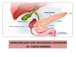
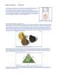
Leslie Kobayashi MD January 31 2012 Anatomy Liver Bile ducts Pancreas Duodenum Transverse colon Anatomy Fundus Body Infundibulum Neck Cystic duct Spiral Valves of ID: 702908

cbd stones risk bile stones cbd bile risk cholecystitis duct complications pregnancy gallbladder increased cystic stone conversion gbw injury rate hida open

Share:

Link:

Embed:

Download Presentation from below link

Download Presentation The PPT/PDF document "The gallbladder, gallstones, and beyond..." is the property of its rightful owner. Permission is granted to download and print the materials on this web site for personal, non-commercial use only, and to display it on your personal computer provided you do not modify the materials and that you retain all copyright notices contained in the materials. By downloading content from our website, you accept the terms of this agreement.


Presentation Transcript

Slide1

The gallbladder, gallstones, and beyond….

Leslie Kobayashi, MDJanuary 31, 2012Slide2

Anatomy

LiverBile ductsPancreasDuodenumTransverse colonSlide3

Anatomy

FundusBodyInfundibulum/Neck

Cystic duct

Spiral Valves of

HeisterSlide4

Anatomy

Triangle of calotBorders: CHD, cystic duct, liver edge

Contents: Cystic artery, node of

CalotSlide5

Ductal Anatomy

Right and Left Hepatic ductsCommon Hepatic ductCystic ductCommon bile ductSlide6

Aberrant anatomy

VascularNormally (>90%) cystic a. arises from RHA

Replaced right hepatic a.

Replaced left hepatic a.Slide7

Bile

500-1500mL produced dailyComposition: water, electrolytes, bile salts, proteins, lipids

Ductal

epithelium products

Alkaline

phosphatase

HCO3

Hepatocyte

products

Bile in conjugated soluble form synthesized from cholesterol

Primarily

cholate

and

chenodeoxycholateSlide8

Bile

95% of bile re-absorbed into the liver via portal vein (enterohepatic circulation)85-90% in terminal ileum via active transport

10-15%

deconjugated

in colon, absorbed passively

5% excreted in stool

Cycles 6-10x daily

80% of bile stored in GB in fasting stateSlide9

GB

Function store and concentrate bileAbsorption: NaCL, H2O occurs rapidly

Secretion: mucus, H+

GB average capacity 30-50mL

Can increase to 300mL with obstruction

Normal ejection 50-70% in 30-40minSlide10

Significance

Do gallbladder problems create a significant healthcare burden?Slide11

YES!

Health burden6.2 Billion$ in US1.8 million ambulatory care visits

Increased 20% since 1980’s

Cholecystectomy

most common elective abdominal procedure in the US

750,000 annually Slide12

Why does that happen?

StonesSlide13

Types of stones

Cholesterol stones (75%)Female fat fertileBlack stones (20%)

Hemolytic diseases (Sickle cell disease)

Cirrhosis

Brown stones (5%)

Infection

PSC

*primarily form in the

ductsSlide14

Cholesterol stones

Low calcium, radiolucentCreated when fractional cholesterol content of bile increased, and with incomplete emptying of GB

Associated with obesity, rapid weight loss, Native American/Hispanic heritage, ↑TG’s, ↓HDL, Spinal cord injurySlide15

Cholesterol stones

Hormonal influenceEstrogen increases lithogenicity

of bile

Increased risk for females

Increased risk in obesity

Progesterone increases SM relaxation and bile stasis, decrease bile salt secretion

Increased risk in pregnancySlide16

Cholesterol stones

Increase risk of stone formationTPNOctreotideCeftriaxone

Decrease risk of stone formation

Statins

?

ursodiol

Slide17

Pigmented stones

Often radiopaque due to calcium bilirubinate, calcium fatty acid soaps and inorganic calcium salts

Two types

Black

BrownSlide18

Pigmented stones

BlackForm in GBBile sterileAssociated with age, hemolytic DO’s, alcoholism, cirrhosis, Gilbert’s syndrome, Cystic fibrosis, pancreatitis and TPN

Cholecystectomy

curativeSlide19

Pigmented stones

BrownForm in ducts as well as GBAlways infected 1

O

with enteric organisms, often associated with

cholangitis

Associated with parasitic infection (liver fluke)

Associated with IBD, duodenal

diverticulae

Will often recur after LC/OCSlide20

Stones: Where do they go?

And what do they do?Slide21

GSP

Choledocho

Cholangitis

Biliary

colic

CholecystitisSlide22

In the gallbladder

Incidence: 10-30% of the populationAsymptomatic (80%)Symptomatic (1-3% per year)No inflammation:

Biliary

colic

+inflammation: acute

cholecystitis

+obstruction :

choledocholithiasis

, GSP+obstruction+inflammation

:

cholangitisSlide23

Biliary colic

History Transient abdominal painOccurs after fatty meals

Exam

Benign

Labs

Normal

Ultrasound

GSSlide24

Hyperechoic

masses, dependent in location

Acoustic shadowingSlide25

Cholecystitis

HistoryProlonged painFeversNausea/emesis

Exam

Fever, tachycardia

RUQ TTP, Murphy’s sign

Labs

Leukocytosis

Mild

↑ LFT’s

Imaging

Ultrasound

HIDASlide26

Cholecystitis

GallstonesObstruction of gallbladder

Obstruction causes inflammation

Inflamed wall is thickened

Edema or emphysema of GBWSlide27

Cholecystitis

Inflammation may or may not be associated with infection 50-70% of bile cultures are positive

E. coli,

Klebsiella

, Streptococcus,

EnterobacterSlide28

Ultrasound

95% sensitivity/specificitySigns of cholecystitis

Gallstones

GBW >3mm

Pericholecystic

fluid

GBW striations or air within GBW

Sonographic

Murphy’s signSlide29

GS with GBW thickening

Normal GBW <3mm

Pericholecystic

fluidSlide30

HIDA

Cholescintigraphy: Injection of Tc99 labeled hydroxyl iminodiacetic

acid

HIDA→hepatocytes→secreted

into bile

Normal visualization of GB, CBD and SB within 30-60 min

+scan if no visualization of GB within 1hr and +uptake in CBD or SBSlide31

HIDA

Rim sign

*Sphincter, ↙CBD

Normal HIDA

Positive HIDASlide32

HIDA

False positives common in fasting patientsUp to 40-60% in critically illCan decrease false+ rate with morphine

↑sphincter of

Oddi

pressure causing preferential filling of the GBSlide33

Cholecystitis: Complications

↑Tension in GBW =↓perfusion →Necrosis of GBWGangrenous/emphysematous

cholecystitis

1% of cases, 3:1 M>F

Conversion rate 30-50%

GB Perforation

Assoc with ↑mortality (~20%)

Gallstone

ileusSlide34

Gallstone ileusSlide35

Complications

Cystic duct obstruction→ HydropsBile is absorbed but GB mucosa continues to secrete mucus

GB tense, filled with

mucinous

fluidSlide36

Complications

Mirrizi’s syndromeImpacted stone in

infundibulum

or CD →External compression of the CBD

0.7-1.4% of patients

Assc

with ↑risk of CBD injury, GB cancerSlide37

What if the stones escape the GB?Slide38

Stones in the CBDSlide39

Choledocholithiasis

History: jaundice, icterus, pruritis, dark urine,

steatorrhea

,

acholic

stools, bleeding

Exam: jaundice,

icterus

, RUQ pain, Murphy’s signLabsElevated LFT’s, INRElevated bilirubin

highest PPV 25-50%

May be normal in up to 30% of patientsSlide40

Choledocholithiasis

ImagingDilated CBD on UTZCBD <5mm risk of stone ~1%

CBD >5mm risk of stone 58%

MRCP

Sensitivity 95%

Specificity 89%Slide41

CBD dilation

Stones within the bile ductSlide42

Cholangitis

History/Exam: similar to choledocholithiasis with sepsis, septic shockLabs/Imaging: similar to

choledocholithiasis

with

leukocytosis

,

bactermia

, ±MSOF

Charcot’s triadReynolds pentad

RUQ pain, fevers, jaundice

Triad +

Δ

MS, shockSlide43

Beyond the CBDSlide44

Gallstone pancreatitis

History: epigastric pain, nausea/emesisExam: RUQ/

epigastric

TTP, SIRS

Labs: amylase/lipase ↑3x

nl

, ±↑LFT’s,

leukocytosis

Imaging: ±CBD dilation, pancreatic edema, necrosis, fluid collectionSlide45

Ranson’s criteria-Alcoholic

First 24hours: Glucose >200Age >55

LDH>350

AST>250

WBC>16k

48 hours

Ca <8

Hct

↓>10

PaO2 <60

BUN↑>5

Base Deficit >4

Sequestration >6LSlide46

Ranson’s criteria-Non-Alcoholic

First 24hours: Glucose >220Age >70

LDH>400

AST>440

WBC>18k

48 hours

Ca <8

Hct

↓>10

PaO2 <60

BUN↑>2

Base

Deficit >5

Sequestration >6LSlide47

Ranson’s criteria

Each category 0 or 1Add up total pointsMortality0-2 <5%

3-4 15%

5-6 40%

7-8 ~100%Slide48

TreatmentsSlide49

Treatment

MedicalSurgicalLap

Open

CBDE

ERCP

sphincterotomy

,

stent

Percutaneous

Cholecystostomy

tubeSlide50

Treating the gallbladderSlide51

Gallstone “Cleanse”

Preparation

Eat a diet high in alkaline-forming foods and low in fats for at least 3-5 days before the cleanse.

Help to gently prepare the liver by having a glass of fresh apple juice every day for 1 week prior to the cleanse. Apple juice helps to dissolve the stones

Ingredients

Epsom salts (Magnesium Sulfate): 4 tablespoons

Olive oil: 1/2 cup or 125 ml

Fresh pink grapefruit: squeeze 1/2 cup (125 ml) juice

Or use 7-8 fresh lemons/limes: squeezed into 1/2 cup juice

1 liter jar with lidSlide52

Medical

Or you could try:IVF hydrationAntibiotics

Bowel restSlide53

Medical

Ursodiol: used asMechanism: supplemental bile acid decreases lithogenicity of bile, dissolve existing stones

Indications: bridge to LC/OC, too sick for OR,

cirrhotics

, PSC, TPN

Efficacy: may ↓LFT’s in PSC/

cirrhotics

, may ↓stones/sludge on UTZ, does not ↓symptoms, prevent need for OR, stones recur after cessation of medicationSlide54

Medical

Diet: Slide55

Treatment

Failure of medical management in acute cholecystitis 32%Recurrence rate of GSP 29-63%

Surgical management results in reduced HLOSSlide56

Treatment

Timing of surgery for acute cholecystitisWithin 48hrs vs

>72hrs no difference in conversion rates, OR time, LOS

Comparing first hospitalization (<7d)

vs

delayed (>6wks)

17.5%

rqr

emergent cholecystectomy for recurrent/

unresolving

sx’s

No difference in conversion rates or CBD injurySlide57

Treatment

Timing of surgery for GSPEarly operation safe with mild pancreatitis Rason’s criteria <3

Increased conversion rate, HLOS, and operative complications in early operation in severe pancreatitis

Ranson’s

criteria

≥3Slide58

Surgical approachesSlide59

Laparoscopic

Port placementUmbilicusSubxiphoid just to the right of the

falciform

at the level of the inferior liver edge

2-3cm below costal margin in

midclavicular

line

Anterior

axillary line, below the fundus

of gallbladder Slide60

Laparoscopic

Retraction and dissection of Triangle of Calot prior to Gallbladder removal from fossa

CD may be clipped, sutured, tied, stapled

Remove gallbladder in

fundus→dome

directionSlide61

Open

Right subcostal incisionMini-

cholecystectomy

(5-8cm) incision associated with equivalent outcomes/complications and less post-op pain, decreased LOS

Dome down dissection technique

Isolate cystic artery/duct and suture

ligateSlide62

Lap vs. open

Conversion rate: 0.18-35% ave 4.7%CBD injury ratesLap 0.2-0.6%

Open 0-0.3%

Complication rate

Lap ~1.2%

Open (bile leak 1%)

LOS: shorter for LapSlide63

Difficult Cholecystectomy

RF’s for conversion Male sexObesity

↑age

Wide short cystic duct

Low surgeon case load

Gangrenous or emphysematous

chole

↑risk of conversion RR 3.2 (CI 2.5-4.2)

No ↑risk of local complications or CBD injurySlide64

Other optionsSlide65

Cholecystostomy tube

Can be transhepatic or transperitoneal no difference in outcomes

Technical success 96-98%

Resolution of symptoms 68-96%

Mortality 3-14%

Complications

Dislodged catheter 16-33%

Bleeding 1.5-1.8%

Recurrent cholecystitis 7-41%Slide66

Clearing the ductSlide67

Natural history of CBD stones

Choledocholithiasis Stones in CBD in 10-15% of symptomatic pt’s55-70% pass spontaneously

GSP20-30% of patients have CBD stones

85-90% pass spontaneously

Symptomatic

cholecystitis

4.6% +IOC at the time of LC

97.8% pass spontaneouslySlide68

Surgical approaches

CBDECan be performed lap or open

Transcystic

or via

choledochotomySlide69

Surgical approaches

CBDEImaging ductFluorscopic

guidance

Choledochoscopy

Clearing duct

Basket, snare, flush

+/- glucagon to relax sphincterSlide70

Surgical approaches

CBDECompletion cholangiogram

Clip, tie or staple cystic duct stump

Close

choledochotomy

over T-tube

+/-drain external

Success rate of duct clearance 75-95%Slide71

ERCP

Efficacy 1 procedure: 71-75% Multiple procedures: 84-95%

Mortality 0.2-0.5%

Complication rate 5-8%

Perforation

Bleeding

Pancreatitis

Cholangitis

Slide72

ComplicationsSlide73

Complications

1-2% of patients will represent with CBD stone following cholecystectomy

Dx

<2yrs post-op = retained stone

Dx

> 2yrs post-op =recurrent stoneSlide74

Other Complications

Ileus Incisional/port site hernia

Wound infection

Abscess

Biloma

/bile leakSlide75

CBD Injury

Strasberg-Bismuth classificationA-CD stump,

fossa

B/C-aberrant RHD

D-lateral injury

E-circumferential injury to major ductSlide76

Special circumstancesSlide77

Pregnancy

Increased risk of stones2-12% have stones0.05-1.2% symptomatic during pregnancy

Risk of stones increased in:

Hispanic

Pre-pregnancy obesity (4x)

Decreased by

EtOH

consumptionSlide78

Pregnancy

Biliary disease the most common non-obstetrical cause of maternal hospitalizationCholecystitis most common 40%

GSP 30%

CBD stone 20%

Biliary

colic 10%Slide79

Pregnancy

If symptomatic risk of recurrence high40%-70% recur prior to deliveryIf symptomatic risk of fetal loss high 10-20%Slide80

Pregnancy

Treatment goalsTreat infectionMaintain nutrition

Prevent contractions/preterm labor

Prevent fetal loss

Prevent maternal morbidity/mortalitySlide81

Pregnancy

Surgical management associated with fewer complications than medical management Contractions equivalent (~30%)Decreased preterm delivery, need for c-section, and recurrent symptoms

Fetal loss with LC 0-5%Slide82

Pregnancy

Ideal timing LC/OC 2nd trimester↓

preterm labor (0% vs. 40%)

fetal loss

risk of fetal malformation

Technically easier

1st

delay to 2

nd

, 3

rd

delay to postpartumSlide83

Pregnancy

ERCP can be performed safely with:Low radiation exposure

Fluoro

time 14sec-3.2min

Radiation exposure 40-310

mrad

Few complications ~7%Slide84

Pregnancy

Operative considerationsPort placement to accommodate uterusHassan vs.

Veress

likely equivalent

insufflation

pressure 10-12Slide85

Cirrhotics

Stones more common in cirrhotics (2x)Diagnosis difficult

Pain nonspecific

Elevated LFT’s nonspecific

Leukocytosis

nonspecific

GBW thickening nonspecific

HIDA may be helpfulSlide86

Cirrhotics

Management differencesIncreased operative risk Morbidity 3x

Conversion 2x

Bleeding 8x

Increased risk with

cholecystostomy

Bleeding

Ascites

/Leak Slide87

Cirrhotics

MortalityOverall acceptable 0.6-0.8%Significantly increased in Child’s C patients (17%)

LC safer than OC

Less bleeding

Shorter OR time

Shorter HLOS

Possibly lower mortality (open mortality 8-25%)Slide88

Other pathology

Acalculous cholecystitis M>F 1.5:1

4-8% of all

cholecystitis

Dx

with UTZ/HIDA

Gallbladder polyps

Gallbladder cancerSlide89

Thank You