/
A Comprehensive Review A Comprehensive Review

A Comprehensive Review - PDF document

naomi
naomi . @naomi
Follow
342 views
Uploaded On 2022-10-27

A Comprehensive Review - PPT Presentation

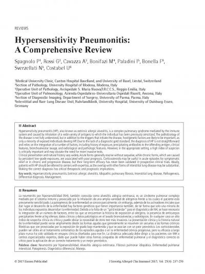
La neumonitis por hipersensibilidad NH también conocida como alveolitis alérgica extrínseca es un síndrome pulmonar complejo mediado por el sistema inmune y provocado por la inhalación de un ID: 960936

chronic x00660069 hypersensitivity lung x00660069 chronic lung hypersensitivity pneumonitis disease acute brosis diagnosis med respir clinical interstitial exposure 146

Share:

Link:

Embed:

Download Presentation from below link

Download Pdf The PPT/PDF document "A Comprehensive Review" is the property of its rightful owner. Permission is granted to download and print the materials on this web site for personal, non-commercial use only, and to display it on your personal computer provided you do not modify the materials and that you retain all copyright notices contained in the materials. By downloading content from our website, you accept the terms of this agreement.


Presentation Transcript

A Comprehensive Review La neumonitis por hipersensibilidad (NH), también conocida como alveolitis alérgica extrínseca, es un síndrome pulmonar complejo mediado por el sistema inmune y provocado por la inhalación de una amplia variedad de alérgenos frente a los cuales el paciente está J Investig Allergol Clin Immunol 2015; Vol. 25(4): 237-250 Hypersensitivity pneumonitis (HP), also known as extrinsic allergic alveolitis, is a complex syndrome that results from repeated inhalation of and sensitization to a wide variety of aerosolized antigens [1]. Clinical presentation and disease course are highly variable, and depend on factors such as intensity and duration of exposure to the causative antigen, the nature of the antigen, and host factors [2]. The immunopathogenesis of HP is poorly understood, although T-cell hyperreactivity and immune complex formation and deposition appear to play a prominent role. Notably, the disease develops in only a minority of individuals exposed to potential disease-causing antigens (ie, the majority of individuals with the same exposure are either sensitized but healthy, or do not even become sensitized), suggesting the existence of a genetic predisposition to HP [3].According to data from registries of interstitial lung diseases (ILDs) in 3 European countries, HP accounts for 4% to 15% of all ILD cases [4], but this �gure falls to 2% according to a population-based study conducted in New Mexico [5]. However, the incidence and prevalence of HP are dif�cult to estimate with precision, mainly because of the number of cases that are misdiagnosed or not recognized, prevalence varies from country to country (and even within countries) owing to geographic, seasonal, and climatic [6]. For instance, farmer’s lung—the prototype of HP with seasonal and geographic variations in incidence—occurs cattle), and in regions with both heavy rainfall and severe winter conditions[7]. It is estimated that between 1% and 19% of farmers exposed to moldy hay develop farmer’s lung [8] and that between 6% and 20% of individuals exposed to bird droppings develop bird fancier’s lung [9]. These 2 conditions are the most common type of HP, and can affect all age groups, Disease DefinitionThere is no universally agreed upon de�nition of HP[10,11]. However, there is consensus on the following key features of ) HP is a pulmonary disease which may or may not be accompanied by systemic manifestations (eg, fever and ) It is caused by the inhalation of an antigen to which the individual is sensitized and hyperresponsive; and ) It is de�ned by exposure to a given antigen, sensitization to this antigen, and the presence of clinical symptoms. Indeed, many exposed individuals develop an antigen-speci�c immune response limited to the presence of serum IgG antibodies and an increased number of lymphocytes in the lung [12], but they es in the lung [12], but they Causative AgentsAgents capable of inducing HP are found in a number of settings, including the workplace, home, and recreational environments. A wide and increasing variety of antigens different types of disease in different settings. In addition, although HP is caused by specific antigens, additional “triggers” (either genetic or environmental) may be needed to induce the disease [14], possibly explaining why, despite the universal and wide distribution of the offending antigens, only a few individuals develop HP. HP-inducing antigens are commonly classi�ed in 5 broad categories represented by disease prototypes (Table 1) [15]. The most common causes of HP are avian antigens and microbial agents [16,17]. However, Table 1. Common Types of Hypersensitivity Pneumonitis According to Major Classes of Antigens Class of AntigensSpeci�c AntigensType of DiseaseSaccharopolyspora rectivirgulaMoldy hay, grainFarmer’s lungAspergillusMoldy hay, grainFarmer’s lungAspergillusMushrooms worker’s lungTrichosporon cutaneumJapanese summer-type HPCheese washer’s lungWoodworker’s lungMycobacterium avium-intracellulareMycobacterium avium-intracellulareMist from pool

water, spraysPigeon breeder’s lung, bird fancier’s lungAvian proteins Feather beds, pillow, duvetsChemical worker’s lung in recent years, a number of exposures (eg, mycobacterial antigens, which are typically encountered in hot tubs, pools, or metal working �uids) have also been recognized as an established cause of HP [18,19]. The latency between exposure and onset of disease is variable, and ranges from months to decades [20], making it challenging for the clinician to detect the type and source of the antigen, particularly in cases of HP is the result of an immunologically induced in�ammation of the lung parenchyma (speci�cally, the disease involves the alveoli, terminal bronchioli, and interstitium) that occurs in [2]. The immunopathogenesis of the disease is likely to be similar, regardless of the causative agent, and both humoral and cellular mechanisms appear to contribute to the development of HP [21]. The genetic basis of HP is poorly understood, and, although some studies suggest that polymorphisms within human leukocyte antigen (HLA) class II genes increase the risk for HP in populations from different genetic backgrounds[22,23], no genetic factors have consistently been associated with the disease. If exposed to agents capable of inducing HP, most individuals develop immune tolerance, and inhalation of the antigen may result in a mild increase in local lymphocytes, without clinical signi�cance. However, according to a “two-hit” hypothesis, the coexistence of inducing factors (eg, antigens) and promoting factors (eg, genetic abnormalities or additional environmental exposures) may lead to the development of an exaggerated immune reaction that results in marked lung in�ammation [2]. In acute HP, lung in�ammation appears to be mediated by immune complexes, as suggested by the presence of high titers of antigen-speci�c precipitating IgG in the serum, and an increase in lung neutrophils primed for an enhanced respiratory burst [24]. Conversely, subacute and chronic HP is characterized by an exaggerated T cell–mediated immune response, in which increased T-cell migration, local proliferation, and decreased apoptosis contribute to the characteristic T-lymphocytic alveolitis [25,26]. This process is dependent on the transcription factor STAT-4 and T-bet, a major regulator of T1 lineage [27]. A number of studies have also shown that the immune response in HP is polarized toward a 1-like pattern of differentiation, which is largely mediated by IL-12 and IFN-γ [26,28]. However, it has also been shown that, Saccharopolyspora rectivirgula T cells display a preferential T17 polarization with differential expression of IL-17A and IL-22 [29], suggesting that this polarization, together with upregulation of T17 signature cytokines, may play an important role in the pathogenesis of HP. The immune processes that lead to persistent disease and progression to �brosis are less clear. However, features associated with chronic HP include an increase in CD4 T cells and in the CD4 ratio, a skewing toward T2 T-cell differentiation and cytokine pro�le as well as an exhaustion T cells [26]. Increased T17 cells following chronic inhalation of aerosolized antigens may also contribute to the development of lung �brosis (by promoting collagen deposition) [30], as shown by the protective effect against the disease of both genetic deletion of IL-17 and antibody-on of IL-17 and antibody-The “Protective” Role of Cigarette SmokingSmoking is less prevalent in patients with HP compared with unaffected controls with similar antigenic exposure [8,32,33]. Moreover, when exposed to high levels of HP antigens, smokers have lower levels of speci�c antibodies to the causative antigen. The mechanisms that account for the “protective” effect of smoking are poorly understood, but nicotine is thought to inhibit macrophage activation and lymphocyte proliferation and function [34]. In fact, mice rectivirgula and simultaneously treated with nicotine have shown a significant decrease in lung in�

;ammation [34]. Although more common in nonsmokers, when it occurs in smokers, HP is associated with a more chronic Disease ClassificationHP has been conventionally classi�ed as acute, subacute, and chronic [36], although there are no widely accepted criteria to distinguish the various forms. In addition, clinical manifestations of each form are highly variable due to a number of factors, including the intensity and duration of exposure, the nature of the antigen, and host factors; yet there may be signi�cant overlap between these forms. The observation of considerable overlap between the clinical manifestations of farmer’s lung (considered the prototype of acute HP) and pigeon breeder’s lung or bird fancier’s lung (considered the prototype of subacute and chronic HP, respectively) suggests that the pattern of antigen exposure may be as important as the antigen itself in determining disease manifestations[11,37]. Although the classi�cation scheme described above is widely used, it applies only to typical cases, and can be misleading when, for instance, acute and chronic disease coexist in the same patient, or when the clinical presentation and disease course are dif�cult to de�ne. In this regard, Selman [38] proposed an alternative classi�cation scheme based primarily on disease behavior, distinguishing between acute nonprogressive and intermittent disease, acute progressive/subacute disease; chronic nonprogressive disease, and chronic progressive disease. Recently, Lacasse and colleagues [39] used data from the HP study and divided patients (n=168) with differing clinical patterns into a limited number of categories (“clusters”). The variables included in this cluster analysis were derived from clinical history, physical examination, blood testing, chest radiograph, high-resolution computed tomography (HRCT), and bronchoalveolar lavage (BAL). A 2-cluster solution best �t the data, with patients in cluster 1 (n=41) having more frequent recurrent systemic symptoms and normal chest radiographs, and those in cluster 2 (n=127) displaying more frequent digital clubbing, hypoxemia, restrictive ventilator defect on pulmonary function tests (PFTs), and �brosis on HRCT scans. The results of this analysis differed considerably from results based on the current classi�cation of HP and, again, subacute HP was particularly dif�cult to de�ne. This new classi�cation scheme, however, needs to be validated prospectively. Various diagnostic criteria have been proposed for HP, but none of these has been validated. As such, diagnosis relies on the integration of a variety of factors, including history of antigen exposure, precipitating antibodies to the offending antigen, clinical features, inhalation challenge, BAL, and radiological and pathologic abnormalities [17]. However, in the appropriate setting, a high index of suspicion remains of critical importance and may obviate the need for more invasive testing [40]. A clinical prediction model for a diagnosis of HP has also been developed (Table 2). If all 6 predictors in the model are present, the probability of having HP is 98% [11].dyspnea, fever, and decreased forced vital capacity (FVC) and oxygen saturation a few hours (8-12) after exposure. Because the magnitude of the attack is unpredictable, the patient should be monitored closely for at least 24 hours. In the appropriate clinical setting, a positive inhalation challenge is virtually diagnostic, although false negative results may occur [46,47]. Owing to a lack of standardization (both in the inhalation protocols and the criteria used to de�ne a positive response) and because of the risk of severe reactions, the test should only be performed in selected Chest radiography is usually the �rst step in the evaluation of a patient with suspected HP. As in other ILDs, the chest radiograph reveals nonspecific findings, particularly in acute and subacute phases of the disease, and it may also be normal[48-50]. However, a variable combination of �ne nodular opa

cities and widespread ground glass opacity (GGO) may be observed. Pulmonary abnormalities associated with chronic HP may be more speci�c; indeed, the upper lobe predominance of �brotic changes (eg, reticular opacities and honeycombing) is almost invariably present in chronic HRCT may either show typical findings, which may be virtually diagnostic of HP in the appropriate clinical setting, or provide important clues that may suggest a correct [49,51]. HRCT �ndings vary widely based on the stages of the disease. Only a few reports have described HRCT abnormalities in acute HP, as HRCT is seldom performed at this stage due to the rapid resolution of symptoms [2]. However, in cases with severe clinical manifestations (eg, acute respiratory failure), acute HP on HRCT scans may resemble the exudative phase of diffuse alveolar damage (DAD)[52, 53]. GGO primarily re�ects the presence of diffuse lymphocytic interstitial in�ltration, and the differential diagnosis with other disorders manifesting with diffuse GGO (eg, opportunistic infections, pulmonary edema, and cellular nonspeci�c interstitial pneumonia [NSIP]) may not Table 2. Clinical Predictors for the Diagnosis of Hypersensitivity VariableExposure to a known offending antigen11.6-129.61.8-11.7Weight loss Source: Adapted from Lacasse et al. [11].Serum can be assayed for precipitating IgG antibodies (precipitins) against various potential antigens [41], but precipitins are only a marker of exposure. Indeed, up to 40% of HP in the absence of clinically signi�cant disease and without long-term sequelae [42,43]. The prevalence of serum precipitins in asymptomatic bird breeders is even higher, probably due to more intense and prolonged exposure to inciting antigens [44]. Nevertheless, in the appropriate clinical setting, a positive test supports the diagnosis of HP. Conversely, the absence of serum precipitins does not rule out HP, primarily because most commercial assays test for only a small fraction of the potential causative antigens. Furthermore, in acute and chronic HP (particularly if the underlying pathology is usual interstitial pneumonia [UIP]), the test tends to lose sensitivity, , Inhalation ChallengeRe-exposure of the patient to the suspected inciting agent after a period of avoidance can reveal a relationship between the exposure being analyzed and the development of symptoms and laboratory and functional abnormalities, thus supporting a diagnosis of HP. A positive test is characterized by cough, Figure 1.Chronic hypersensitivity pneumonitis. High-resolution computed tomography showing a reticular pattern with subpleural distribution and distortion of the lung parenchyma suggestive of established brosis. attenuation suggests an acute exacerbation in this patient who presented with acute respiratory failure. be straightforward[54-56]. Furthermore, GGO superimposed on a background of chronic changes may be observed in acute exacerbation of chronic HP or in chronic cases following intense exposure to antigens (Figure 1) [57]. Conversely, HRCT abnormalities observed in the subacute phase of the disease may be more speci�c, and include poorly de�ned nodules, GGO, and areas of decreased attenuation (Figure(FigurePoorly defined nodules may be the predominant or only abnormality in patients with subacute HP. They are less than mm in diameter and are generally numerous and have a typical centrilobular distribution, although few nodules with an atypical distribution may sometimes be seen. The nodules may be seen throughout the lungs, although they typically predominate in the mid to upper zones. Centrilobular nodules produce a characteristic appearance that narrows the differential diagnosis to either smoking- or occupation-related lung disorders, thus making the patient’s history critical for securing a �nal diagnosis. However, in smoking-related ILD the nodules are typically more patchy and less widespread [49,58-60]. Areas of decreased attenuation are distributed in a lobular fashion, mirroring the air trapping on expiratory CT that is caused by antigen deposition

in the small airways (Figures 1,3). Although this HRCT pattern may be observed in up to 90% of patients, it is usually limited in extent and less pronounced than in obliterative bronchiolitis associated with other conditions. The variable combination of areas of decreased attenuation, GGO, and normal lung may produce the so-called head-cheese pattern, which is highly suggestive of HP, although it may also be observed in respiratory bronchiolitis-associated ILD [51,61-64]. Coexisting thin-walled lung cysts have been reported in 13% of patients with subacute HP [49,58,59,65], and are believed to be caused by partial bronchiolar obstruction by peribronchiolar lymphocytic in�ltration. These cysts are usually few in number, range in size from 3 to 25 mm, and resemble those seen in lymphocytic interstitial pneumonia (LIP). However, LIP is often associated with other conditions, such as connective tissue diseases or lymphatic disorders (eg, human immunodeficiency virus Consolidation is an unusual �nding, and may represent foci of organizing pneumonia or superimposed infection. Irregular nodules larger than 10 mm in diameter may also represent focal areas of organizing pneumonia [49,63]. HP classically occurs in nonsmokers; yet, it may be associated with emphysema. one-third of cases) and prominent �nding in patients with farmer’s lung. Emphysema associated with chronic air�ow obstruction may be related to massive intermittent exposure, whereas chronic low-level exposure, as in bird fancier’s lung, s lung, Figure 2.Subacute hypersensitivity pneumonitis. High-resolution computed tomography showing profuse poorly dened, relatively low attenuation nodules and ground glass opacity in the middle lung zones. Figure 3.Chronic hypersensitivity pneumonitis. A, Coronal reformatted computed tomography (CT) image obtained at suspended inspiration shows a reticular pattern predominant in the lower lobes; areas of decreased attenuation are also seen (arrows); B, The intensity and extent of the areas of decreased attenuation increase on expiratory CT (arrows) indicating small airway involvement, an almost invariable nding in hypersensitivity pneumonitis. In this case, CT ndings led clinicians to re-evaluate the case and discover a previously unappreciated exposure to avian antigens. AB The chronic stage of HP is characterized by fibrotic changes, although evidence of active disease (eg, superimposed centrilobular �uffy nodules and GGO) may still be present. HRCT �ndings include intralobular and interlobular septal thickening, traction bronchiectasis, and honeycombinghoneycombingOften, but not invariably, the �brotic abnormalities show mid to upper lung zone predominance. Patients with HP may exhibit histologic and imaging features of NSIP or UIP, and thus the radiological overlap with idiopathic pulmonary �brosis (IPF) and idiopathic NSIP may be substantial (Figure(FigureImaging features that favor HP over IPF and idiopathic NSIP centrilobular nodules, and conspicuous air trappingnodules, and conspicuous air trappingSilva and co-workers [73] assessed the accuracy of thin-section CT in distinguishing chronic HP from IPF and NSIP [73]. Lobular areas of mosaic attenuation were seen signi�cantly more frequently in patients with chronic HP (80% of cases) than in IPF (43%) or NSIP (34%). Similarly, centrilobular nodules were more common in patients with chronic HP (56%) than in those with IPF (15%) or NSIP (14%). Finally, thin-walled cysts were also more common in patients with chronic HP (39%) than in those with IPF (0%) or NSIP (12%). Notably, the frequency of honeycombing was similar in chronic HP (64%) and IPF (67%) [73]. On the other hand, Sverzellati and co-workers [74] showed that 12% of biopsy-proven IPF cases resembled chronic HP on HRCT, thus suggesting that a diagnosis of IPF should not be excluded based on HRCT appearance alone. Widespread areas of mosaic attenuation with lobular areas of decreased attenuation and vascularity associated with parenchymal distortion and fibrosis that resemble chronic HP may also be observed in

fibrotic Histologic findings Acute HPHistologic studies in acute HP are scanty as lung biopsy is generally not necessary for diagnosis. In the few reported cases, the main abnormalities were �brin deposition and neutrophils (mostly interstitial and sometimes with features of capillaritis), variably associated with �ndings of subacute HP (see below) [77]. Figure 4 illustrates a case of acute HP. The differential diagnosis includes any cause of acute lung injury and infections in particular should be carefully excluded. The variable combination of �brin, neutrophils, cellular in�ltrates, and tiny granulomas may be suf�cient to raise the suspicion of acute HP, but a compatible clinical scenario remains of critical The classic “histologic triad” of subacute HP includes interstitial in�ltrate, cellular bronchiolitis, and poorly formed granulomas [78-81] (Figure 5). This triad is present in up to 75% of cases, and any of these features may be the predominant one [1]. The interstitial infiltrate is mainly composed of lymphocytes and plasma cells, which account for the striking cellularity appreciated at low magni�cation. Descriptively, many cases can be classi�ed as having a cellular NSIP pattern. The in�ltrate tends to be more pronounced in the centrilobular regions, thus leading to cellular bronchiolitis; occasionally, bronchiolitis may be isolated without an associated interstitial infiltrate. Features of chronic bronchiolar damage are Figure 4.Transbronchial biopsy in a young farmer who presented with fever and severe dyspnea shortly after working in a cowshed. The biopsy shows intra-alveolar brin (A) associated with cellular interstitial inltrate consisting of lymphocytes, plasma cells and neutrophils (B). A single tiny granuloma is seen (C). The patient recovered promptly after institution of steroid therapy (hematoxylin-eosin, original magnication x100). ABC frequently present, including peribronchiolar metaplasia, bronchiolectasis, and bronchiolar wall �brosis. Accordingly, the histologic boundaries between subacute and chronic HP may be blurred. An indirect feature of bronchiolitis, which is present in some cases of HP and quite characteristic, is a microscopic obstructive pneumonia consisting of focal accumulation of foamy macrophages in the peribronchiolar airspaces. The typical granulomas of HP are present in about 80% of surgical biopsies, and consist of loose collections of clefts (Figure 6), or other nonspeci�c cytoplasmic inclusions, such as Schaumann bodies and oxalate crystals. The latter are birefringent and should not be misinterpreted as exogenous material. Interstitial Schaumann bodies may be the only evidence of pre-existing granulomas. Although traditionally considered exclusively interstitial, granulomas in HP may also be intra-alveolar [81]. Occasionally, well-formed granulomas can be found, but numerous compact granulomas are not a feature of HP and their presence should suggest other diagnostic possibilities. Other �ndings frequently present in HP are foci of organizing pneumonia and lymphoid follicles, sometimes with germinal centers. Eosinophils can be present as well, but they are never prominent. When present, the histologic triad described above strongly supports the diagnosis of HP. However, because drug reactions and autoimmune diseases may occasionally have a similar histology, the diagnosis always requires a compatible clinical setting. When the histologic �ndings are less compelling (for example, when the biopsy shows only cellular NSIP [82]), the support from In chronic stages, HP may be particularly dif�cult to diagnose, and pathology plays a key role in this regard. Histologically, chronic HP may overlap with other ILDs, including �brosing NSIP [82], airway-centered interstitial fibrosis [83], and UIP [45,84-90]. In particular, chronic HP may have a combination of �ndings characteristic of UIP, including patchy �brosis with subpleural/paraseptal distribution, �broblastic foci,

and honeycombing. The main ancillary features for differentiating chronic HP from IPF are centrilobular fibrosis/inflammation (sometimes with “bridging” �brosis, consisting of a �brotic net connecting bronchioles with each other and with pleural/septal regions), a signi�cant lymphoid/plasmacytic in�ltrate (particularly outside the �brotic areas), and small granulomas/giant cells (Figure7). While these �ndings are often evident, in some cases they are subtle and need to be carefully searched for. A variable combination of the ancillary �ndings mentioned above is present in the vast majority of cases of chronic HP, particularly if biopsies are taken from at least 2 different lobes [85], with only rare cases being histologically indistinguishable from IPF. Similarly to other forms of �brotic lung disease, chronic HP can undergo acute exacerbation [91-94], consisting histologically of areas of acute lung injury (DAD or organizing pneumonia) superimposed on chronic changes. A histologic pattern of UIP is associated with an increased risk of acute risk of acute Diagnostic Role of Bronchoscopic TechniquesBronchoalveolar lavageBAL is the most sensitive tool to detect alveolitis in patients suspected of having HP [95]. The total cell yield is usually very high, more than 20 million from a BAL of mL total instillation [1], and the most typical pattern is a marked lymphocyte-rich alveolitischronic (20% and often chronic 50% of the total cells recovered) [96]. Lymphocyte count is usually higher than 50% in subacute HP ( Figure 8), and accompanied by an increase of CD8 T cells [96]. The presence of mast cells, plasma cells, and foamy macrophages in the BAL are additional features in support of a diagnosis of HP. In patients with chronic HP under corticosteroid treatment, the BAL lymphocytosis is less marked, while neutrophil count tends Figure 6.Granulomas of hypersensitivity pneumonitis frequently consist of scattered giant cells with cholesterol clefts, as in this case (hematoxylin-eosin, original magnication x200). Figure 5.characterized by a cellular inltrate with centrilobular accentuation. A few small granulomas can be seen even at low magnication (hematoxylin-eosin, original magnication x40). to increase (particularly in patients with a UIP pattern of lung �brosis), but a cutoff level of 30% for lymphocytes con�dently differentiates chronic HP from IPF [45,97].Not infrequently, transbronchial biopsy may reveal some—and sometimes all—of the typical histologic �ndings of HP (Figure 9). In the appropriate clinical setting, this may be suf�cient to establish a diagnosis. Transbronchial cryobiopsy is an innovative technique that provides larger samples of lung parenchyma [98], thus increasing the sensitivity of transbronchial biopsy in the diagnosis of ILDs, including HP, Acute HP, which generally results from intense exposure to an inciting agent, is characterized by �u-like symptoms (eg, Figure 7.Histologic features of chronic hypersensitivity pneumonitis. Figure A, illustrates a case of chronic hypersensitivity pneumonitis with a usual interstitial pneumonia (UIP) pattern characterized by patchy brosis and broblastic foci indistinguishable from the UIP seen in idiopathic pulmonary brosis (hematoxylin-eosin, original magnication x40). Other areas of the same biopsy show some degree of cellularity (B, hematoxylin-eosin, original magnication x40) and a few interstitial giant cells (C, hematoxylin-eosin, original magnication x200) that suggest the correct diagnosis. Another example of chronic hypersensitivity pneumonitis is shown in panel D (hematoxylin-eosin, original magnication x40). In this case, the key diagnostic feature is the presence of a brotic net linking bronchioles with each other and with the periphery of the lobule (bridging brosis). Figure 8.Bronchoalveolar lavage uid showing some foamy macrophages and lymphocytic alveolitis (hematoxylin-eosin, original magnication ACBD intermittent, does not prog

ress, and tends to improve following antigen avoidance [2]. Subacute HP results from prolonged low-level antigen exposure, and is characterized by an insidious onset of cough, dyspnea, fatigue, and weight loss that develop over several weeks to a few months. PFTs generally reveal a restrictive or mixed (obstructive and restrictive) ventilator defect accompanied by a reduced diffusing capacity of the lung for carbon monoxide (DLCO). As with other ILDs, the main role of PFTs in HP is to assess disease severity at baseline, evolution over time and response to treatment, although functional data from follow-up studies are limited [2]. Broadly speaking, subacute HP is a progressive disease, with cough and dyspnea becoming persistent and often requiring corticosteroid therapy along with antigen avoidance. An unrecognized and untreated subacute episode may progress to chronic disease, but it is unclear how often this occurs. Chronic HP often has an insidious onset over a period of months to years with a slowly progressive cough, exertional dyspnea, fatigue, and Removal of the offending agent at this stage results in only partial improvement, and steroid therapy is often required. Digital clubbing occurs frequently in advanced disease, and may be predictive of clinical worsening[101]. Chronic HP may progress to end-stage �brosis and pulmonary hypertension, which are associated with increased mortality[102,103]. Due to the insidious presentation and the absence of recognizable acute episodes, chronic forms that progress to irreversible �brosis may be mistaken for other forms of ILD, particularly IPF [74,104]. In a recent study by Morell and colleaguesstudy by Morell and colleaguesalmost half (20/46) of the patients diagnosed with IPF based on the 2011 guidelines were subsequently diagnosed with chronic HP after additional testing, including the administration of a standardized questionnaire designed to look for occult exposure, inhalation challenge to the putative antigen, BAL, and surgical lung biopsy. The authors observed that most of these cases were due to exposure to occult avian antigens from Avoidance of exposure to a suspected or confirmed causative agent is the cornerstone of HP management and a major determinant of prognosis, as progression is largely, though not invariably, preventable with appropriate antigen avoidance. On the other hand, a number of studies have shown that farmers with HP may not progress, even if they do not change their employment [105,106], suggesting that the phenotypic expression of the disease is likely to depend on a complex interaction between environmental and host/[107]. In cases in which elimination of antigen exposure does not result in full recovery, treatment with systemic corticosteroids may be warranted. Corticosteroids may be indicated for acute symptomatic relief and in patients with subacute progressive and chronic disease, but they do not appear to impact on the long-term outcome [108]. There is no universally agreed upon treatment regimen. A reasonable kg/d (up to a maximum daily dose of 60 mg) for 1 to 2 weeks in acute HP or for 4 to 8 weeks in subacute/chronic HP, followed chills, fever, sweating, myalgias, and headache), which start a few hours after exposure, peak within 6 to 24 hours, and last for symptoms such as cough, chest tightness, and dyspnea are common but not universal. Physical examination usually reveals diffuse �ne crackles. However, acute HP may also be associated with wheezing and bronchial hyperresponsiveness but with a normal chest radiograph; in such cases, the main differential diagnosis is occupational asthma. A characteristic clinical sign of HP is represented by isolated, short, high-pitched end-inspiratory sound (squawks), which were �rst described in 1967 [99]. Although a high-pitched inspiratory wheeze may also occur in other airway diseases (eg, bronchiolitis obliterans associated with rheumatoid disease), squawks in HP are characterized by higher frequency, shorter duration, and later onset. It has been postulated that this sound is produced by the rapid oscillation of small airways, which open late in inspiration,

and re�ects widespread bronchiolar [100]. During symptomatic episodes, PFT usually reveals a restrictive ventilatory defect, but an obstructive pattern is found in some cases. In general, the acute form is Figure 9.Transbronchial biopsy showing cellular interstitial inltrate (A, hematoxylin-eosin, original magnication x25) and a few interstitial giant cells with cholesterol clefts (B, hematoxylin-eosin, original magnication x200). In the correct clinical context, this combination of ndings can be AB by a gradual taper to off or a maintenance dose of approximately 10mg/d[38]. Long-term treatment should be guided by clinical response, pulmonary function, and radiographic improvement. Progressive pulmonary �brosis that characterizes chronic advanced HP does not respond to treatment, and lung transplantation should be considered in such cases. Determining the inciting antigen is also critically important in patients with chronic HP. In a recent study of 142 such cases, inability to identify the offending agent was a signi�cant predictor of shortened survival, even after adjusting for other important variables, such as age, presence of �brosis, FVC, DLCO, and smoking history, with median survival dropping identified [109]. Yet, identifying the causative exposure remains challenging, particularly in chronic forms of HP, and this was possible in only approximately half of the cases in 2 of the largest series published to date [109,110]. The main reasons for missed diagnosis include lack of a clear temporal relationship between antigen exposure and the onset of symptoms and inadequate questioning of patients about persistent low-level exposure (eg, feather pillows). HRCT studies provide very similar mortality estimates when comparing patients with and without radiological evidence of �brosis, and demonstrate a dose-response effect of the severity of �brosis on mortality [69,111,112]. However, the prognosis of �brotic HP remains better than that of IPF, even if the causative agent is not identi�ed [109,113]. Patients with acute HP, if correctly and timely diagnosed and treated, generally have an excellent prognosis. Conversely, patients with subacute/chronic disease, particularly those with bird fancier’s lung (due to the high levels of bird antigens that can be detected in the home environment for prolonged cleanup [114]) may progress to pulmonary �brosis and die within few years of the diagnosis [115]. Factors associated with worse prognosis include duration of exposure (eg, individuals than those with a longer exposure) [116]; a histologic pattern of either �brotic NSIP or UIP [45,93]; digital clubbing [101]; older age [117]; greater intensity of exposure [118]; and greater severity of traction bronchiectasis and increased extent of honeycombing on HRCT [69].Hypersensitivity pneumonitis is a complex pulmonary syndrome characterized by diffuse in�ammation of lung parenchyma and airways in response to the inhalation of antigens to which the individual has been previously sensitized. The disease has protean clinical manifestations and substantial overlap with other ILDs. Disease pathogenesis is not fully understood and identi�cation and removal of the offending agents remains the cornerstone of treatment and a major determinant of prognosis. Finally, and perhaps most importantly, chronic HP should always be considered and excluded before making a diagnosis of idiopathic �brotic ILD. The diagnosis of HP is often difficult and effective treatments in progressive forms are lacking. There is an urgent need for an expert consensus statement in order to improve disease de�nition, establish acceptable and validated diagnostic criteria, de�ne factors that affect both the occurrence and natural history of the disease, and develop a battery of standardized and easily available antigens to be used in clinical and research settings. As with other ILDs, diagnosis and management of HP requires multidisciplinary expertise, for instance for interpreting HRCT 

60069;ndings and deciding whether histological confirmation of the diagnosis is needed. Occupational physicians should be part of the multilevel discussion. Novel emerging data from genomics, epigenetics, and proteomics will hopefully provide new candidate biomarkers to predict disease outcome, or drive therapeutic decisions. To this end, the establishment of multicenter collaborative networks, including tissue and imaging repository, is essential.The authors declare that no funding was received for the present study.The authors declare that they have no con�icts of interest related to the present study. Costabel U, Bonella F, Guzman J. Chronic hypersensitivity pneumonitis. Clin Chest Med. 2012;33:15163.Selman M, Pardo A, King TE Jr. Hypersensitivity pneumonitis: insights in diagnosis and pathobiology. Am J Respir Crit Care Med. 2012;186:314-24.Spagnolo P, Richeldi L, du Bois RM. Environmental triggers and susceptibility factors in idiopathic granulomatous diseases. Semin Respir Crit Care Med. 2008;29:610-9.Thomeer MJ, Costabe U, Rizzato G, Poletti V, Demedts M. European countries. Eur Respir J Suppl. 2001;32:114s-118s.Coultas DB, Zumwalt RE, Black WC, Sobonya RE. The epidemiology of interstitial lung diseases. Am J Respir Crit Care Med. 1994;150:967-72.Grant IW, Blyth W, Wardrop VE, Gordon RM, Pearson JC, Mair A. Prevalence of farmer's lung in Scotland: a pilot survey. Br Med J. 1972;1:530-4.Terho EO, Heinonen OP, Lammi S, Laukkanen V. Incidence of clinically conrmed farmer's lung in Finland and its relation to meteorological factors. Eur J Respir Dis Suppl. 1987;152:47-56.Depierre A, Dalphin JC, Pernet D, Dubiez A, Faucompré C, Breton JL. Epidemiological study of farmer’s lung in ve districts of the French Doubs province. Thorax. 1988;43:429- Rodrguez de Castro F, Carrillo T, Castillo R, Blanco C, Díaz F, Cuevas M. Relationships between characteristics of exposure to pigeon antigens. Clinical manifestations and humoral immune response. Chest. 1993;103:1059-63.Fink JN, Ortega HG, Reynolds HY, Cormier YF, Fan LL, Franks TJ, Kreiss K, Kunkel S, Lynch D, Quirce S, Rose C, Schleimer RP, Schuyler MR, Selman M, Trout D, Yoshizawa Y. Needs and opportunities for research in hypersensitivity pneumonitis. Am J Respir Crit Care. Med 2005;171:792-8.Lacasse Y, Selman M, Costabel U, Dalphin JC, Ando M, Morell F, Erkinjuntti-Pekkanen R, Muller N, Colby TV, Schuyler M, Cormier Y; HP Study Group. Clinical diagnosis of hypersensitivity pneumonitis. Am J Respir.Crit Care Med. Cormier Y, Bélanger J, Beaudoin J, Laviolette M, Beaudoin R, Hebert J. Abnormal bronchoalveolar lavage in asymptomatic dairy farmers. Study of lymphocytes. Am Rev Respir Dis. Cormier Y, Létourneau L, Racine G. Signicance of precipitins and asymptomatic lymphocytic alveolitis: a 20-yr follow-up. Eur Respir J. 2004;23:523-5.Lacasse Y, Girard M, Cormier Y. Recent advances in hypersensitivity pneumonitis. Chest 2012;142:208-17.Lacasse Y, Assayag E, Cormier Y. Myths and controversies in hypersensitivity pneumonitis. Semin Respir Crit Care Med. Hanak V, Golbin JM, Ryu JH. Causes and presenting features in 85 consecutive patients with hypersensitivity pneumonitis. Mayo Clin Proc. 2007;82:812-6.Glazer CS. Chronic hypersensitivity pneumonitis: important considerations in the work-up of this brotic lung disease. Curr Opin Pulm Med. 2015;21:171-7.Glazer C, Martyny JW, Rose CS. Hot tub associated granulomatous lung disease from mycobacterial aerosols. Clin Pulm Med. 2008; 15:138-44.Gupta A, Rosenman KD. Hypersensitivity pneumonitis due to metal working uids: sporadic or under reported? Am J Ind Med. 2006;49:423-33.Morell F, Roger A, Reyes L, Cruz MJ, Murio C, Muñoz X. Bird fancier’s lung: a series of 86 patients. Medicine (Baltimore) Selman M, Buendía-Roldán I. Immunopathology, diagnosis, and management of hypersensitivity pneumonitis. Semin Respir Crit Care Med. 2012;33:543-54.Ando M, Hirayama K, Soda K, Okubo R, Araki S, Sasazuki T. HLA-DQw3 in Japanese summer-type hypersensitivity pneumonitis induced by Trichosporon cutaneum. Am Rev Respir Dis. 1989;140:948-50.Camarena A, Juárez A, Mejía M, Estrada A, Carrillo G, Falfán R, Z

uñiga J, Navarro C, Granados J, Selman M. Major histocompatibility complex and tumor necrosis factor-alpha polymorphisms in pigeon breeder’s disease. Am J Respir Crit Care Med. 2001;163:1528-33.Vogelmeier C, Krombach F, Münzing S, König G, Mazur G, Beinert T, Fruhmann G. Activation of blood neutrophils in acute episodes of farmer’s lung. Am Rev Respir Dis. 1993;148:396-Laamme C, Israël-Assayag E, Cormier Y. Apoptosis of bronchoalveolar lavage lymphocytes in hypersensitivity pneumonitis. Eur Respir J. 2003;21:225-31.Barrera L, Mendoza F, Zuñiga J, Estrada A, Zamora AC, Melendro EI, Ramírez R, Pardo A, Selman M. Functional diversity of T-cell subpopulations in subacute and chronic hypersensitivity pneumonitis. Am J Respir Crit Care Med. Glimcher LH. Trawling for treasure: tales of T-bet. Nat Immunol. Yamasaki H, Ando M, Brazer W, Center DM, Cruikshank WW. Polarized type 1 cytokine prole in bronchoalveolar lavage T cells of patients with hypersensitivity pneumonitis. J Immunol. Simonian PL, Roark CL, Wehrmann F, Lanham AK, Diaz del Valle F, Born WK, O’Brien RL, Fontenot AP. Th17-polarized immune and lung brosis. J Immunol. 2009;182:657-65.Simonian PL, Roark CL, Born WK, O’Brien RL, Fontenot AP. Gammadelta T cells and Th17 cytokines in hypersensitivity pneumonitis and lung brosis. Transl Res. 2009;154:222-7.Joshi AD, Fong DJ, Oak SR, Trujillo G, Flaherty KR, Martinez FJ, Hogaboam CM. Interleukin-17-mediated pneumonitis. Am J Respir Crit Care Med. 2009;179:705-16Warren CPW. Extrinsic allergic alveolitis: a disease commoner in non-smokers. Thorax. 1977;32:567-9.Arima K, Ando M, Ito K, Sakata T, Yamaguchi T, Araki S, Futatsuka M. Effect of cigarette smoking on prevalence of summer-type hypersensitivity pneumonitis caused by Trichosporon cutaneum. Arch Environ Health. 1992;47:274-8.Blanchet MR, Israël-Assayag E, Cormier Y. Inhibitory effect of and in vitro. Am J Respir Crit Care Med. 2004;169:903-9.Ohtsuka Y, Munakata M, Tanimura K, Ukita H, Kusaka H, Masaki Y, Doi I, Ohe M, Amishima M, Homma Y, Kawakami Y. Smoking promotes insidious and chronic farmer’s lung disease, and deteriorates the clinical outcome. Intern Med. Richerson HB, Bernstein IL, Fink JN, Hunninghake GW, Novey HS, Reed CE, Salvaggio JE, Schuyler MR, Schwartz HJ, Stechschulte DJ. Report of the Subcommittee on Hypersensitivity Pneumonitis. Guidelines for the clinical evaluation of hypersensitivity pneumonitis. J Allergy Clin Girard M, Lacasse Y, Cormier Y. Hypersensitivity pneumonitis. Allergy. 2009;64:322-34.Selman M. Hypersensitivity pneumonitis. In: Schwarz MI, King TE, editors. Interstitial lung disease. Shelton (CT): People’s Medical Publishing House-USA; 2011. p. 597-635.Lacasse Y, Selman M, Costabel U, Dalphin JC, Morell F, Erkinjuntti-Pekkanen R, Mueller NL, Colby TV, Schuyler M, Jomphe V, Cormier Y; HP Study Group. Classication of hypersensitivity pneumonitis: a hypothesis. Int Arch Allergy Immunol. 2009;149:161-6.Jacobsen G, Schlünssen V, Schaumburg I, Sigsgaard T. Increased incidence of respiratory symptoms among female woodworkers exposed to dry wood. Eur Respir J. Fenoglio CM, Reboux G, Sudre B, Mercier M, Roussel S, Cordier JF, Piarroux R, Dalphin JC. Diagnostic value of serum precipitins to mould antigens in active hypersensitivity pneumonitis. Eur.Respir J. 2007;29:706-12 Cormier Y, Létourneau L, Racine G. Signicance of precipitins and asymptomatic lymphocytic alveolitis: a 20-yr follow-up. Eur Respir J. 2004;23:523-5.Cormier Y, Belanger J, Durand P. Factors inuencing the development of serum precipitins to farmer’s lung antigen in Quebec dairy farmers. Thorax. 1985;40:138-42.Fink JN, Schlueter DP, Sosman AJ, Unger GF, Barboriak JJ, Rimm AA, Arkins JA, Dhaliwal KS. Clinical survey of pigeon breeders. Chest. 1972;62:277-81.Ohtani Y, Saiki S, Kitaichi M, Usui Y, Inase N, Costabel U, Yoshizawa Y. Chronic bird fancier’s lung: histopathological and clinical correlation. An application of the 2002 ATS/pneumonias. Thorax. 2005; 60:665-71.Ramírez-Venegas A, Sansores RH, Pérez-Padilla R, Carrillo G, Selman M. Utility of a provocation test for diagnosis of chronic pigeon breeder’

;s disease. Am J Respir Crit Care Med. Ohtani Y, Kojima K, Sumi Y, Sawada M, Inase N, Miyake S, Yoshizawa Y. Inhalation provocation tests in chronic bird fancier’s lung. Chest. 2000;118:1382-9.Mönkäre S, Ikonen M, Haahtela T. Radiologic ndings in farmer's lung. Prognosis and correlation to lung function. Chest. 1985;87:460-6.Silva CI, Churg A, Müller NL. HP: spectrum of high resolution CT and pathologic ndings. AJR Am J Roentgenol. 2007;188:334-Lacasse Y, Cormier Y. Hypersensitivity pneumonitis. Orphanet J Rare Dis. 2006;1:25Glazer CS, Rose CS, Lynch DA. Clinical and radiologic manifestations of hypersensitivity pneumonitis. J Thorac Schwarz MI, Albert RK, “Imitators” of the ARDS: implications for diagnosis and treatment. Chest. 2004;125:1530-5.Tasaka S, Kanazawa M, Kawai C, Soejima K, Yamaguchi K, Takata A, Torikata C, Hata J. Fatal diffuse alveolar damage from bird fanciers’ lung. Respiration. 1997;64:307-9.Hewitt MG, Miller WT Jr, Reilly TJ, Simpson S. The relative frequencies of widespread ground-glass opacity: a retrospective cohort. Eur J Radiol. 2014;83:1970-6.El-Sherief AH, Gilman MD, Healey TT, Tambouret RH, Shepard JA, Abbott GF, Wu CC. Clear vision through the haze: a practical approach to ground-glass opacities. Curr Probl Diagn Radiol. 2014;43:140-58.Battista G, Sassi C, Zompatori M, Palamarini D, Canini R. Ground-glass opacity: interpretation of high resolution CT ndings. Radiol Med. 2003;106:425-42.Dickson SD, Tankersley MS. Fatal hypersensitivity pneumonitis from exposure to Fusarium vasinfectum in a home environment: a case report. Int Arch Allergy Immunol. 2015;166:150-3.Patel RA, Sellami D, Gotway MB, Golden JA, Webb WR. Hypersensitivity pneumonitis: patterns on high-resolution CT.J Comput Assist Tomogr. 2000;24:965-70.Hirschman JV, Pipavath SN, Godwin Jd. Hypersensitivity pneumonitis: a historical, clinical, and radiologic review. Radiographics. 2009;29:1921-38.Glazer CS, Martyny JW, Lee B, Sanchez TL, Sells TM, Newman LS, Murphy J, Heifets L, Rose CS. Nontuberculous mycobacteria in aerosol droplets and bulk water samples from therapy pools and hot tubs. J Occup Environ Hyg. 2007;4:831-40.Webb WR. Thin-section CT of the secondary pulmonary lobule: anatomy and the image—the 2004 Fleischner Lecture. Stern EJ, Müller NL, Swensen SJ, Hartman TE. CT mosaic pattern of lung attenuation: etiologies and terminology. J Thorac Imaging. 1995;10:294-7.Ito T, Sugino K, Satoh D, Muramatsu Y, Sano G, Sato K, Sakaguchi S, Isobe K, Sakamoto S, Takai Y, Hata Y, Shibuya K, Uekusa T, Kurosaki A, Homma S. Bird fancier’s lung pneumonia. Intern Med. 2010;49:2605-8.Verma G, Jamieson F, Chedore P, Hwang D, Boerner S, Geddie WR, Chapman KR, Marras TK. Hot tub lung mimicking classic acute and chronic hypersensitivity pneumonitis: two casa reports. Can Respir J. 2007;14:354-6.Souza CA, Müller NL, Flint J, Wright JL, Churg A. Idiopathic pulmonary brosis: spectrum of high-resolution CT ndings. AJR Am J Roentgenol. 2005;185:1531-9.Lynch DA, Travis WD, Müller NL, Galvin JR, Hansell DM, Grenier PA, King TE Jr. Idiopathic interstitial pneumonias: CT features. Radiology. 2005;236:10-21.Johkoh T, Müller NL, Pickford HA, Hartman TE, Ichikado K, Akira M, Honda O, Nakamura H. Lymphocytic interstitial pneumonia: thin section CT ndings in 22 patients. Radiology. Dalphin JC, Manzoni P, Ranfaing E, Reboux G. Bronchial involvement in hypersensitivity pneumonitis. Presse Med. Walsh SL, Sverzellati N, Devaraj A, Wells AU, Hansell DM. Chronic hypersensitivity pneumonitis: high resolution computed tomography patterns and pulmonary function indices as prognostic determinants. Eur Radiol. 2012;22:1672-Zompatori M, Calabrò E, Chetta A, Chiari G, Olivieri D. Chronic Diagnostic role of high resolution Computed Tomography (HRCT). Radiol Med. 2003;106:135-46.Travis WD, Hunninghake G, King TE Jr, Lynch DA, Colby TV, Galvin JR, Brown KK, Chung MP, Cordier JF, du Bois RM, Flaherty KR, Franks TJ, Hansell DM, Hartman TE, Kazerooni EA, Kim DS, Kitaichi M, Koyama T, Martinez FJ, Nagai S, Midthun DE, Müller NL, Nicholson AG, Raghu G, Selman M, Wells A. Idiopathic nonspecic interstitial pneumonia: rep

ort of an American Thoracic Society project. Am J Respir Crit Care Med. Lynch DA, Newell JD, Logan PM, King TE Jr, Müller NL. Can CT distinguish hypersensitivity pneumonitis from idiopathic pulmonary brosis? AJR Am J Roentgenol. 1995;165:807-11.Silva CI, Müller NL, Lynch DA, Curran-Everett D, Brown KK, Lee KS, Chung MP, Churg A. Chronic HP: differentiation from idiopathic pulmonary brosis and NSIP using CT. Radiology. Sverzellati N, Wells AU, Tommasetti S, Desai SR, Copley SJ, Aziz ZA, Zompatori M, Chilosi M, Nicholson AG, Poletti V, Hansell DM. Biopsy-proved idiopathic pulmonary brosis: spectrum of nondiagnostic thin-section CT diagnosis. Radiology. Spagnolo P, Sverzellati N, Wells AU, Hansell DM. Imaging aspects of the diagnosis of sarcoidosis. Eur Radiol. Jeong YJ, Lee KS, Chung MP, Han J, Ichikado K. Chronic Hypersensitivity pneumonitis and pulmonary sarcoidosis: resolution computed tomography. Semin Ultrasound CT MR. Hariri LP, Mino-Kenudson M, Shea B, Digumarthy S, Onozato M, Yagi Y, Fraire AE, Matsubara O, Mark EJ. Distinct hypersensitivity pneumonitis. Hum Pathol. 2012;43:660-8.Coleman A, Colby TV. Histologic diagnosis of extrinsic allergic alveolitis. Am J Surg Pathol. 1988;2:514-8.Myers JL. Hypersensitivity pneumonia: the role of lung biopsy in diagnosis and management. Mod Pathol. 2012;25:S58-S67.Grunes D, Beasley MB. Hypersensitivity pneumonitis: a review and update of histologic ndings. J Clin Pathol. 2013;66:888-Castonguay MC, Ryu JH, Yi E, Tazelaar HD. Granulomas and giant cells in hypersensitivity pneumonitis. Hum Pathol. Vourlekis JS, Schwarz MI, Cool CD, Tuder RM, King TE, Brown KK. Nonspecic interstitial pneumonitis as the sole histologic expression of hypersensitivity pneumonitis. Am J Med. Churg A, Myers J, Suarez T, Gaxiola M, Estrada A, Mejia M, Selman M. Airway-centered interstitial brosis: a distinct form of aggressive diffuse lung disease. Am J Surg Pathol. Churg A, Muller N, Flint J, Wright JL. Chronic hypersensitivity pneumonitis. Am J Surg Pathol. 2006;30:201-8.Trahan S, Hanak V, Ryu JH, Myers JL. Role of surgical lung biopsy in separating chronic hypersensitivity pneumonia from usual interstitial pneumonia/idiopathic pulmonary brosis. Chest. 2008;134:126-32.Takemura T, Akashi T, Ohtani Y, Inase N, Yoshizawa Y. Pathology of hypersensitivity pneumonitis. Curr Opin Pulm Med. 2008;14:440-56.Churg A, Sin DD, Everett D, Brown K, Cool C. Pathologic patterns and survival in chronic hypersensitivity pneumonitis. Am J Surg Pathol. 2009;33:1765-70.Akashi T, Takemura T, Ando N, Eishi Y, Kitagawa M, Takizawa T, Koike M, Ohtani Y, Miyazaki Y, Inase N, Yoshizawa Y. pulmonary brosis/usual interstitial pneumonia. Am J Clin Pathol. 2009;131:405-15.Takemura T, Akashi T, Kamiya H, Ikushima S, Ando T, Oritsu M, Sawahata M, Ogura T. Pathological differentiation of chronic brosis/usual interstitial pneumonia. Histopathology. Morell F, Villar A, Montero MÁ, Muñoz X, Colby TV, Pipvath S, Cruz MJ, Raghu G. Chronic hypersensitivity pneumonitis in patients diagnosed with idiopathic pulmonary brosis: a prospective case-cohort study. Lancet Respir Med 2013;1:685-Park IN, Kim DS, Shim TS, Lim CM, Lee SD, Koh Y, Kim WS, Kim WD, Jang SJ, Colby TV. Acute exacerbation of interstitial pneumonia other than idiopathic pulmonary brosis. Chest. Churg A, Müller NL, Silva CI, Wright JL. Acute exacerbation (acute lung injury of unknown cause) in UIP and other forms of brotic interstitial pneumonias. Am J Surg Pathol. Miyazaki Y, Tateishi T, Akashi T, Ohtani Y, Inase N, Yoshizawa Y. Clinical predictors and histologic appearance of acute exacerbation in chronic hypersensitivity pneumonitis. Chest. Olson AL, Huie TJ, Groshong SD, Cosgrove GP, Janssen WJ, Schwarz MI, Brown KK, Frankel SK Acute exacerbation of brotic hypersensitivity pneumonitis. A case series. Chest. Reynolds HY. Present status of bronchoalveolar lavage in interstitial lung disease. Curr Opin Pulm Med. 2009;15:479-Cordeiro CR, Jines JC, Alfaro T, Ferreira AJ. Bronchoalveolar lavage in occupational lung diseases. Semin Respir Crit Care Med. 2007;28:504-13.Ohshimo S, Bonella F, Cui A, Beume M, Kohno N, Guzman J,

Costabel U. Signicance of bronchoalveolar lavage for the diagnosis of idiopathic pulmonary brosis. Am J Respir Crit Care Med. 2009;179:1043-7.Casoni GL, Tomassetti S, Cavazza A, Colby TV, Dubini A, Ryu JH, Carretta E, Tantalocco P, Piciucchi S, Ravaglia C, Gurioli C, Romagnoli M, Gurioli C, Chilosi M, Poletti V. Transbronchial lung cryobiopsy in the diagnosis of brotic interstitial lung diseases. PLoS One. 2014;9:e86716.Forgacs P. Crackles and wheezes. Lancet. 1967;2:203-5.Earis JE, Marsh K, Pearson MG, Ogilvie CM. The inspiratory broses. Thorax. 1982;37:923-6.Sansores R, Salas J, Chapela R, Barquin N, Selman M. Clubbing in hypersensitivity pneumonitis. Its prevalence and possible prognostic role. Arch Intern Med. 1990;150:1849-51.Vourlekis JS, Schwarz MI, Cherniack RM, Curran-Everett D, Cool CD, Tuder RM, King TE Jr, Brown KK. The effect of pulmonary brosis on survival in patients with hypersensitivity pneumonitis. Am J Med. 2004;116:662-8.Koschel DS, Cardoso C, Wiedemann B, Höffken G, Halank M. Pulmonary hypertension in chronic hypersensitivity pneumonitis. Lung. 2012;190:295-302.Spagnolo P, Sverzellati N, Rossi G, Cavazza A, Tzouvelekis A, Crestani B, Vancheri C. Idiopathic pulmonary brosis: an update. Ann Med. 2015;47:15-27.Cormier Y, Bélanger J. Long-term physiologic outcome after acute farmer's lung. Chest. 1985;87:796-800.Braun SR, doPico GA, Tsiatis A, Horvath E, Dickie HA, Rankin J. Farmer's lung disease: long-term clinical and physiologic outcome. Am Rev Respir Dis. 1979;119:185-91Spagnolo P, Grunewald J, du Bois RM. Genetic determinants of pulmonary brosis: evolving concepts. Lancet Respir Med. Kokkarinen JI, Tukiainen HO, Terho EO. Effect of corticosteroid lung. Am Rev Respir Dis. 1992;145:3-5.Fernández Pérez ER, Swigris JJ, Forssén AV, Tourin O, Solomon JJ, Huie TJ, Olson AL, Brown KK. Identifying an inciting antigen is associated with improved survival in patients with chronic hypersensitivity pneumonitis. Chest. 2013;144:1644-51. Ryerson CJ, Vittinghoff E, Ley B, Lee JS, Mooney JJ, Jones KD, Elicker BM, Wolters PJ, Koth LL, King TE Jr, Collard HR. Predicting survival across chronic interstitial lung disease: the ILD-GAP model. Chest. 2014;145:723-8.Hanak V, Golbin JM, Hartman TE, Ryu JH. High-resolution CT hypersensitivity pneumonitis. Chest. 2008;134:133-8.Mooney JJ, Elicker BM, Urbania TH, Agarwal MR, Ryerson CJ, Nguyen ML, Woodruff PG, Jones KD, Collard HR, King TE, Koth LL. Radiographic brosis score predicts survival in hypersensitivity pneumonitis. Chest. 2013; 144:586-92.Raghu G, Collard HR, Egan JJ, Martinez FJ, Behr J, Brown KK, Colby TV, Cordier JF, Flaherty KR, Lasky JA, Lynch DA, Ryu JH, Swigris JJ, Wells AU, Ancochea J, Bouros D, Carvalho C, Costabel U, Ebina M, Hansell DM, Johkoh T, Kim DS, King TE Jr, Kondoh Y, Myers J, Müller NL, Nicholson AG, Richeldi L, Selman M, Dudden RF, Griss BS, Protzko SL, Schünemann HJ; ATS/ERS/JRS/ALAT Committee on Idiopathic Pulmonary Fibrosis. An ofcial ATS/ERS/JRS/ALAT statement: idiopathic pulmonary brosis: evidence-based guidelines for diagnosis and management. Am J Respir Crit Care Med. 2011;183:788-Craig TJ, Hershey J, Engler RJ, Davis W, Carpenter GB, Salata K. Bird antigen persistence in the home environment after removal of the bird. Ann Allergy. 1992;69:510-2 Pérez-Padilla R, Salas J, Chapela R, Sánchez M, Carrillo G, Pérez R, Sansores R, Gaxiola M, Selman M. Mortality in Mexican patients with chronic pigeon breeder's lung compared with those with usual interstitial pneumonia. Am Rev Respir Dis. de Gracia J, Morell F, Boll JM, Curull V, Orriols R. Time of pneumonitis. Respir Med. 1989;83:139-43.Allen DH, Williams GV, Woolcock AJ. Bird breeder's hypersensitivity pneumonitis: progress studies of lung function after cessation of exposure to the provoking antigen. Am Rev Respir Dis. 1976;114:555-66.Schmidt CD, Jensen RL, Christensen LT, Crapo RO, Davis JJ. Longitudinal pulmonary function changes in pigeon breeders. Chest. 1988;93:359-63. Spagnolo P, et al.J Investig Allergol Clin Immunol 2015; Vol. 25(4): 237-250 Hypersensitivity Pneumonitis: A ReviewJ Investig Allergol Clin Immunol 2015; Vol. 25(4): 2