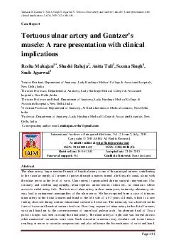
/
Mahajan R, Raheja S, Tuli A, Singh S, Agarwal S. Tortuous ulnar artery Mahajan R, Raheja S, Tuli A, Singh S, Agarwal S. Tortuous ulnar artery

Mahajan R, Raheja S, Tuli A, Singh S, Agarwal S. Tortuous ulnar artery - PDF document

natalia-silvester
natalia-silvester . @natalia-silvester
Follow
402 views
Uploaded On 2016-08-14

Mahajan R, Raheja S, Tuli A, Singh S, Agarwal S. Tortuous ulnar artery - PPT Presentation

Plain Chest Radiography Heart Views 2008 9 246 10Valsecchi O Vassileva A Musumeci G et al Failure of transradial approach during coronary interventions Anatomic considerations Cathet Car ID: 446507

Plain Chest Radiography. Heart Views

Share:

Link:

Embed:

Download Presentation from below link

Download Pdf The PPT/PDF document "Mahajan R, Raheja S, Tuli A, Singh S, Ag..." is the property of its rightful owner. Permission is granted to download and print the materials on this web site for personal, non-commercial use only, and to display it on your personal computer provided you do not modify the materials and that you retain all copyright notices contained in the materials. By downloading content from our website, you accept the terms of this agreement.


Presentation Transcript

Mahajan R, Raheja S, Tuli A, Singh S, Agarwal S. Tortuous ulnar artery and Gantzer’s muscle: A rare presentation with clinical implications. IAIM, 2015; 2(7): 141-146.  Plain Chest Radiography. Heart Views, 2008; 9: 24-6. 10.Valsecchi O, Vassileva A, Musumeci G, et al. Failure of trans-radial approach during coronary interventions: Anatomic considerations. Cathet Cardiovasc Interv, 2006; 67: 870-8. 11.Layton KF, Kallmes DF, Kaufmann TJ. Use of the ulnar artery as an alternative access site for cerebral angiography. AJNR Am J Neuroradiology, 2006; 27(10): 2073-4. 12.Liu W, Lagaac R, Pettigrew GJ, Callaghan CJ. Outcomes after ulnar-basilic arteriovenous fistula formation. Ann Vasc Surg, 2013; 27(2): 232-7. doi: 10.1016/j.avsg.2012.04.014. Epub 2012 Sep 12. 13.Alizadeh M, Ghafari A, Akbari S. Mid forearm superficial ulnar artery cephalic vein fistula in hemodialysis patient: A case report. Journal of Nephrology and Renal Transplantation, 2012; 4(2): 2-4. 14.Bender M, Whitelaw S, Sakarwala A, Joseph D. The Hypothenar Ulnar Artery: A Case Series of Aneurysm, Tortuosity and Thrombosis. Journal for Vascular Ultrasound, 2009; 33(3): 138-42. Mahajan R, Raheja S, Tuli A, Singh S, Agarwal S. Tortuous ulnar artery and Gantzer’s muscle: A rare presentation with clinical implications. IAIM, 2015; 2(7): 141-146. \n test, significant anatomic variations such as loops, tortuous configurations, stenoses, hypoplasia, and aberrant origin, and vasospasm leading to radial artery access failure or failure to achieve coronary artery cannulation [10]. Other possible considerations for PCI include pulsation and diameter of ulnar artery being more than that of radial artery. The ulnar artery is also used as an alternative access site for cerebral angiography in patients with occluded abdominal aorta or the common iliac or femoral arteries where transradial approach is unsuccessful, which may be useful to neuroradiologists who encounter difficulty with other arterial access sites [11]. Ulnar artery is used alternative to radial artery for creating arterio-venous fistulas in patients undergoing dialysis.The ulnar-basilic and ulnar-cephalic arterio-venous fistulas can provide a substitute to upper-arm fistulas, especially in those with previously functioning ipsilateral radio-cephalic arterio-venous fistulas [12, 13]. Thus, Duplex ultrasound should be used ideally for evaluation and follow-up of distal ulnar artery abnormalities in experienced vascular laboratories as bruits or thrills over the ulnar artery may indicate pseudoaneurysmal dilation, tortuosity which are commonly seen in the ulnar artery [14]. Conclusion An understanding of variations in the regional anatomy is essential for surgeons, orthopedicians, cardiologists and neuroradiologists for preventing failure of surgical procedures. The knowledge of the variant tortuous course of ulnar artery which has been reported here, is important in procedures like cardiac catheterizations, arterial grafting and other angiographic procedures. Anatomical variants like Gantzer’s muscle can cause compression not visible on imaging and need to be considered for successful surgical decompressions. References 1.Standring S, Borley NR, Collins P, Crossman AR, Gatzoulis MA, Healy JC, et al. Gray's Anatomy: The Anatomical Basis of Clinical Practice. Forearm, wrist and hand. 40th edition. London: Churchill Livingstone, 2008; p. 852, 893-4. 2.Emel E, Guzey FK, Alatas I. Guyon's Canal Syndrome Due to Tortuous Ulnar Artery: A Case Report. Turk Neurosurg, 2003; 13: 107-10. 3.Jose RM, Bragg T, Srivastava S. Ulnar nerve compression in Guyon’s canal in the presence of a tortuous ulnar artery. J Hand Surg Br, 2006; 31(2): 200–2. 4.Zeeshan M, Ahmed F, Kanwal D, Khalid QSB, Ahmed MN. Guyon's canal syndrome due to tortuous ulnar artery with DeQuervain stenosing tenosynovitis, ligamentous injuries and dorsal intercalated segmental instability syndrome, a rare presentation: A case report. Cases Journal, 2009; 2: 9390. doi: 10.186/1757-1626-2-9390. 5.S. R. Dodds. Aneurysmal Arterial Disease. 2001. http://www.ehow.com/ about _6633259_ tortuous-artery_.html 6.Yoshii S, Ikeda K, Murakami H. Ulnar nerve compression secondary to ulnar artery true aneurysm at Guyon's canal. J Neurosurg Sci, 1999; 43: 295-7. 7.Marie I, Herve F, Primard E, Cailleux N, Levesque H. Long-term follow-up of hypothenar hammer syndrome: A series of 47 patients. Medicine (Baltimore), 2007; 86(6): 334–43. 8.Taarnhoj NCBB, Munch IC, Sander B, Kessel L, Hougaard JL, Kyvik K. Straight versustortuous retinal arteries in relation to blood pressure and genetics. British Journal of Ophthalmology, 2008; 92: 1055–60. 9.Bhat V, Muzrakchi AA. Meandering Vessels: A Sign of Arterial Tortuosity on Mahajan R, Raheja S, Tuli A, Singh S, Agarwal S. Tortuous ulnar artery and Gantzer’s muscle: A rare presentation with clinical implications. IAIM, 2015; 2(7): 141-146.  Discussion In the present case, an isolated presentation of a tortuous ulnar artery was seen in the distal forearm and hand of left side. It was not associated with any other lesion in the surrounding structures which is similar to observation by other authors [2, 3] but is in contrast to findings by another researcher who noticed other incidental findings along with the tortuous ulnar artery [4]. In the present case, tortuous course of the ulnar artery was seen in a male cadaver without any aneurysmal dilatation which is in disagreement with findings of few investigators who believed that men and those with a family history of tortuous blood vessels are much more likely to experience aneurysms and blood clots because of tortuous arteries [5, 6]. The present case demonstrates the maximum number of bends in the ulnar artery in the proximal hand distal to Guyon’s canal which is comparable to observations of researchers who established the fact that in Hypothenar Hammer Syndrome, the ulnar artery is damaged over a 2 cm segment just distal to Guyon's canal because of its superficial position and with palmar trauma, the at-risk ulnar artery segment is compressed against the adjacent hamulus which has been compared to a hammer-on-anvil phenomenum. Manual labour requiring constant hammering or repetitive traumatic activities may damage the ulnar artery leading to its tortuosity resulting in “hypothenar hammer syndrome” because of involvement of ulnar nerve [7]. In this case, the Gantzer’s muscle runs distally and obliquely across the ulnar artery from medial to lateral side underneath the flexor digitorum superficialis to join the flexor pollicis longus muscle arising from the radius. As per our knowledge, such unusual tortuous course of ulnar artery in the presence of Gantzer’s muscle has not been reported in modern literature. Knowledge of this variation is very important to surgeons in appropriately planning the operative procedures. Also, this occasional head of flexor pollicis longus (Gantzer’s muscle) has to be borne in mind during anterior approaches to the proximal radius and the elbow joint, as also during a decompressive fasciotomy for compartment syndrome of the forearm. The deficiencies in the normal ongoing repair of arteries cause the development of tortuosity and blood tends to pool and clot in certain regions and bends of the arterial pathway. Tortuous arteries may be genetic or caused by other health conditions, such as thickening of the arteries, atherosclerosis, hypertension or simple aging. The thickening of arteries or high blood pressure can force arteries to pave alternate twisted pathways [8, 9] which is consistent with the present case who is an elderly male and could be suffering from atherosclerotic changes, in addition to the compression of the artery from the overlying muscle belly of the accessory head of flexor pollicis longus coercing the artery to follow a tortuous path. Clinical implications Tortuous blood vessels make upper limb surgeries difficult and dangerous. CT scans can often depict dense tortuous artery regions as solid masses. The looping and winding of such arteries can lead physicians to attempt to take a biopsy of what they may consider a tumor. Tortuous arteries also make surgeries meant to open or widen blocked arteries difficult. There is always a danger of puncture or perforation of arterial wall when vessels are extremely winding and constructed of thinner walls. The "accordion effect" of the crumpling tortuous arteries can complicate both diagnosis and treatment of various conditions. The ulnar artery is usually considered for Percutaneous Coronary Intervention (PCI) as radial approach does not seem suitable for 5–15% of patients undergoing cardiac catheterization for reasons including an abnormal Allen’s Mahajan R, Raheja S, Tuli A, Singh S, Agarwal S. Tortuous ulnar artery and Gantzer’s muscle: A rare presentation with clinical implications. IAIM, 2015; 2(7): 141-146.  cm distal to the lower border of flexor retinaculum with convexity towards the lateral aspect. The fifth curvature was seen 2.5 cm distal to the lower border of flexor retinaculum with convexity towards the medial side with the apex of the bend just touching the hypothenar eminence. The sixth twist in the vessel was seen at the commencement of the superficial palmar arch with convexity towards the lateral aspect. The seventh curvature was seen in the proximal part of the common palmar digital artery supplying the ring and the little fingers with convexity towards the medial aspect (Figure - ). The thickness of the wall was normal throughout the length of the vessel and no aneurysmal changes were seen. The ulnar artery of right side was normal and no other anatomical variations were seen. Figure - 1 : Distal forearm and hand of left side showing tortuous ulnar artery. Black arrows (1, 2, 3, 4, 5, 6, 7) show loops and bends at different levels. Figure - 2 : Cubital fossa and forearm of left side showing ulnar artery (red arrows) being crossed by accessory head of flexor pollicis longus (blue arrows). Mahajan R, Raheja S, Tuli A, Singh S, Agarwal S. Tortuous ulnar artery and Gantzer’s muscle: A rare presentation with clinical implications. IAIM, 2015; 2(7): 141-146.  An understanding of variations in the regional anatomy is essential for surgeons, cardiologists and neuroradiologists for preventing failure of surgical procedures. Key words Tortuosity, Ulnar artery, Gantzer’s muscle, Guyon’s canal, Surgery. Introduction The ulnar artery, the larger terminal branch of the brachial artery is one of the major arteries contributing to the vascular supply of forearm. It begins 1cm distal to the flexion crease of the elbow and reaches the medial side of the forearm midway between elbow and wrist. In the forearm the artery initially lies on brachialis, deep to pronator teres, flexor carpi radialis, palmaris longus and flexor digitorum superficialis. It subsequently lies on flexor digitorum profundus, between flexor carpi ulnaris and flexor digitorum superficialis, and is covered by the skin, superficial and deep fasciae. The median nerve is a medial relation for approximately 2.5 cm distal to the elbow, and then crosses the artery, from which it is separated by the ulnar head of pronator teres. The ulnar nerve lies medial to the distal two-thirds of the artery, which supplies the nerve throughout its length. The palmar cutaneous branch of the ulnar nerve descends along the ulnar artery to reach the hand. The ulnar artery crosses the flexor retinaculum lateral to the ulnar nerve and pisiform bone to enter the hand. The ulnar artery is accompanied throughout its length by venae comitantes. At the wrist the ulnar artery is covered by skin, fasciae and palmaris brevis. It lies between the superficial and main parts of the flexor retinaculum, lateral to the ulnar nerve and pisiform in the Guyon's canal which is a fibro-osseous tunnel situated along the anteromedial aspect of wrist joint [1]. Lesions of ulnar artery such as aneurysms, tortuosity, aberrancy, etc. occurring in the Guyon’s canal at the level of wrist may lead to entrapment neuropathies of the ulnar nerve which runs along with it. A tortuous artery is one that twists and winds in a meandering path. Ulnar artery is approached during surgical interventions such as, Coronary angiography, Cerebral angiography, Ulnar-cephalic arteriovenous fistula where access to radial artery fails. The tortuous arteries may complicate medical procedures. Tortuous ulnar artery is a rare presentation seen during routine cadaveric dissections. We have reported here an accidental finding of tortuous ulnar artery in the distal forearm, wrist and hand. Case report On routine educational cadaveric dissection for undergraduate students, a tortuous ulnar artery was observed in the distal forearm and hand of the left side of a 62 years old male cadaver. The artery was dissected out carefully to see its further course. The tortuosity was appreciated for a length of about 15 cm in the distal forearm, wrist and proximal hand. There were twists and bends at various levels up to the commencement of superficial palmar arch (Figure - 1). An accessory head of flexor pollicis longus (AHFPL) known as the Gantzer’s muscle was seen taking origin from the common flexor origin at the medial epicondyle and crossing the ulnar artery in the proximal forearm about 14cm distal to the origin of the muscle from the medial epicondyle. This muscle head joined the main belly of flexor pollicis longus arising from anterior surface of radius (Figure - 2). The tortuosity in the form of loops and bends was seen distal to this chiasma formation between AHFPL and ulnar artery. The first curvature in the tortuous course of the artery was seen about 8 cm proximal to the upper border of flexor retinaculum with convexity towards the lateral side. The second bend was seen about 3 cm proximal to the upper border of flexor retinaculum with convexity towards the lateral side. The third bend was seen just distal to the Guyon’s canal with convexity towards the medial side. The fourth curvature was seen 1.5 Mahajan R, Raheja S, Tuli A, Singh S, Agarwal S. Tortuous ulnar artery and Gantzer’s muscle: A rare presentation with clinical implications. IA IM, 2015; 2(7): 141   Case Report Tortuous ulnar artery and Gantzer’s muscle: A rare presentation with clinical implications Reeha Mahajan1* , Shashi Raheja Sneh Agarwal5 Senior Resident, Department of Anatomy, Lady Hardinge Medical College & New Delhi, India Director Professor, Department of Anatomy, Lady Hardinge Medical College & Associated hospitals, New Delhi, India Director Professor and Head, Department of Anatomy, Lady Hardinge Medical College & Associated ho spitals, New Delhi, India 4 Assistant Professor, Department of Anatomy, All India Institute of Medical sciences, New Delhi, India Professor, Department of Anatomy, Lady Hardinge Medical College & Associated ho Delhi, India Corresponding author email: mahajan.reeha@gmail.com International Archives of Inte Received on: Source of support: Abstract The ulnar artery, larger terminal branch of brachial artery is one of the principal arteries contributing to the vascular supply of forearm. It passes through a narrow tunnel, the Guyon’s canal, along with the ulnar nerve at the level of wrist. Ulnar artery is appr coronary and cerebral angiography, ulnar access to radial artery fails. The lesions of ulnar artery such as aneurysms, tortuosity, aberrancy, etc. may lead to entrapment neuropathies of the ulnar nerve. We ulnar artery in the distal forearm and hand of the left side of a 62 years old male, which is a rare finding observed during routine educational cadaveric dissection. form of twists and bends at various levels in 15cm long segment of ulnar artery in the distal forearm, wrist and hand up to the commencement of superficial palmar arch. An aberrant head of flexor pollicis longus was seen c rossing the ulnar artery. The ulnar artery of right side was normal and no other anatomical variations were seen. resulting in nerve or vascular compressions and also influence the biomechanics of wr Mahajan R, Raheja S, Tuli A, Singh S, Agarwal S. Tortuous ulnar artery and Gantzer’s muscle: A rare presentation with IM, 2015; 2(7): 141 -146. Tortuous ulnar artery and Gantzer’s muscle: A rare presentation with clinical , Shashi Raheja , Anita Tuli , Seema Singh Senior Resident, Department of Anatomy, Lady Hardinge Medical College & Associated hospi Director Professor, Department of Anatomy, Lady Hardinge Medical College & Associated Head, Department of Anatomy, Lady Hardinge Medical College & spitals, New Delhi, India Assistant Professor, Department of Anatomy, All India Institute of Medical sciences, New Delhi, Professor, Department of Anatomy, Lady Hardinge Medical College & Associated ho mahajan.reeha@gmail.com International Archives of Inte grated Medicine, Vol. 2, Issue 7, July Copy right © 2015, IAIM, All Rights Reserved. Available online at http://iaimjournal.com/ ISSN: 2394-0026 (P) ISSN: 2394- 0034 (O) Received on: 16-06-2015 Accepted on: 27 Source of support: Nil Conflict of interest: artery, larger terminal branch of brachial artery is one of the principal arteries contributing to the vascular supply of forearm. It passes through a narrow tunnel, the Guyon’s canal, along with the ulnar nerve at the level of wrist. Ulnar artery is appr oached during surgical interventions like, coronary and cerebral angiography, ulnar - cephalic arteriovenous fistula etc., in situations where access to radial artery fails. The lesions of ulnar artery such as aneurysms, tortuosity, aberrancy, etc. to entrapment neuropathies of the ulnar nerve. We have report ed here ulnar artery in the distal forearm and hand of the left side of a 62 years old male, which is a rare finding observed during routine educational cadaveric dissection. T he tortuosity was observed in the form of twists and bends at various levels in 15cm long segment of ulnar artery in the distal forearm, to the commencement of superficial palmar arch. An aberrant head of flexor rossing the ulnar artery. The ulnar artery of right side was normal and no other anatomical variations were seen. Such muscular variations may simulate soft tissue tumors resulting in nerve or vascular compressions and also influence the biomechanics of wr Mahajan R, Raheja S, Tuli A, Singh S, Agarwal S. Tortuous ulnar artery and Gantzer’s muscle: A rare presentation with  Tortuous ulnar artery and Gantzer’s muscle: A rare presentation with clinical , Seema Singh , Associated hospi tals, Director Professor, Department of Anatomy, Lady Hardinge Medical College & Associated Head, Department of Anatomy, Lady Hardinge Medical College & Assistant Professor, Department of Anatomy, All India Institute of Medical sciences, New Delhi, Professor, Department of Anatomy, Lady Hardinge Medical College & Associated ho spitals, New grated Medicine, Vol. 2, Issue 7, July , 2015. Copy right © 2015, IAIM, All Rights Reserved. http://iaimjournal.com/ 0034 (O) 27 -06-2015 Conflict of interest: None declared. artery, larger terminal branch of brachial artery is one of the principal arteries contributing to the vascular supply of forearm. It passes through a narrow tunnel, the Guyon’s canal, along with oached during surgical interventions like, cephalic arteriovenous fistula etc., in situations where access to radial artery fails. The lesions of ulnar artery such as aneurysms, tortuosity, aberrancy, etc. ed here a case of tortuous ulnar artery in the distal forearm and hand of the left side of a 62 years old male, which is a rare he tortuosity was observed in the form of twists and bends at various levels in 15cm long segment of ulnar artery in the distal forearm, to the commencement of superficial palmar arch. An aberrant head of flexor rossing the ulnar artery. The ulnar artery of right side was normal and no Such muscular variations may simulate soft tissue tumors resulting in nerve or vascular compressions and also influence the biomechanics of wr ist and hand.