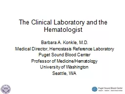
/
VOL 17 NO 3  SUMMER 2004    CLINICAL LABORATORY SCIENCE165 VOL 17 NO 3  SUMMER 2004    CLINICAL LABORATORY SCIENCE165

VOL 17 NO 3 SUMMER 2004 CLINICAL LABORATORY SCIENCE165 - PDF document

norah
norah . @norah
Follow
342 views
Uploaded On 2022-08-16

VOL 17 NO 3 SUMMER 2004 CLINICAL LABORATORY SCIENCE165 - PPT Presentation

The Focus section seeks to publish relevant and timely continuingeducation for clinical laboratory practitioners Section editors topicsand authors are selected in advance to cover current areas of ID: 936236

cells anemia marrow aplastic anemia cells aplastic marrow bone patients cell acquired blood pnh stem clinical hematopoietic failure laboratory

Share:

Link:

Embed:

Download Presentation from below link

Download Pdf The PPT/PDF document "VOL 17 NO 3 SUMMER 2004 CLINICAL LAB..." is the property of its rightful owner. Permission is granted to download and print the materials on this web site for personal, non-commercial use only, and to display it on your personal computer provided you do not modify the materials and that you retain all copyright notices contained in the materials. By downloading content from our website, you accept the terms of this agreement.


Presentation Transcript

VOL 17, NO 3 SUMMER 2004 CLINICAL LABORATORY SCIENCE165 The Focus section seeks to publish relevant and timely continuingeducation for clinical laboratory practitioners. Section editors, topics,and authors are selected in advance to cover current areas of interest in FOCUS: BONE MARROW FAILURE ANEMIASAcquired Aplastic Anemia ELAINE M KEOHANEAcquired aplastic anemia (AA) is a disorder characterized bya profound deficit of hematopoietic stem and progenitor cells,bone marrow hypocellularity, and peripheral blood pancy-topenia. It primarily affects children, young adults, and thoseover 60 years of age. The majority of cases are idiopathic;however, idiosyncratic reactions to some drugs, chemicals,and viruses have been implicated in its etiology. An autoim-mune T-cell reaction likely causes the stem cell depletion,but the precise mechanism, as well as the eliciting and targetantigens, is unknown. Symptoms vary from severe life-threat-ening cytopenias to moderate or non-severe disease that doesnot require transfusion support. The peripheral blood typi-cally exhibits pancytopenia, reticulocytopenia, and nor-mocytic or macrocytic erythrocytes. The bone marrow ishypocellular and may exhibit dysplasia of the erythrocyte 166VOL 17, NO 3 SUMMER 2004 CLINICAL LABORATORY SCIENCEacquired AA.1,2,3,4 The incidence of AAas a complication of drug therapy israre. A genetic predisposition to idio-syncratic reactions may exist due toindividual differences in metabolic orimmune response pathways. Suttonand others found a higher than ex-pected frequency of glutathione S-transferase (GST) gene deletions inAA. GSTT1 null and GSTM1/GSTT1 double null genotypes werefound in 30% and 22% of Caucasianswith AA, respectively. GST deficiencymay hinder the biometabolism of somechemical toxins and increase the riskof development of AA.AA may occur as a rare complication inpregnancy, autoimmune disease, and insome viral infections, such as Epstein-Barr virus and human immunodefi-ciency virus.1,2 As many as 2% to 10%of patients have a history of acute hepa-titis one to three months prior to pre-sentation with severe AA; however, testsfor hepatitis A, B, and C are negative.It is important to recognize hepatitis-associated AA syndrome for its poorprognosis and high mortality rate.Quantitative and qualitative deficiencyof hematopoietic stem and progenitorcells cause the peripheral blood pan-cytopenia and hypocellular bone mar-row in acquired AA.7,8 In aplasia fol-lowing cytotoxic chemotherapy andradiation, as well as in benzene toxic-ity, there is direct damage to the DNAor proteins of hematopoietic stem cellsand progenitor cells, resulting in celldeath. However, in idiopathic AA, vi-ral associations, and idiosyncratic re-actions to drugs or chemicals, bonemarrow failure is thought to be the re-sult of a T-cell-mediated autoimmuneattack against hematopoietic stem andFOCUS: BONE MARROW FAILURE ANEMIASprogenitor cells. It is likely that theoffending antigen in this autoimmunereaction is expressed on stem and earlyprogenitor cells, but the precise mecha-nism for this reaction, as well as theidentity of the inciting and target an-tigens, remains unknown.PATHOPHYSIOLOGYHematopoietic stem cells are severelydecreased in the bone marrow of AApatients as evidenced by a greater thanten-fold reduction in CD34+ cells de-tected by flow cytometry (mean of557/mL in AA compared to 5,867/mLin normal controls), and diminishedcolony formation and growth of long-term culture initiating cells in vitro.7,8Stem cell numbers remain low despiteelevated serum growth factors.10 Stro-mal cells in AA produce normal or in-creased growth factors and support thegrowth of normal CD34+ cells in cul-ture.10,11,12 Further evidence of a func-tional stroma is demonstrated clinicallyby the successful engraftment of bonemarrow transplants in AA patients.An autoimmune pathophysiol

ogy of AAwas first proposed in the late 1960s whenpatients had an unexpected improve-ment in cell counts after unsuccessfulmarrow transplantation.13 Mathe andothers suggested that the improvementmight be due to the antilymphocyteglobulin used in the immunosuppressiveconditioning regimen required for trans-plant. Since then, there has been increas-ing laboratory and clinical evidence insupport of an autoimmune pathophysi-ology of the bone marrow failure. Thisis indirectly inferred from the fact thatthe majority of patients with AA respondto immunosuppressive therapy with im-provement of cell counts. In laboratoryexperiments, bone marrow cells from AApatients inhibit hematopoietic colonyformation.14 Furthermore, T-cells in ac-quired AA produce increased amountsTable 1. Agents infrequently associated with acquired aplastic anemiaDrugsNonsteroidal anti-inflammatory agents (butazones, indomethacin, diclofinac, naproxen, piroxicam)Antiarthritics (gold salts, D-penicillamine)Antithyroids (carbimazole, thiouracil)Antibiotics (chloramphenicol, sulphonamides)Anticonvulsants (carbamazepine, phenytoin)Antidepressants (phenothiazine, dothiepin)Antiprotozoals (chloroquine, quinacrine)Antidiabetes drugs (chlorpropamide, tolbutamide)Occupational and environmental exposuresBenzene*InsecticidesPetrochemicalsLubricating agents*Benzene is more commonly associated with acquired AA than the other agents listed,but the incidence varies by the amount and duration of exposure. The incidence ofacquired AA is estimated at 1 in 10,000 individuals exposed at air concentrations of 10to 20 ppm, and increases to 1 in 100 at concentrations� 100 ppm. VOL 17, NO 3 SUMMER 2004 CLINICAL LABORATORY SCIENCE167of interferon- (INF-) and tumor necrosis factor- (TNF-),known inhibitors of hematopoiesis.15,16 In AA patients, there isan increase in the number of cytotoxic CD8+ cells in the bloodand bone marrow, as well as an increase in their expression ofthe HLA-DR activation marker and cytoplasmic INF-measured by flow cytometry.17 These findings were shown toreverse after immunosuppressive therapy.17 Nimer and othersfound that HLA-DR2 has a 1.9-fold higher incidence in AAthan in the general population.18 However, the significance ofthat finding in terms of pathophysiology, treatment, or prog-nosis is unclear.Patients with AA have a greater proportion of apoptoticCD34+ cells compared to normal controls when stained withfluorescent 7-amino actinomycin D and measured by flowcytometry, and these cells have a higher expression of Fasreceptors.19,20 Since INF- and TNF- induce apoptosisthrough Fas receptor signaling, this pathway may be impor-tant in the destruction of stem cells in AA.20 Further, geneexpression profiling of CD34+ cells using GeneChip analy-sis demonstrates a marked increase in expression of apoptoticgenes in AA compared to normal individuals.21Various mechanisms may initiate the autoimmune responsein AA such as:9,17¥antigen modification by drugs, chemicals or their metabolites,¥alteration of self-proteins,¥viral-induced aberrant protein expression,¥production of novel fusion proteins due to undetectedchromosome alterations,¥cross-reactivity of drugs, chemicals, or viruses with selfantigens, or¥exposure of cryptic antigens by tissue damage.The offending antigen in acquired AA may also be a mem-brane glycosylphosphatidylinositol (GPI)-anchored proteinor the GPI anchor itself.22,23 Approximately one-third of AApatients experience an expansion of clones that lack the GPIanchor and its associated proteins, the defect in paroxysmalnocturnal hemoglobinuria (PNH).22 GPI-negative PNHclones are able to proliferate in the marrow of acquired AApatients and apparently are not affected by the immune de-struction and apoptosis experienced by the other progenitorcells in the marrow.22CLINICAL FINDINGSSevere AA can be rapidly fatal, w

hile the clinical course of non-severe aplastic anemia may be asymptomatic and transfusion-independent. Patients with AA do not exhibit splenomegalyand hepatomegaly. The most common symptoms at presenta-tion are bleeding due to thrombocytopenia and fatigue, dysp-nea, and pallor due to anemia. Bruising, petechiae, epistaxis,bleeding gums, excessive menses, retinal hemorrhages, intesti-nal bleeding, and rarely cerebral hemorrhage may occur as mani-festations of the thrombocytopenia. Fever and infection, due toneutropenia, are infrequent at presentation, but may occur laterin the disease. Prolonged neutropenia can result in fatal bacte-rial sepsis and systemic fungal infections.2,24LABORATORY FINDINGSPeripheral bloodTable 2 summarizes the peripheral blood findings in acquiredAA. Platelet, white blood cell, and red blood cell counts aredecreased, but initially only one or two of the cell lines maybe affected. The hemoglobin is below 10 gm/dL, and thereticulocyte count and reticulocyte production index aredecreased reflecting the inability of the bone marrow to ad-equately respond to the anemia. The absolute neutrophilcount is decreased, but the absolute lymphocyte count isusually normal. The mean cell volume is normal or increased.The peripheral blood film has decreased numbers of plate-lets, neutrophils, and monocytes. Blasts and bands are char-acteristically absent, and neutrophils may have toxic granu-lation. The red blood cells can be normocytic or macrocyticwithout other morphologic abnormalities. The platelets arenormal in appearance.FOCUS: BONE MARROW FAILURE ANEMIASTable 2. Peripheral blood findings in acquiredaplastic anemiaDecreasedAbsolute neutrophil countHemoglobinPlatelet countReticulocyte countReticulocyte production indexWhite blood cell countIncreased or normalMean cell volumeBlood smearDecreased neutrophils, monocytes, and plateletsNormocytic or macrocytic red blood cellsToxic granulation of neutrophils may be present 168VOL 17, NO 3 SUMMER 2004 CLINICAL LABORATORY SCIENCEBone marrowBone marrow aspirates and biopsies are hypocellular withprominent fat cells, and patchy cellularity (Figure 1). A bonemarrow biopsy is required for accurate assessment of cellu-larity. Blast, granulocytic, and megakaryocytic cells are de-creased or absent, and reticulin staining is normal. Lympho-cytes, plasma cells, and macrophages may be present. Al-though dyserythropoiesis may be found, there is no dyspla-sia of the granulocytes or megakaryocytes.ClassificationAcquired AA is classified as non-severe aplastic anemia(NSAA), also called moderate aplastic anemia (MAA), se-vere aplastic anemia (SAA), and very severe aplastic anemia(VSAA) based on cell counts and bone marrow cellularity.Table 3 depicts the criteria for each category.25,26 Classifica-tion of AA patients by severity of disease is important toguide treatment decisions.Other laboratory findingsThere is a marked decrease in CD34+ cells measured byflow cytometry, and their quantity may remain low evenafter hematopoietic recovery.7,8 Serum levels of erythro-poietin and other growth factors are elevated.10 Liver func-tion tests may be abnormal if the AA was preceded byacute hepatitis.Chromosomal abnormalities are infrequent in AA at presenta-tion, and their development is considered by some as a reasonto exclude a diagnosis of AA.2,27,28 As many as 26% of AA pa-tients develop an abnormal karyotype over the course of theirdisease.27,29 The most common abnormalities found includeabnormalities of chromosome 7, trisomy 8, and abnormalitiesof chromosome 13.2,27,28 Chromosome abnormalities in someAA patients may disappear and reappear during the course ofthe disease, the significance of which is uncertain. Evaluation ofchromosomes is often difficult since hypocellular bone mar-rows with decreased proliferation potential may yield fewmetaphases for analysis. Kar

yotyping of peripheral blood lym-phocytes may be more successful. The incidence of chromo-some abnormalities may be higher with the use of more sensi-tive techniques such as the interphase fluorescent in situ hy-bridization (FISH) using probes for specific chromosomes.2,5,29Differential diagnosisIt is important to distinguish AA from similar conditions sothat the appropriate treatment can be implemented. PNH andAA may have similar features of pancytopenia, macrocytosis,and bone marrow hypocellularity; however, patients who presentwith primary PNH have reticulocytosis and clinical and bio-chemical evidence of hemolysis. Due to an acquired clonalmutation, PNH cells lack the GPI anchor on their surface, andare characteristically deficient in GPI-linked proteins such asFOCUS: BONE MARROW FAILURE ANEMIASTable 3. Classification of acquired aplastic anemiaNon-severe aplastic anemia (NSAA) or moderate aplastic anemia (MAA)Presence of at least two of the following:Hemoglobin 10 g/dLPlatelets: 20-50 x 10/LNeutrophils 0.5-1.5 x 10/LSevere aplastic anemia (SAA)Bone marrow cellularity 25%, or 25% to 50% with 30% residual hematopoietic cells, and presence of at least two of the following: Neutrophils 0.5 x 10/L Platelets 20 x 10/L Reticulocytes 20 x 10/LVery severe aplastic anemia (VSAA)Includes criteria of SAA plus:Neutrophils 0.2 x 10/L Figure 1. Hypocellular bone marrow aspirate inaplastic anemia, 100X magnification, Tetrachrome-Giemsa stain. Note prominent fat cells.Photo courtesy of Dr Peter Maslak, Memorial Sloan KetteringCancer Center, New York. VOL 17, NO 3 SUMMER 2004 CLINICAL LABORATORY SCIENCE169CD55 and CD59.2,22 This leads to increased susceptibility tocomplement-mediated hemolysis. Flow cytometry is the mostsensitive method for detection of PNH cells, and the classicalsucrose hemolysis test and Ham test for complement-mediatedhemolysis may be positive if a sufficient number of PNH eryth-rocytes are present in the peripheral blood.2, 24There is a strong association between AA and PNH. Bonemarrow failure is a major clinical manifestation of PNH 24,29Conversely, expansion of GPI-negative PNH clones and de-velopment of hemolytic PNH occurs in approximately 10%to 25% of AA patients. 22,29 Wang and others detected PNHneutrophils in the peripheral blood of 88.6% of newly diag-nosed AA patients using a sensitive flow cytometric techniquethat analyzed at least 10 cells in the granulocyte gate.30 Theappearance of PNH cells in acquired AA is sometimes tran-sient, and the significance of this finding is uncertain. AA andPNH may be a single entity that can present either as primaryAA or primary PNH.29 Nevertheless, it is important to testfor PNH cells in acquired AA due to the complications ofhemolysis and thrombosis associated with the disorder.Acquired AA is also similar to myelodysplastic syndrome(MDS) in that both can have pancytopenia, macrocytosis, anddyserythropoiesis. Although the bone marrow in MDS is usu-ally hypercellular, 20% of MDS cases have a hypocellular bonemarrow at presentation.1,2 MDS usually has chromosomal ab-normalities, however, they may not always be present.24,27 MDShas additional features that are absent from acquired AA in-cluding dyspoiesis of the granulocytic and megakaryocytic cells,increased blasts, and increased reticulin in the bone marrow.Approximately 20% of patients with acquired AA progress toMDS during the course of their disease.29 Although this pro-gression is often preceded by the appearance of chromosomeabnormalities, the underlying mechanism for the clonal evo-lution to MDS is unknown.27,29AA may also be similar to hypocellular acute leukemia, how-ever, acute leukemia has increased blasts and reticulin in thebone marrow. Hairy cell leukemia (HCL) also has pancy-topenia, but the bone marrow in HCL is fibrotic, the circulat-ing hairy cells co-express CD20, CD11

c, CD25, and CD103,and most patients have splenomegaly. Decreased levels of vi-tamin B12 and/or folate can identify the pancytopenia associ-ated with megaloblastic anemia. Finally, acquired AA may besimilar to Fanconi anemia, a rare congenital aplasia most of-ten diagnosed in childhood, but occasionally presenting inadults. A genetic test for mitomycin-C induced chromosomebreakage is characteristic of Fanconi anemia.1,24TREATMENTThe treatment rationale in acquired AA is twofold, either 1)replacement of the deficient or damaged stem cells in the bonemarrow by transplantation, or 2) suppression of the autoim-mune reaction against the stem cells. Hematopoietic stem celltransplantation (HSCT) is the preferred therapy for patientsunder 30 to 40 years of age that have a HLA identical sib-ling.2,24 For approximately 70% of SAA patients, HSCT isnot an option because of age or the lack of an appropriatedonor. In these cases immunosuppression therapy (IST) is thestandard initial treatment.2,24 The purpose of IST is to pre-vent the T-cell attack on the hematopoietic cells by decreasingthe number of activated T-cells and inhibiting their function.31Combined IST using antithymocyte globulin (ATG) andcyclosporine (CSA) is preferred since the combination has agreater response rate than either agent alone.2,31 ATG resultsin the cytolysis and reduction of T-cells by recognition of theirsurface antigens, while CSA inhibits T cell activation andcytokine release.31 IST may take months or years for improve-ment in cytopenias and independence from transfusions, andrelapses are frequent. 31 Some patients may need to continueCSA therapy for a prolonged period, and even after a hemato-logic recovery, stem cell numbers remain low.2,31,32 The rea-sons for the incomplete recovery with IST may be due to in-trinsic defects in the stem cell, continued inhibition of thestem cells by lymphocytes, or irreversible stem cell loss.32For those patients who are not initially responsive to IST, orthose who relapse after treatment, a second or third ISTcourse may be used. If the patient is still not responsive,and the patient is young, HLA-matched unrelated bonemarrow transplant may be an option; however, survival isnot optimal with matched unrelated donors.Supportive care for thrombocytopenia and anemia is providedby platelet transfusions when the platelet count falls below 10x 10/L (20 x 10/L in febrile patients) and red cell transfu-sions to alleviate anemia-related symptoms. Antibiotics andantifungal drugs are used prophylactically in patients withprolonged neutropenia. The use of erythropoietin (rHuEPO)and other growth factors as primary treatments is not recom-mended due to lack of efficacy and serious side effects.2,10,24However, a short course of G-CSF may improve the neutro-phil count in severely neutropenic patients with infections whoare not responding to antibiotics. Single agent corticosteroidtherapy is not recommended because low doses are not effec-tive and high doses result in excessive toxicity.2,24FOCUS: BONE MARROW FAILURE ANEMIAS 170VOL 17, NO 3 SUMMER 2004 CLINICAL LABORATORY SCIENCEPatients with NSAA who are transfusion-independent do notrequire treatment, but are periodically monitored for theirblood cell counts and presence of abnormal cells. IST is rec-ommended once the patient becomes transfusion-dependent.Other immunosuppressive agents under investigation for treat-ment of acquired AA include oxymetholone, ATG/CSA in com-bination with mycophenolate mofetil or G-CSF, rapamycin,and monoclonal antibodies to the IL-2 receptor. 2,24,33PROGNOSISSurvival rate is poor when patients are treated only with trans-fusions and antibiotics.24 Approximately 75% to 90% ofyoung patients receiving HSCT from a HLA identical sib-ling have long-term survival.24,29 Evolution of leukemia andmyelodysplasia after HCST occurs infrequently, an

d is likelydue to the conditioning regimen required for the transplant.29Approximately 60% to 80% of patients respond to IST, anda functional cure is obtained in 50% of responding pa-tients.24,27,29,34 Approximately 10% to 25% of patients de-velop hemolytic PNH, and 10% to 20% progress to MDSor acute leukemia after IST.22,29 One study reported a 42%combined risk of developing PNH or MDS.35 AA patientswho develop PNH have a better prognosis than those whodevelop MDS or leukemia. The development of MDS andacute leukemia is usually preceded by the development ofchromosomal abnormalities. Patients who develop mono-somy 7 have a higher likelihood of developing leukemia.24,27Patients with trisomy 8 have a better prognosis, but mayrequire long term CSA to maintain normal counts.24,27Peripheral blood granulocytes and monocytes of AA patientshave a progressive shortening of their telomeres that mayplay a role in the evolution of MDS in these patients.36CONCLUSIONMajor progress has occurred in the elucidation of the patho-physiology of acquired AA as well as the protocols for itstreatment. However, many questions remain about the trig-gering event for the autoimmune reaction, the inciting andtarget antigens, and the nature of the autoimmune response.Further research on the pathophysiology may lead to morespecific therapeutic strategies and may further elucidate theinterrelationship between acquired AA, PNH, and MDS.REFERENCES1.Young NS, Maciejewski JP. Aplastic anemia. In: Hoffman R, Benz Jr.EJ, Shattil SJ and others, editors. Hematology. Basic principles andpractice, 3rd ed. New York: Churchill Livingstone; 2000. p 297-331.2.Marsh JCW, Ball SE, Darbyshire P, and others. Guidelines for thediagnosis and management of acquired aplastic anemia. Br JHaematol 2003;123:782-801.3.Muir KR, Chilvers CED, Harriss C, and others. The role of occupa-tional and environmental exposure in the aetiology of acquired se-vere aplastic anemia: a case control investigation. Br J Haematol2003;123:906-14.4.Smith MT. Overview of benzene-induced aplastic anaemia. Eur JHaematol Suppl 1996;60:107-10.5.Sutton JF, Stacey M, Kearns WG, and others. Increased risk for aplas-tic anemia and myelodysplastic syndrome in individuals lacking glu-tathione S-transferase genes. Pediatr Blood Cancer 2004;42:122-6.6.Brown KE, Tisdale J, Barrett AJ, and others. Hepatitis-associatedaplastic anemia. N Engl J Med 1997;336:1059-64.7.Maciejewski JP, Anderson S, Katevas P, and others. Phenotypic andfunctional analysis of bone marrow progenitor cell compartments inbone marrow failure. Br J Haematol 1994;87:227-34.8.Maciejewski JP, Selleri C, Sato T, and others. A severe and consistentdeficit in marrow and circulating primitive hematopoietic cells (longterm culture-initiating cells) in acquired aplastic anemia. Blood1996;88:1983-91.9.Young NS, Maciejewski J. The pathophysiology of acquired aplasticanemia. N Eng J Med 1997;336:1365-72.10.Marsh JCW. Hematopoietic growth factors in the pathogenesis andfor the treatment of aplastic anemia. Semin Hematol 2000;37:81-90.11.Marsh JC, Chang J, Testa NG, and others. In vitro assessment ofmarrow Òstem cellÓ and stromal cell function in aplastic anemia. BrJ Haematol 1991;78:258-67.12.Novitzky N, Jacobs P. Immunosuppressive therapy in bone marrowaplasia: the stroma functions normally to support hematopoiesis.Exp Hematol 1995;23:1472-7.13.Mathe G, Amiel JL, Schwarzenberg L, and others. Bone marrowgraft in man after conditioning by antilymphocytic serum. Br MedJ 1970;2:131-6.14.Kagan WA, Ascensao JA, Pahwa RN, and others. Aplastic anemia:presence in human bone marrow of cells that suppress myelopoiesis.Proc Natl Acad Sci USA 1976:73:2890-4.15.Zoumbos NC, Gascon P, Djeu JY, and others. Interferon is a media-tor of hematopoietic suppression in aplastic anemia in vitro andpossibly in vivo. Proc Natl Acad Sci USA 1985;82:188-92.16.Sel

leri C, Sato T, Anderson S, and others. Interferon-gamma andtumor necrosis factorÐalpha suppress both early and late stages ofhematopoiesis and induce programmed cell death. J Cell Physiol1995;165:538-46.17.Young NS. Hematopoietic cell destruction by immune mechanismsin acquired aplastic anemia. Semin Hematol 2000;37:3-14.18.Nimer SD, Ireland P, Meshkinpour A, and others. An increased HLADR2 frequency seen in aplastic anemia patients. Blood 1994;84:923-7.19.Philpott NJ, Scopes J, Marsh JC, and others. Increased apoptosis inaplastic anemia bone marrow progenitor cells: possible pathophysi-ologic significance. Exp Hematol 1995;23:1642-8.20.Maciejewski JP, Selleri C, Sato T, and others. Increased expression ofFas antigen on bone marrow CD34 sup+ cells of patients with aplas-tic anemia. Br J Haematol 1995.91:245-52.21.Zeng W, Chen G, Kajigaya S, and others. Gene expression profilingin CD34 cells to identify differences between aplastic anemia pa-tients and healthy volunteers. Blood 2004;103:325-32.22.Kinoshita T, Inoue N. Relationship between aplastic anemia and par-FOCUS: BONE MARROW FAILURE ANEMIAS VOL 17, NO 3 SUMMER 2004 CLINICAL LABORATORY SCIENCE171oxysmal nocturnal hemoglobinuria. Int J Hematol 2002;75:117-22.23.Karadimitris A, Luzzatto L. The cellular pathogenesis of paroxysmalnocturnal haemoglobinuria. Leukemia 2001;15:1148-52.24.Young NS. Acquired aplastic anemia. Ann Intern Med 2002;136:534-46.25.International agranulocytosis and aplastic anemia study. Incidenceof aplastic anemia: Relevance of diagnostic criteria. Blood1987;70:1718-21.26.Bacigalupo A, Hows J, Gluckman E, and others. Bone marrow trans-plantation (BMT) versus immunosuppression for the treatment ofsevere aplastic anaemia (SAA): a report of the EMBT SAA workingparty. Br J Haematol 1988;70:177-82.27.Maciejewski JP, Risitano A, Sloand EM, and others. Distinct clini-cal outcomes for cytogenetic abnormalities evolving from aplasticanemia. Blood 2002;99:3129-35.28.Keung Y-K, Pettenati MJ, Cruz JM, and others. Bone marrow cytoge-netic abnormalities of aplastic anemia. Am J Hematol 2001;66:167-71.29.Socie G, Rosenfeld S, Frickhofen N, and others. Late clonal diseasesof treated aplastic anemia. Semin Hematol 2000;37:91-101.30.Wang H, Chuhjo T, Yamazaki H, and others. Relative increase ofgranulocytes with a paroxysmal nocturnal hemoglobinuria pheno-FOCUS: BONE MARROW FAILURE ANEMIAStype in aplastic anaemia patients: the high prevalence at diagnosis.Eur J Haematol 2001;66:200-5.31.Frickhofen N, Rosenfeld SJ. Immunosuppressive treatment of aplasticanemia with antithymocyte globulin and cyclospoine. SeminHematol 2000;37:56-68.32.Maciejewski JP, Kim S, Sloand E, and others. Sustained long-termhematologic recovery despite a marked quantitative defect in thestem cell compartment with aplastic anemia after immunosuppres-sive therapy. Am J Hematol 2000;65:123-31.33.Maciejewski JP, Sloand EM, Nunez O, and others. Recombinanthumanized anti-IL-2 receptor antibody (daclizumab) produces re-sponses in patients with moderate aplastic anemia. Blood2003;102:3584-6.34.Rosenfeld S, Follman D, Nunez O, and others. Antithymocyte globu-lin and cyclosporine for severe aplastic anemia. Association betweenhematologic response and long-term outcome. JAMA2003;289:1130-5.35.Tichelli A, Gratwohl A, Nissen C, and others. Late clonal complica-tions in severe aplastic anemia. Leuk Lymphoma 1994;12:167-75.36.Ball SE, Gibson FM, Rizzo S, and others. Progressive telomere short-ening in aplastic anemia. Blood 1998;91:3582-92. American Society for Clinical Laboratory Science $35/member and $55/non-member; multi-user discounts availableFor more information or to purchase, visit the ASCLS OnlineStore, www.ascls.org,click on Education and follow link to Store, then click on CDs for Learning,or call the ASCLS Education Office at 301-657-2768 for ordering information. on CD from AS