/
A- and V-Patterns and Oblique Muscle Overaction A- and V-Patterns and Oblique Muscle Overaction

A- and V-Patterns and Oblique Muscle Overaction - PDF document

olivia-moreira
olivia-moreira . @olivia-moreira
Follow
414 views
Uploaded On 2015-09-13

A- and V-Patterns and Oblique Muscle Overaction - PPT Presentation

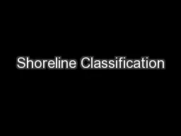
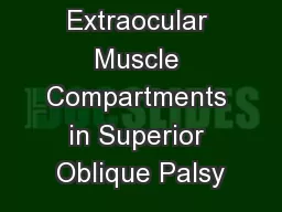
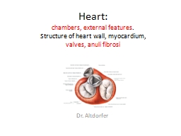
18 A and VPatterns Clinical Features Etiology Management with Horizontal Rectus Muscle Offsets Inferior Oblique Overaction Etiology Clinical Features Differential Diagnosis Management Superior Obliq ID: 127888

18 A- and V-Patterns Clinical Features Etiology Management with

Share:

Link:

Embed:

Download Presentation from below link

Download Pdf The PPT/PDF document "A- and V-Patterns and Oblique Muscle Ove..." is the property of its rightful owner. Permission is granted to download and print the materials on this web site for personal, non-commercial use only, and to display it on your personal computer provided you do not modify the materials and that you retain all copyright notices contained in the materials. By downloading content from our website, you accept the terms of this agreement.


Presentation Transcript

18 A- and V-Patterns and Oblique Muscle Overaction A- and V-Patterns Clinical Features Etiology Management with Horizontal Rectus Muscle Offsets Inferior Oblique Overaction Etiology Clinical Features Differential Diagnosis Management Superior Oblique Overaction Etiology Clinical Features Differential Diagnosis Management Superior Oblique Weakening Procedures Complications A- and V-patterns of strabismus are changes in the horizontal deviation as the patient looks up and down. They are usually associated with oblique dysfunction, either overaction or paresis. Primary oblique muscle overaction is when an oblique muscle is too strong for its antagonist and there is no known cause. Secondary oblique overaction is caused by a paresis of the antagonist muscle. A superior oblique paresis results in inferior oblique overaction, and inferior oblique paresis results in superior oblique overaction. This chapter covers the management of A- and V- patterns and primary oblique muscle overaction. A- and V-Patterns CLINICAL FEATURES �A-patterns are defined as increasing divergence in down gaze (10 prism diopters [PD]), whereas Vpatterns are �increased divergence (15 prism diopters) in up gaze. The type of A- or V-pattern helps identify the cause. Superior oblique paresis produces a V-pattern, arrow subtype, with convergence in down gaze. The arrow pattern subtype indicates a lack of abduction in down gaze, the field of action of the superior oblique muscles. Inferior oblique overaction, on the other hand, has a V-pattern, Y subtype, with increased abduction in up gaze. The Y-pattern occurs because the field of action of the inferior oblique muscles is up gaze and they are abductors. Lambda subtype is typically associated with superior oblique overaction, with increased abduction in down gaze, because the field of action is in down gaze. These patterns usually require bilateral oblique dysfunction to become manifest. ETIOLOGY Oblique muscle overaction or paresis usually causes A- and V-patterns, but primary patterns without significant oblique dysfunction may occur. Inferior oblique overaction and superior oblique paresis result in V-patterns. The oblique muscles are abductors so inferior oblique overaction causes excessive abduction in up gaze causing a V- pattern. Likewise, superior oblique paresis causes a lack of abduction in down gaze and a V-pattern. Superior oblique overaction or rarely inferior oblique paresis can cause A-patterns. Patterns can also be primary or occur in association with cranial facial abnormalities. Patterns in these cases probably represent vertical displacement of the horizontal rectus muscles and/or horizontal displacement of vertical rectus muscles. MANAGEMENT WITH HORIZONTAL RECTUS MUSCLE OFFSETS The first step in the management of A- and V-patterns is to determine if there is associated oblique muscle overaction or paresis. If there is significant oblique dysfunction then surgically address the oblique muscles. Primary A- and V- patterns with normal oblique muscle function can be managed by offsetting the horizontal rectus muscles. The horizontal rectus offset procedure works by moving the horizontal rectus muscles up (supraplaced) or down (infraplaced) to change the vector of the muscle's force. For example, if the medial rectus muscles were infraplaced, they would tighten in up gaze and loosen in down gaze, which would collapse a V-pattern. Conversely, supraplacement would loosen the medial rectus in up gaze and tighten them in down gaze and collapse an A-pattern. Vertically transposing the horizontal rectus muscles, however, does not significantly change the muscle's force in primary position. Fig. 18-1 shows a method for remembering which direction to move the horizontal rectus muscles for A- and V- patterns. Move the medial recti to the apex of the letter A or V, and move the lateral recti toward the open part of the letter. For a V-pattern, move the medial rectus muscles inferiorly and lateral rectus muscles superiorly. For an A- pattern, move the medial rectus superiorly and the lateral rectus inferiorly. As a rule, a one-half tendon width of vertical displacement results in 10 to 15 PD of pattern correction. A full tendon width vertical displacement produces approximately 20 to 25 PD of correction and is reserved for extremely large patterns. Fig. 18-2 shows a full tendon width inferior transfer of the medial rectus muscle. Inferior Oblique Overaction ETIOLOGY Inferior oblique overaction may be primary and of unknown etiology, or secondary to a congenital superior oblique palsy, as covered in Chapter 19. Primary inferior oblique overaction is commonly associated with congenital esotropia, with the oblique overaction usually presenting after 1 year of age. In addition to congenital eso tropia, primary inferior oblique muscle overaction may be associated with exotropia or occur as an isolated inferior oblique overaction without other strabismus. CLINICAL FEATURES Primary inferior oblique overaction is usually bilateral; however, it can be very asymmetric. Clinical signs of primary inferior oblique overaction include an upshoot of the adducting eye on versions, a V-pattern, and extorsion on indirect ophthalmoscopy. If the overaction is symmetric, there will be no significant hypertropia in primary position. Children with primary inferior oblique overaction do not experience subjective extorsion because they have learned to sensorially adapt at a young age. Bilateral inferior oblique overaction produces a left hypertropia in right gaze, a right hypertropia in left gaze, and little or no vertical deviation in primary position (Fig. 18-3). Distinguishing primary inferior oblique overaction from overaction associated with secondary superior oblique muscle paresis is largely dependent on the head-tilt test as described in Chapter 19. If the head-tilt test is negative this indicates a primary inferior oblique overaction, whereas a positive head-tilt test (increasing hypertropia with tilt to the side of the inferior oblique overaction) indicates a superior oblique paresis. Differentiating the V-pattern subtype is also helpful. A Y- pattern subtype indicates primary inferior oblique overaction, whereas an arrow pattern indicates a superior oblique paresis. Clinical signs that differentiate primary inferior oblique overaction from superior oblique paresis are listed in Table 18-1. Unilateral inferior overaction is associated with an ipsilateral hypertropia (usually 5 to 15 PD) in primary position that increases in the opposite gaze to the overacting muscle. A left inferior oblique overaction results in a left hypertropia in primary position, which increases in right gaze. A V-pattern may be present but it will be relatively small (usually 5 to 10 PD). Unilateral primary inferior oblique overaction is unusual and many cases may actually represent very asymmetric bilateral inferior oblique overaction. DIFFERENTIAL DIAGNOSIS Inferior oblique overaction is characterized by an upshoot of the adducting eye on versions, but there are other causes for an upshoot of the adducting eye. Dissociated vertical deviation (DVD) can become manifest since the adducting eye is blocked by the nasal bridge, which can mimic inferior oblique overaction. Clinical signs that indicate inferior oblique overaction include the presence of a V-pattern, objective extorsion, and a true hypertropia that increases in lateral gaze (e.g., right hyper increases in left gaze). DVD, on the other hand, is associated with a hypertropia that is the same on lateral gaze, and the hypertropia is not a true tropia since there is no corresponding hypotropia of the fellow eye. It is important to remember that DVD and inferior oblique overaction often coexist, making the distinction and relative contribution of each difficult to ascertain. The presence of a tight lateral rectus muscle can also cause an upshoot of the eye in adduction, which can look like inferior oblique overaction. Such an upshoot is often associated with Duane's co-contraction syndrome, and to a lesser extent, in patients with large-angle intermittent exotropia. A tight or co-contracting lateral rectus muscle acts as a leash to pull the eye up, since it is adducted and slightly elevated. In these patients with a tight or co-contracting lateral rectus muscle, there is often an X-pattern with both an upshoot and downshoot when the eye is adducted. MANAGEMENT If significant inferior oblique overaction is associated with horizontal strabismus, inferior oblique overaction should be surgically addressed with the horizontal strabismus. Do not alter the amount of horizontal surgery because of oblique surgery. Weakening procedures of oblique muscles do not significantly change the horizontal deviation in primary position. In general, surgery is indicated for inferior oblique overaction of +2 or more. If the inferior oblique overaction is bilateral yet asymmetric, bilateral inferior oblique weakening procedures should be performed even when one eye displays only +1 overaction to avoid unmasking of the minimally overacting inferior oblique. If amblyopia is present (amblyopia greater than 2 lines difference), it is advisable to restrict surgery to the amblyopic eye. The surgical management of inferior oblique overaction is based on either weakening the inferior oblique muscle by diminishing the muscle tension or changing the vector of mechanical function of the muscle by moving the insertion site. Techniques such as myectomy (Fig. 18-4, A) and recession (Fig. 18-4, B) act by decreasing the inferior oblique muscle tone. Inferior oblique recession with anteriorization (Fig. 18-4, C) diminishes muscle tension and changes the functional insertion. Anteriorizing the inferior oblique muscle insertion anterior to the eyeball's equator changes the inferior oblique muscle from an elevator to more of a depressor. The more the inferior oblique muscle is anteriorized the more it becomes a depressor. Severe inferior oblique overaction is treated with a full anteriorization to the inferior rectus insertion, whereas mild overaction is managed with less of an anterization. This author suggests using the "graded anteriorization" protocol listed in Box 18-1. Graded inferior oblique anteriorization works extremely well for both primary inferior oblique overaction and inferior oblique overaction associated with superior oblique paresis. It is important to note that full anteriorization of the inferior oblique can be performed with a "J" deformity of the new insertion, which can limit ocular elevation and a produce a postoperative hypotropia worse in up gaze. The J deformity is created when the posterior fibers of the inferior oblique are anteriorized parallel or in front of the inferior rectus insertion. Because of this potential complication, the full anteriorization with a J deformity should be avoided for most circumstances. Some have advocated full anteriorization with J deformity for patients with severe (+4) bilateral inferior oblique overaction or bilateral severe inferior oblique overaction with significant DVD. Superior Oblique Overaction ETIOLOGY The cause of primary superior oblique overaction is unknown. Superior oblique overaction may be associated with a paresis of the contralateral inferior rectus muscle causing the yoke superior oblique to overact, or rarely an ipsilateral inferior oblique paresis. CLINICAL FEATURES Patients with superior oblique overaction show a downshoot of the adducting eye on versions and divergence in down gaze, which produces the characteristic A-pattern (Fig. 18-5). The A-pattern associated with superior oblique overaction is usually a lambda pattern (l). In addition to the horizontal and vertical changes, superior oblique overaction also causes intorsion, which increases in down gaze. Since primary superior oblique overaction is congenital, patients adapt to the intorted position and almost never experience subjective torsion in free view or by Maddox rod testing. Indirect ophthalmoscopy, however, will reveal objective intorsion. Bilateral superior oblique overaction is associated with a small or no hypertropia in primary position, since the bilateral vertical deviations cancel each other. However, reversing hypertropias in side gaze are typical, with a right hypertropia to right gaze and a left hypertropia to left gaze. The lambda type of A-pattern is often quite large measuring greater than 20 PD (see Fig. 18-5). If the bilateral superior oblique overaction is asymmetric, this will cause a hypertropia in primary position. Like inferior oblique overaction, most cases of superior oblique overaction are bilateral with varying degrees of asymmetry. There maybe some limitation to elevation in adduction of both eyes associated with tight superior oblique muscles. Unilateral superior oblique overaction will result in a hypertropia contralateral to the overacting superior oblique. The hypertropia increases on horizontal versions to the opposite side of the overacting muscle. Note that a small A- pattern less than 10 PD is typical of unilateral superior oblique overaction. There maybe some limitation to elevation in adduction associated with a tight superior oblique muscle. DIFFERENTIAL DIAGNOSIS Brown's syndrome can usually be differentiated from primary superior oblique overaction, on a clinical basis even without forced ductions (also see Chapter 20). In contrast to superior oblique overaction where the hypertropia is greatest in down gaze, Brown's syndrome is associated with a restriction of elevation thus causing a hypertropia that is greatest in up gaze. Also, instead of an A-pattern as in the case of superior oblique overaction, Brown's syndrome is associated with a Y-pattern with an exotropia in up gaze. Standard forced duction testing, rotating the eye up and in, shows restriction in Brown's syndrome and no restriction in superior oblique overaction. Exaggerated forced ductions as described by Guyton, may show mild tightness of the superior oblique in patients with superior oblique overaction. Inferior oblique paresis is another entity that may be confused with primary superior oblique overaction. Inferior oblique paresis is rare; it can be congenital or acquired. Patients with inferior oblique paresis show ipsilateral superior oblique overaction, but unlike primary superior oblique overaction, inferior oblique paresis is associated with a positive head-tilt test, and a hypertropia that is greatest when the patient looks up and in. Inferior oblique muscle paresis looks like a Brown's syndrome with ipsilateral superior oblique overaction and a small A-pattern. Table 18-2 compares the clinical features of superior oblique overaction, Brown's syndrome and inferior oblique paresis. MANAGEMENT Superior oblique overaction of +2 or more can be dissociating and is reason to consider superior oblique surgery especially if a significant A-pattern is present. If superior oblique overaction is associated with a horizontal strabismus, perform the oblique surgery at the same time as the horizontal surgery. Do not change the amount of horizontal surgery because of simultaneous oblique muscle surgery, since weakening the superior oblique muscles does not significantly change the horizontal alignment in primary position. If the amount of superior oblique overaction is relatively small in comparison to the amount of A-pattern, then consider supraplacement of the medial rectus or infraplacement of the lateral rectus muscles in addition to weakening the superior oblique muscles. Caution should be exercised when considering superior oblique weakening procedures such as tenotomy and tenectomy in patients with binocular fusion, especially those with high-grade stereopsis. Patients with fusion are at risk for developing postoperative diplopia, if a consecutive superior oblique paresis occurs postoperatively. Patients with intermittent exotropia and Apattern superior oblique overaction are in this danger category, since these patients usually have high-grade binocular fusion. If superior oblique overaction and A-pattern is present in a fusing patient, consider vertical off sets of the horizontal muscles, or the Wright superior oblique tendon expander rather than a superior oblique tenotomy or tenectomy to avoid postoperative superior oblique paresis. Superior oblique weakening will exacerbate dissociated vertical deviation (DVD). If DVD and superior oblique overaction coexists, consider the following options. In patients with significant DVD (greater than 8 PD) and significant superior oblique overaction (+2 to +3) with a large A-pattern, perform simultaneous superior rectus recession and Wright superior oblique tendon expander procedure. If there is a small DVD () superior oblique overaction, then use the silicone expander but undercorrect by 1 mm to avoid exacerbating the DVD. If horizontal rectus surgery is planed and the superior oblique overaction and the DVD is mild, vertically shift the horizontal rectus muscles to correct the A-pattern and leave the superior oblique muscles alone. Patients with unilateral superior oblique overactionoften have a significant vertical deviation in primary position. If the deviation is less than 8 to 10 PD, a superior oblique tendon weakening procedure (tenotomy, tenectomy, or Wright tendon expander) alone will correct the hypertropia in primary position. Deviations larger than 8 to 10 PD, however, will not be corrected by the single superior oblique weakening procedure, and a surgery on a vertical rectus muscle should be added to the surgical plan. Bilateral superior oblique overaction is usually associated with a large A-pattern and requires bilateral su perior oblique weakening procedures. Both the superior oblique tenotomy and the superior oblique tendon expander procedure will eliminate the A-pattern, as well as the superior oblique overaction as seen on versions. If the overaction is asymmetric, perform a larger tendon expander on the side with more overaction. Do not expect asymmetric expander procedures to correct more than 6 to 8 PD of vertical deviation. If a vertical deviation in primary position is more than 8 to 10 PD, consider adding a vertical rectus muscle to the surgical plan. Patients with unilateral inferior oblique paresis will show ipsilateral superior oblique overaction and a large contralateral hypertropia primary position. The hypertropia increases when the patient looks up and in. An ipsilateral superior oblique weakening procedure and a contralateral superior rectus recession best manages these cases. Superior Oblique Weakening Procedures Controversy remains regarding the best superior oblique weakening procedure. Berke's superior oblique tenotomy, performing a nasal incision and nasal superior oblique tenotomy, is the long-standing classic. The drawback with this incision is that intermuscular septum along the nasal aspect of the superior rectus muscle is disrupted, and it can often lead to scarring and even fat adherence. Parks recommends performing the initial conjunctival incision temporal to superior rectus muscle, then reflecting the incision nasally and performing the superior oblique tenotomy nasally, leaving the intermuscular septum intact. This has resulted in a more controlled tenotomy with less scarring. It also helps prevent secondary superior oblique paresis since the fascial connections around the nasal aspect of the tendon remained more or less intact and prevents large separations of the tendon. Others have recommended superior oblique recession.Superior oblique recession, however, changes the insertion characteristics since it moves the insertion nasal and anterior to the original insertion site. This often creates an anterior leash and limits depression of the operated eye. Other procedures include posterior tenectomy and tenotomy, which involve removing the posterior fibers of the superior oblique but leaving the anterior fibers to keep the intorsion function intact. These procedures have only mild weakening effects and often result in under correction of the superior oblique overaction. The Wright superior oblique tendon expander procedure was developed to allow for a controlled weakening of the superior oblique tendon. Surgery consists of performing a tenotomy, then suturing a segment of 240 silicone retinal band between the cut ends of the tendon to elongate the tendon (Fig. 18-6). The minimal length of the silicone expander should be approximately 4 mm and 1 mm added for each degree of superior oblique overaction to a maximum of 7 mm. It is important to approach the superior oblique tendon per Parks' superior oblique tenotomy with the conjunctival incision being made temporal to the superior rectus muscle; then the conjunctival opening is reflected nasally to expose the nasal aspect of the superior oblique tendon. By performing the initial conjunctival incision temporal to the superior rectus muscle, the nasal intermuscular septum can be left intact, thus preventing the silicone expander from scarring the sclera. The author has reoperated on one superior oblique expander to treat a secondary dissociated vertical deviation and found the expander to be encapsulated, but not adherent to the sclera. The silicone expander has the advantage of promoting a quantitative slackening of the superior oblique tendon while maintaining the functional integrity of the tendon. Additionally, the superior oblique tendon procedure is reversible since the cut ends of the tendon can be found since they are attached to the silicone segment. The surgeon can revise the surgery by increasing or decreasing the length of the silicone tendon expander. Some have advocated a suture bridge between cut ends of the tendon instead of a firm silicone band. The suture bridge between the cut ends of the tendon does not hold the tendon ends apart and does not prevent postoperative scar contracture from reducing the tendon separation. It is this author's experience that even with superior oblique tenotomy or tenectomy, the cut ends of the tendon can occasionally grow back together and a residual superior oblique overaction will occur several weeks after surgery. COMPLICATIONS The most common error when performing superior oblique weakening procedures is missing some of the posterior tendon fibers, which causes residual superior oblique overaction. The surgeon can verify a complete superior oblique tenotomy by using the Guyton exaggerated forced duction procedure after severing the superior oblique tendon. A difficult complication of free tenotomy is a consecutive superior oblique palsy. One approach for correcting this is to reunite the tendon ends. It is technically difficult to find the cut ends of the tendon and exploration can lead to scarring and fat adherence. Mild unilateral superior oblique underaction with inferior oblique overaction can usually be managed by an ipsilateral inferior oblique weakening procedure. Severe superior oblique underaction associated with down gaze esotropia and extorsion, on the other hand, usually requires reuniting of the tendon ends. Fat adherence and nasal scarring is a complication resulting from surgical technique. Careful dissection and the use of the Parks temporal conjunctival approach to a nasal tenotomy can avoid this. Another uncommon but important technical error is mistaking the superior rectus muscle for the superior oblique tendon. At first thought, this may appear to be difficult to do; however, if part of the superior rectus is hooked and pulled into the surgical field, the muscle will blanch taking on the appearance of a tendon. Inadvertent cutting of part or all of the superior rectus is a well-known complication of superior oblique surgery. It is important to perform careful dissection with avoidance of hemorrhage, so fascial tissue planes can be correctly identified. Superior oblique surgery requires a skilled surgeon who is familiar with the anatomic relationships of the superior oblique and superior rectus. Suggested Readings Apt L, Call NB: Inferior oblique muscle recession, Am J Ophthalmol 95:95, 1978. Berke RN: Tenotomy of the superior oblique for hypertropia, Trans Am Ophthalmol Soc 44:304-342, 1946. Bremer DL, Rogers GL, Quick LD: Primary position hypotropia after anterior transposition of the inferior oblique, Arch Ophthalmol 104:229-232, 1986. Elliot L, Nankin J: Anterior transposition of the inferior oblique, J Pediatr Ophthalmol Strabismus 18:35, 1981. Guyton DL: Clinical assessment of ocular torsion, Am Orthopt J 33:7, 1983. Stager DR, Weakley DR, Stager D: Anterior transposition of the inferior oblique: anatomic assessment of the neurovascular bundle, Arch Ophthalmol 110:360, 1992. Wright KW, Min BM, Park C: Comparison of superior oblique tendon expander to superior oblique tenotomy for the management of superior oblique overaction and Brown syndrome, J Pediatr Ophthalmol Strabismus 29:92-99, 1992. Wright KW: Current approaches to inferior oblique muscle surgery. In Hoyt CS (editor): Focal points 1986: clinical modules for ophthalmologists, Am Acad Ophthalmol 1, 1986. Wright KW: Superior oblique silicone expander for Brown's syndrome and superior oblique overaction, J Pediatr Ophthalmol Strabismus 28:101-107, 1991. 18-1 Primary inferior oblique overaction versus superior oblique palsy Clinical sign Primary overaction Secondary overaction Inferior oblique overaction Yes Yes V-pattern Yes (Y-pattern) Yes (arrow pattern) Head-tilt test Negative Positive Subjective torsion No Yes (except in congenital superior oblique palsies) Objective extor- sion (fundus examination) Yes Yes Underaction of ipsilateral superior oblique muscle No (minimal if any) Yes . 18-1 Graded Anteriorization Protocol Full anteriorization for +4 overaction 1 mm posterior to inferior rectus insertion for +3 overaction 3 to 4 mm posterior to inferior rectus insertion for +2 overaction 4 mm posterior and 2 mm lateral to inferior rectus insertion for +1 overaction. 18-2 Differential diagnosis and superior oblique overaction Brown's syndrome Primary superior oblique overaction Inferior oblique paresis Bilateral involvement Rare Common Unusual Pattern down gaze) A (convergence in up gaze) Superior oblique overaction No Yes Yes Inferior oblique underaction Yes Minimal or moderate Yes Standard forced ductions Head-tilt test Negative Negative Positive Torsion None to slight intorsion in up gaze Intorsion (increasing in down gaze) Intorsion (increasing in up gaze) Greatest vertical deviation Up gaze Down gaze Up gaze 18-1 Diagram of how to transpose the horizontal rectus muscles for A- and V-patterns. For V-patterns, move the medial rectus muscles to the apex (down) and the lateral rectus muscles up. For A-patterns, move the medial rectus muscles to the apex (up) and the lateral rectus muscles in the opposite direction (down). (From Wright KW: Color atlas of ophthalmic surgery, Philadelphia, 1991, JB Lippincott.) 18-2 Drawing of a medial rectus muscle recession with infraplacement. "A" represents the distance of the medial rectus insertion from the limbus. "B" represents the amount of recession and "A + B" indicates the measurement from the limbus to the inferior pole of the rectus muscle. When performing supra- or infra-placements it is easy to miscalculate the recession; therefore these measurements are important. (From Wright KW: Color atlas of ophthalmic surgery, Philadelphia, 1991, JB Lippincott.) 18-3 A, Primary bilateral inferior oblique overaction with left hypertropia in right gaze, and the right hypertropia in left gaze. The left hypertropia in primary position is because of the asymmetry with the left inferior oblique overacting more than the right. B, Composite photograph of patient with esotropia and primary inferior oblique overaction. There is a small left hypertropia in primary position that increases in right gaze. In left gaze there is a right hypertropia. There is bilateral inferior oblique overaction, the left eye overacting more than the right eye. Also note that the V-pattern changes more from primary position to up gaze than primary position to down gaze. This is indicative of primary inferior oblique overaction and a Y-pattern subtype. 18-4 A, Drawing of inferior oblique myectomy. The hash marks on the inferior oblique indicate removal of 10 mm of inferior oblique muscle. B, Diagram of inferior oblique recession. The inferior oblique muscle is disinserted, then reattached close to the inferior rectus muscle. This produces slack in the inferior oblique muscle, thus weakening the inferior oblique. C, Diagram of inferior oblique anteriorization procedure. Inferior oblique muscle is disinserted, then reattached to the globe at the inferior rectus insertion. The posterior fibers of the inferior oblique are, in this case, sutured anteriorly and this is a full anteriorization procedure. In most cases, the author prefers placing the posterior fibers close to the inferior rectus border; thus it becomes a partial anteriorization. The partial anteriorization procedure reduces the complication of induced postoperative hypotropia. (From Wright KW: Color atlas of ophthalmic surgery, ���Philadelphia, 1991, JB Lippincott.);A;B;C 18-5 A, Bilateral superior oblique overaction with right overacting slightly more than left. This causes a small left hypertropia in primary position with a right hypertropia in right gaze and a larger left hypertropia in left gaze. B, Nine- position composite photograph of patient with bilateral superior oblique overaction and A-pattern. Note that there is bilateral superior oblique overaction resulting in a left hypertropia in left gaze and a right hypertropia in right gaze. An A-pattern is present, which is a lambda subtype. Most of the change in horizontal deviation occurs between primary position and down gaze. 18-6 Wright's superior oblique tendon expander for lengthening the superior oblique tendon. A, Segment of a 40 or 240 retinal silicone band is sutured between the cut ends of the superior oblique tendon. B, The silicone segment has been tied in place to elongate the superior oblique tendon. See color plate. Major Points For a V-pattern move the medial rectus muscles inferiorly and lateral rectus muscles superiorly, and for an A- pattern move the medial rectus superiorly and the lateral rectus inferiorly. Distinguishing primary inferior oblique overaction from overaction associated with secondary superior oblique muscle paresis is largely dependent on the head-tilt test. Bilateral inferior oblique overaction produces a left hypertropia in right gaze, a right hypertropia in left gaze, and little or no vertical deviation in primary position. Unilateral or asymmetric bilateral inferior overaction is associated with a hypertropia (usually 5 to 15 PD) in primary position, which increases in the opposite field of gaze to the overacting muscle. Caution should be taken when considering superior oblique weakening procedures such as tenotomy or tenectomy in patients with fusion especially those with high-grade stereopsis. Superior oblique weakening will exacerbate dissociated vertical deviation (DVD). The most common error when performing superior oblique weakening procedures is missing some of the posterior tendon fibers, which causes residual superior oblique overaction.