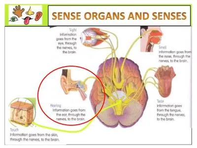
Nancy Sanderson MSN RN Lecture 4 Feature concept Sensory perception Ability to understand and interact through senses Sight Hearing Smell Taste Touch Copyright 2013 by Mosby an imprint of Elsevier Inc ID: 359394
Download Presentation The PPT/PDF document "NRS103 Head, Ears, Nose, and Throat Chap..." is the property of its rightful owner. Permission is granted to download and print the materials on this web site for personal, non-commercial use only, and to display it on your personal computer provided you do not modify the materials and that you retain all copyright notices contained in the materials. By downloading content from our website, you accept the terms of this agreement.
Slide1
NRS103 Head, Ears, Nose, and Throat Chapter 10
Nancy Sanderson MSN, RNLecture 4Slide2
Feature concept: Sensory perception
Ability to understand and interact through senses:Sight
HearingSmellTasteTouchCopyright © 2013 by Mosby, an imprint of Elsevier Inc.
2
Concept Overview Slide3
Recent head trauma?Loss of consciousness?
Headaches? I.e.: Sinus, migraines, neurologicalUse of a helmet when appropriate? I.e.: occupation, contact sports, cycling, and skateboarding.
Review of symptoms (ROS): HeadSlide4
Head
Head and neck contain multiple structures:
Skull encloses brain.Facial structures include eyes, ears, nose, and mouth.Neck structures include: Upper portion of spineEsophagus
Trachea
Thyroid gland
Arteries
Veins
Lymph nodes
Copyright © 2013 by Mosby, an imprint of Elsevier Inc.
4Slide5
Anatomy and Physiology:
HeadSkull is a bony structure that protects brain and upper spinal cord:
Contains special senses of vision, hearing, smell, taste.Comprises six bones fused at sutures.Covered by scalp tissue typically covered with hair.Face comprises 14 bones:Mandible articulates with temporal bone to form temporomandibular joint.
Facial muscles innervated by cranial nerves
V (trigeminal) and VII (facial).
Copyright © 2013 by Mosby, an imprint of Elsevier Inc.
5Slide6
Copyright © 2013 by Mosby, an imprint of Elsevier Inc.
6Slide7
Inspect and palpate:
Inspect the head for size shape and skin characteristics. I.e.: Head in relation to neck and shoulders for size and shape.Normocephalic
term designating that the skull is symmetric and appropriately proportioned for the size of the body. Versus microcephaly and macrocephaly. Inspect facial features for size, symmetry, movement, skin characteristics, and facial features. Face & Head InspectionSlide8
All facial features should appear symmetric with a calm facial expression. Facial bones should be symmetric and proportional to the size of the head.
Palpate the structures of the skull for contour and, tenderness and intactness. Palpation takes place when there is suspected injury, observed irregularity or abnormality or reported pain. The skull should be symmetric, and feel firm without tenderness.
Palpate the bony structures of the face, jaw, and movement of the jaw, temporal arteries for: tenderness, jaw pain, clicking, pulsation of artery. Continued: Face & Head InspectionSlide9
Asymmetrical facial expressionSlide10
TMJ full range of motion 5/5 muscle strength Slide11
Anatomy and Physiology:
Eyes – External Ocular Structures
External ocular structures:External eye is composed of eyebrows, upper and lower eyelids, eyelashes, conjunctivae, and lacrimal glands.
Palpebral fissure is opening between eyelids.
Conjunctivae are two thin, transparent mucous membranes, between eyelids and eyeball.
Bulbar conjunctiva covers scleral surface of eyeball.
Palpebral conjunctiva lines eyelids and contains blood vessels, nerves, hair follicles, and sebaceous glands.
Copyright © 2013 by Mosby, an imprint of Elsevier Inc.
11Slide12Slide13
External image of the eye:Slide14
Visual pathway of the eyeSlide15
Inspect the conjunctiva of the eye for color, drainage and lesions. This is done by gently pulling done the lower eye lid. No drainage, redness or swelling should be noted.
Inspecting the sclera of the eye commonly referred to as the whites of the eye for erythema, jaundice, or lesions. Sclera should be white and clear although in darker pigmented individuals the sclera may have a slight yellowing to the sclera.
Inspection of the eye: ROSSlide16
Cont.: Inspection of scleraSlide17
Cont.: Subconjunctival hemorrhageSlide18
Snellen Chart distance vision cranial nerve II
Patient sits or stands twenty feet from the chart. Patient reads the line of the smallest letters that are possible for them to read. Test the other eye in the same manner then both eyes. Document findings according to the fraction printed on the Snellen chart. You must also document if the patient is wearing corrective lenses or contacts at the time.
Visual Acuity: CNIISlide19
Pupils for size, shape, reaction to light, accommodation and consensual reaction. Determine the pupil size with a pupil gauge Using a pen light approaching from the side and shining the light directly into the pupil observing for a reaction of the pupil to the light. Consensual reaction the constriction of the iris and the pupil of one eye when a light is shown in the opposite eye.
Pupils: Inspection, CNIISlide20
Accommodation ask the patient to fix her gaze on an pen light or finger, observe the eyes as you move the object in and away from the face. The pupils will dilate and constrict when focusing on objects near and far as a simultaneous response, consensual response.
PERRLA- pupils equal round reacts to light and accommodating.
Pupils:cont. PERRLA Slide21
Six muscles attaching the eyeball to the orbit of the eye. These muscles are stimulated by three cranial nerves.
CNVI-abducens- innervates the lateral rectus muscle for lateral movement. Abducts the eye.
CNVI-trochlear- innervates the superior oblique muscles moving the eye down and in. CNIII- ocular motor- innervates all the rest of the eye moving superior, inferior, medial, rectus and the inferior oblique muscles.Extraocular Muscles: CNIV & VISlide22
Extra ocular muscles function test: EOM- hence extraocular movement. The patient to move or follow the eyes keeping head still through the six cardinal positions of gaze.
Extra Ocular MusclesSlide23
Stand 15 inches away from the patient and off to the side of the patient, shine the light beam on the pupil and look for the orange glow in the pupil. Normally: light reflex. Abnormal: absent light reflex (may be due to opacity of the lens, i.e. cataract)
Red reflex present bilaterally.
Light ReflexSlide24
Anatomy and Physiology:
Ears
Ear is a sensory organ for hearing and maintaining equilibriumDivided into three sections:External earMiddle earInner ear
Copyright © 2013 by Mosby, an imprint of Elsevier Inc.
24Slide25
Anatomy and Physiology:
External Ear
External earAuricle or pinna and external auditory ear canal composed of cartilage and skin.Helix is prominent outer rim.Concha is deep cavity in front of external auditory meatus.Lobule is bottom portion of ear.
Auricle serves three main functions:
Collection and focus of sound waves.
Location and direction of sound.
Protection of external ear from water and dirt.
Copyright © 2013 by Mosby, an imprint of Elsevier Inc.
25Slide26
Copyright © 2013 by Mosby, an imprint of Elsevier Inc.
26Slide27
Copyright © 2013 by Mosby, an imprint of Elsevier Inc.
27Slide28
Sound waves strike the tympanic membrane
Vibrations transmit through the auditory ossicles (malleus, incus, stapes) to oval window (Conductive hearing loss to this point)Vibrations travel to cochlea and then to the round window
(Sensory hearing loss at this point)CN VIII (acoustic)Nerve sends message to brainPhysiologySlide29
How is your hearing?
Use of hearing aid? Taking ototoxic drugs? Have you had any trouble with your ears or balance?
Are you having any vertigo? (feeling as if the room is spinning, different from dizzy)Are you having any tinnitus? (musical ringing in the ear)Does anyone smoke in your household?Increased risk of otitis media in children
ROS: EarSlide30
Use an otoscope with the largest ear speculum that the canal will accommodate
Position the patient’s head so that you can see through the scope
Straighten the ear canal be grasping the auricle firmly and pull it upward, backward and slightly away from the headBrace your hand against the patient’s faceInsert the speculum gently into the ear canal, directing it somewhat down and forward
Examining the Ear Canal and DrumSlide31
Use an
otoscope with the largest ear speculum that the canal will accommodate
Position the patient’s head so that you can see through the scopeStraighten the ear canal be grasping the auricle firmly and pull it upward, backward and slightly away from the headBrace your hand against the patient’s faceInsert the speculum gently into the ear canal, directing it somewhat down and forward
Examining the Ear Canal and DrumSlide32
Identify the handle of the malleus
Identify the short process of the malleusInspect the pars flaccida and Pars tensa
NormalShiny, transparent, pearly gray, slight concave, non-bulging, no perforationTM gray and intact bilaterally
without erythema, bulging,
or retraction.
Inspect the EardrumSlide33
Estimating Hearing
Occlude one ear of your patientStand 1-2 feet behind patientWhisper a word (i.e. 88)
Repeat with other earGross hearing intact by whisper test. Auditory AcuitySlide34
Auditory Acuity
Weber Test
Tap against palm and place midline vertex of headNormal: Hears equally in both earsConductive hearing loss- best in impaired ear
Sensorineural hearing loss- only in normal earSlide35
Auditory Acuity
Rinne
TestTap against palm and place on mastoid process. When no longer hears place 1-2 cm from ear until no longer hearsSensorineural hearing loss- heard longer thru air, but less than 2:1 ratio
Normal air conduction (ac) is
2 times longer
than bone conduction (
bc
)Slide36
Nose and Paranasal Sinuses
Allergic RhinitisItchingSwelling
RhinorrheaSneezingTearing eyesLater- stuffy nose, coughing, decreased smell, sore throat, dark circles under eyesSlide37
Inspect the anterior and inferior surfaces of the nose
Note any asymmetry or deformity
Inspect for dischargeTest patencyPress on each nostril one at a time and have the patient breath inPalpate for any masses, lesions or tendernessNose symmetrical midline. No deformities or skin lesions. Nares patent bilaterally.
Nose and Sinuses, ROS:Slide38
Inspect the inside of the nose
Inspect vestibule, septum and turbinates
Color of nasal mucosaForeign bodyDischarge (note color: clear, yellow, green, bloody)Masses, lesions, polypsSeptum: deviation, perforation, bleeding
Turbinates
: color, swelling,
exudate
, polyps
Normally no swelling,
mucoid
drainage; redder than oral mucosa
Septum without deviation, perforation, or bleeding.
Turbinates
pink, without dc, edema,
exudate
, or polyp.
Nose and Sinuses ………Slide39
Palpate for sinus tenderness
Press up on the frontal sinuses from under the bony brows (avoid pressure on the eyes)Press up on the maxillary sinuses
Normal: pt will feel pressure but no pain with palpationFrontal and maxillary sinuses nontender to palpation
Nose and SinusesSlide40
ROS: Sore throat
Sore tongueBleeding from the gumsTooth pain
HoarsenessMouth and Pharynx: Slide41
Inspect lips
ColorMoisture
Lumps UlcersCrackingSymmetrySwelling (edema)Inspect oral mucosa (inside of mouth)
With good light and a tongue blade, inspect for color, ulcers, white patches, and nodules.
Lips pink and moist without cracking or lesions. Buccal mucosa pink without nodules or lesions.
Mouth and PharynxSlide42
Teeth/Gums
Inspect for missing teeth, caries, conditions, discolorationNote the color of the gums
NormalPinkMargins without swellingNo bleedingTeeth white, straight, evenly spaced, clean and free of decay. Gums pink without swelling or bleeding.
Mouth and PharynxSlide43
Pharynx
With the patient’s mouth open, have the patient say “ah”As the patient says “ah” check the rise of the soft palate (CN X)
Gag reflex (CN IX , X)If needed press a tongue blade firmly down upon the midpoint of the arched tongueInspect the soft palate, anterior and posterior pillars, uvula, tonsils and pharynxNote color, swelling, ulceration
Tonsillar enlargement
Exudate
Breath odor (halitosis)
Soft palpate pink, rises midline. Tonsils pink without enlargement or exudate. Pharyngeal wall pink without exudate or lesions. No halitosis noted.
Mouth and Pharynx …Slide44Slide45
Neck
ROSNeck pain?Lumps or swelling?
History of neck surgery?History of neck trauma?Slide46
Neck Inspection & Palpation
Inspect for:
Head positionNeck muscle symmetryMasses or scarsAbnormal pulsations
Neck supple & symmetrical
Without masses, scars, or abnormal pulsationsSlide47
Trachea
Inspect
Should be midlinePalpateFor tracheal shiftPlace finger in sternal notch and slip to each side.
Trachea midline.Slide48
Cervical Lymph Nodes
Palpate the lymph nodes
Use the pads of your index and middle fingers with a gentle rotary motion.
Preauricular
Posterior auricular
Occipital
Tonsilar
Submandibular
Submental
Superficial cervical
Posterior cervical
Deep cervical chain
supraclavicularSlide49
Cervical Lymph Nodes
Note location, size, shape, delimitation, mobility, consistency and tenderness.
Lymph nodes normally nonpalpable in healthy adultsSmall, soft, mobile, discrete, non-tender nodes (shotty) may be found in normal persons. Enlarged (>1cm) firm, tender, and freely moveable often indicates infection.Hard, non-tender, and fixed often indicates malignancy.
Enlarged supraclavicular node, especially on left, suggests possible metastasis from thorax or abdomen
No lymphadenopathy noted or lymph nodes nonpalpable.Slide50
Thyroid Gland
Inspect first then
palpateAssess for:EnlargementGoiter
Consistency
Symmetry
Nodules
Movement
Thyroid nonpalpable
GoiterSlide51
Summery
PERRLA (pupils are equal round and react to light and accommodation)
Hearing (equal sound –whisper test; finger rubbing; Weber's best)Nose (normal appearance; symmetry; no discolored drainage)Mouth (check for malocclusion; tonsils; dental caries)Throat (Range of motion; palpate [use care]
sides & midline all should be midline &
nontender
)