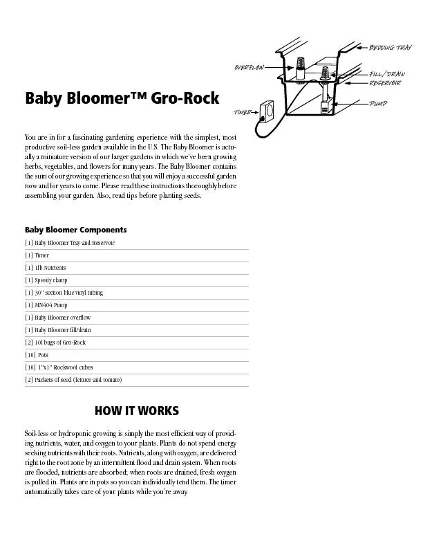
ENDOPLASMIC RETICULUM ER Rough ER Smooth ER Nuclear envelope Nucleolus Chromatin Plasma membrane Ribosomes Golgi apparatus Lysosome Mitochondrion Peroxisome Microvilli Microtubules ID: 724254
Download Presentation The PPT/PDF document "Review Unit 3 and 4 Figure 6.8a" is the property of its rightful owner. Permission is granted to download and print the materials on this web site for personal, non-commercial use only, and to display it on your personal computer provided you do not modify the materials and that you retain all copyright notices contained in the materials. By downloading content from our website, you accept the terms of this agreement.
Slide1
Review Unit 3 and 4Slide2
Figure 6.8a
ENDOPLASMIC RETICULUM (ER)
Rough
ER
Smooth
ER
Nuclear
envelope
Nucleolus
Chromatin
Plasma
membrane
Ribosomes
Golgi apparatus
Lysosome
Mitochondrion
Peroxisome
Microvilli
Microtubules
Intermediate filaments
Microfilaments
Centrosome
CYTOSKELETON:
Flagellum
NUCLEUSSlide3
NUCLEUS
Nuclear
envelope
Nucleolus
Chromatin
Golgi
apparatus
Mitochondrion
Peroxisome
Plasma membrane
Cell wall
Wall of adjacent cell
Plasmodesmata
Chloroplast
Microtubules
Intermediate
filaments
Microfilaments
CYTOSKELETON
Central vacuole
Ribosomes
Smooth
endoplasmic
reticulum
Rough
endoplasmic
reticulum
Figure 6.8cSlide4
Figure 6.UN01a
Nucleus
(ER)Slide5
Figure 6.UN01b
(Nuclear
envelope)Slide6
Figure 6.UN01cSlide7
Endosymbiont Theory
Mitochondria and chloroplasts have their own DNA in circular loops like prokaryotes
Both have a double membrane (inner from original prokaryote and outer from cell
Both are similar in size and structure to bacteriaBoth have ribosomes similar in structure and size to prokaryotesSlide8
Phospholipid
bilayer - What molecules can get through directly? Slide9
Figure 7.19
Passive transport
Active transport
Diffusion
Facilitated diffusion
ATPSlide10
Managing water balance
Cell survival depends on balancing water uptake & loss
freshwater
balanced
saltwaterSlide11
Figure 7.UN03
0.03
M
sucrose
0.02
M glucose
“Cell”
“Environment”
0.01
M sucrose0.01 M glucose0.01
M fructoseSlide12
2005-2006
Cell
(compared to beaker)
hypertonic or hypotonic
Beaker
(compared to cell) hypertonic or hypotonicWhich way does the water flow? in or out of cell
.05 M
.03 M
Osmosis… Slide13
Water Potential
Water Potential =
Y = Ys +
YpYs = -iCRTi = The number of particles the molecule will make in water; for NaCl this would be 2; for sucrose or glucose, this number is 1 C = Molar concentration R = Pressure constant = 0.0831 liter bar/mole K T = Temperature in degrees Kelvin = 273 + °C of solution Click here to see an entire page of water potential problems!Slide14
Water
Potential and Solution Potential
Sample Problem
The molar concentration of a sugar solution in an open beaker has been determined to be 0.3M. Calculate the solute potential at 27 degrees Celsius. Round your answer to the nearest tenths. Slide15
Q3
Solute potential= –
iCRT
i
= 1C= 0.3R = Pressure constant = 0.0831T= 27 +273=300KSolute concentration= -7.5
If a baby carrot with a water potential of -5.2 bars is put into this solution, what will happen? Why?Slide16
Answer…..
If a baby carrot with a water potential of -5.2 bars is put into this solution, what will happen? Why
?
Water moves from areas of higher water potential to areas of lower water potential (towards the more negative number!)So water moves……..out of the carrot!!! The carrot is hypotonic to the solutionSlide17
Water Potential – another example
The value for water potential in root tissue was found to be -3.3 bars. If you take the root tissue and place it in a .1 M solution of sucrose at 20 C in an open beaker, what is the water potential of the solution and in which direction will the net flow of water be? (answer is on the next slide) Slide18
Water Potential
The value for water potential in root tissue was found to be -3.3 bars. If you take the root tissue and place it in a .1 M solution of sucrose at 20 C in an open beaker, what is the water potential of the solution and in which direction will the net flow of water be?
Roots = -3.3 bars
.1M sucrose solution = -2.4 bars (calculate this!)Water moves from the sucrose solution into the roots (from high water potential to low water potential!)Slide19
Figure 9.2
Light
energy
ECOSYSTEM
Photosynthesis
in chloroplasts
Cellular respiration
in mitochondria
CO
2 H2O
O2
Organicmolecules
ATP powers
most cellular work
ATP
Heat
energySlide20
Cellular respiration –
Watch this VideoSlide21
Cellular
respiration –
2 ATP
2 ATP
~36 ATP
+
+
~40 ATPSlide22
Electron Transport Chain (oxygen as an electron acceptor and the generation of ATP)!!!!Slide23
Pyruvate is a branching point
Pyruvate
O
2
O
2
mitochondria
Krebs cycle
aerobic respiration
fermentation
anaerobic
respirationSlide24
Photosynthesis –
WATCH THIS VIDEO (7 minutes)Slide25Slide26
Light: absorption spectra
Photosynthesis gets energy by
absorbing
wavelengths of lightchlorophyll
a absorbs best in red & blue wavelengths & least in greenaccessory pigments with different structures absorb light of different wavelengthschlorophyll b, carotenoids, xanthophylls
Why are
plants green?Slide27
Figure 10.UN02
Primary
acceptor
Primary
acceptor
Cytochrome
complex
NADP
reductase
Photosystem II
Photosystem I
ATP
Pq
Pc
Fd
NADP
+ H
NADPH
H
2
O
O
2
Electron transport
chain
Electron transport
chainSlide28
Light
Light
Reactions:
Photosystem
II
Electron transport chain
Photosystem IElectron transport chain
NADP
ADP
+ P i
RuBPATP
NADPH
3-Phosphoglycerate
Calvin
Cycle
G3P
Starch
(storage)
Sucrose (export)
Chloroplast
H
2
O
CO
2
O
2
Figure 10.22Slide29
Watch Bozeman Biology Photosynthesis and Respiration
Cellular Respiration Video
Photosynthesis Video