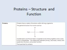
Introducing proteins Proteins are a diverse group of large and complex polymer molecules made up of long chains of amino acids They have a wide range of biological roles including structural ID: 706840
Download Presentation The PPT/PDF document "Proteins – Structure and Function" is the property of its rightful owner. Permission is granted to download and print the materials on this web site for personal, non-commercial use only, and to display it on your personal computer provided you do not modify the materials and that you retain all copyright notices contained in the materials. By downloading content from our website, you accept the terms of this agreement.
Slide1
Proteins – Structure and FunctionSlide2
Introducing proteins
Proteins
are a diverse group of large and complex polymer molecules, made up of long chains of
amino acids
.
They have a wide range of biological roles, including:
structural: proteins are the main component of body tissues, such as muscle, skin, ligaments and hair
catalytic: all enzymes are proteins, catalyzing many biochemical reactions
signalling: many hormones and receptors are proteins
immunological: all antibodies are proteins.Slide3
The general structure of amino acids
All amino acids have the same general structure: the only difference between each one is the nature of the
R group
.
The R group therefore defines an amino acid.
amino
group
carboxylic acid group
R group
The R group represents a side chain from the central ‘alpha’ carbon atom, and can be anything from a simple hydrogen atom to a more complex ring structure.Slide4
The 20 naturally-occurring amino acidsSlide5
Proteins – Structure and FunctionSlide6
Peptide bonds and dipeptidesSlide7
Polypeptides
When more amino acids are added to a dipeptide, a
polypeptide
chain is formed.
A protein consists of one or more polypeptide chains folded into a highly specific 3D shape.
There are up to four levels of structure in a protein:
primary, secondary, tertiary and quaternary. Each of these play an important role in the overall structure and function of the protein.Slide8
Portion of polypeptide chain
Amino acids
Peptide bond
- helix
Hydrogen bonds
Hydrogen bonds form
Primary structure
- the sequence of amino acids in a polypeptide chain
Secondary structure
- the folding of the polypeptide chain into an
α
-helix or a
β
-pleated sheet.Slide9
- pleated sheet
Portion of polypeptide chain
Hydrogen bonds
Hydrogen bonds form
Secondary structure
- the folding of the polypeptide chain into an
α
-helix or a
β
-pleated sheet.Slide10
- helices
- pleated sheet
Amorphous regions
Tertiary structure
- the secondary structures fold up to form a very precise three-dimensional structureSlide11
C
O
-
+
HN
Hydrogen bonds
bonds to moleculebonds to moleculeShared electrons spend longer at these atoms, forming a slight negative charge hydrogen bond
High temperatures and altered pH can split these bonds
Bonds responsible for the tertiary structure…Hydrogen bonds form between these polar groupsSlide12
C
O
-
+
HN
O
HH
Ionic bondsbond to moleculebond to moleculeBasic groupAcidic group
Ionic bondIonic bonds can be split by changing the pH
Bonds responsible for the tertiary structure…Slide13
HS
CH
2
SH
CH2
SCH2
SCH2
Disulphide bondsDisulphide bonds can be split by reducing agentscysteine R group
disulphide bond(covalent)Bonds responsible for the tertiary structure…Slide14
Polar and Non Polar
The arrangement
of
the atoms in some molecules is such that one end of the molecule has a positive electrical charge and the other side has a negative charge. If this is the case, the molecule is called a polar molecule, meaning that it has electrical poles. Otherwise, it is called a non-polar molecule.
Whether molecules are polar or non-polar determines if they will mix to form a solution or that they don't mix well together. Also, polar molecules are water soluble, while non-polar molecules are fat soluble.Slide15
CH(CH
3
)
2
CH2
van der Waal’s forces
These forces can be split by a rise in temperaturePhenylalanine R groupValine R groupWeak van der Waals’ force of attraction
These are weak forces of attraction between non-polar groupsWater excluded from these hydrophobic side chains helps keep the side chains togetherBonds responsible for the tertiary structure…Slide16
CH
3
CH
2
SH
CH2
C-OO
CH
2HS
CH3
CH2
+
HN
NH
CH
2
OH
CH
2
C
NH
2
O
+
NH
3
(CH
2
)
4
CH
2
OH
3
4
1
7
5
2
8
11
14
13
10
12
9
6
15
16
17
18
19
20
21
22
23
24
25
26
27
28
29
30
31
32
33
34
35
36
37
38
Lysine
Tyrosine
Asparagine
Serine
Alanine
Cysteine
Aspartate
Histidine
Basic R group
Acidic R group
Polar R group
Polar R group
Polar R group
Polar R group
Non-polar R group
Non-polar R groupSlide17
CH
3
CH
2
S
CH
2
C
O
O-
CH
2
S
CH
3
CH
2
HN
HN
CH
2
HO
CH
2
C
NH
2
O
+
NH
3
(CH
2
)
4
CH
2
HO
3
4
1
7
5
2
8
11
14
13
10
12
9
6
15
16
17
18
19
20
21
22
23
24
25
26
27
28
29
30
31
32
33
34
35
36
37
38
Ionic bond
Hydrogen bonds
Disulphide bond
van
der
Waal’s forcesSlide18
H
2
0
H
2
0
H20H20
H20H20H20H
20H20H20H2
0H20H20H2
0H
2
0
H
2
0
H
2
0
H
2
0
H
2
0
H
2
0
H
2
0
H
2
0
H
2
0
H
2
0
H
2
0
1
5
11
2
3
4
6
7
8
9
10
12
13
1
5
11
2
3
4
6
7
8
9
10
12
13
NOTE: the cell is an
aqueous
environment
Hydrophilic R groups
Hydrophobic R groups
Globular
proteins form a spherical mass with a specific 3-D shape (tertiary and quaternary structure)
They fold up so that
hydrophilic
groups are on the outside and
hydrophobic
groups are inside the molecule Slide19
Quaternary structure
This is where two or more polypeptide chains are joined to form a protein,
e.g. haemoglobin
collagenSlide20
-chain subunit
-chain subunit
Haem
groups
Haemoglobin is an example of a
globular
protein with quaternary structure
4 polypeptide chains
2
-subunits
2 -subunits
4
haem
prosthetic groupsSlide21
Fibrous proteins
Fibrous protein molecules form long chains or fibres
(they have primary, secondary, tertiary and quaternary structure)
Their fibrous nature makes them insoluble in water...
... this makes them useful for structure and support
Collagen
found in skin, teeth, bones, tendons, blood vessel walls
Fibres form a triple-helix of polypeptide chains
These chains are held together by hydrogen bonds
Polypeptide chains
Hydrogen bondsSlide22
The structure of proteinsSlide23
Protein structureSlide24
Denaturing proteins
If the bonds that maintain a protein’s shape are broken, the protein will stop working properly and is
denatured
.
Changes in temperature, pH or salt concentration can all denature a protein, although the specific conditions will vary from protein to protein.
Fibrous proteins lose their structural strength when denatured, whereas globular proteins become insoluble and inactive.
denaturation: bonds brokenSlide25
Biuret test for proteinsSlide26
PROTEINSdingbats – say what you seeSlide27Slide28
NNNNNNNNNNN
MSlide29
S
2-Slide30
-LINSlide31
- jSlide32
- PASlide33
-k +ySlide34
C
TSlide35
+
arySlide36Slide37Slide38Slide39
+
ary
-d