/
Inguinal canal Inguinal canal

Inguinal canal - PDF document

phoebe
phoebe . @phoebe
Follow
345 views
Uploaded On 2022-10-29

Inguinal canal - PPT Presentation

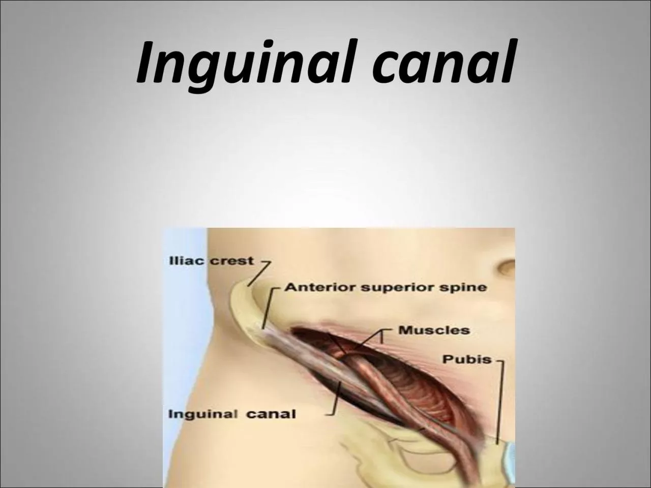
Inguinal Canal It is an oblique passage through the lower part of the anterior abdominal wall Present in both sexes It allows structures to pass to and from the testis to the abdomen in ID: 961684

fascia inguinal testis wall inguinal fascia wall testis canal ring spermatic abdominal scrotum hernia vaginalis superficial nerve oblique part

Share:

Link:

Embed:

Download Presentation from below link

Download Pdf The PPT/PDF document "Inguinal canal" is the property of its rightful owner. Permission is granted to download and print the materials on this web site for personal, non-commercial use only, and to display it on your personal computer provided you do not modify the materials and that you retain all copyright notices contained in the materials. By downloading content from our website, you accept the terms of this agreement.


Presentation Transcript

Inguinal canal Inguinal Canal • It is an oblique passage through the lower part of the anterior abdominal wall • Present in both sexes • It allows structures to pass to and from the testis to the abdomen in males • In fema

les it permits the passage of the round ligament of the uterus from the uterus to the labium majus • Transmits ilioinguinal nerve in both sexes Inguinal Canal • It is about 1 ½ inches or 4 cm long in the adults • Extends

from the deep inguinal ring downward and medially to the superficial inguinal ring • Lies parallel to and immediately above the inguinal ligament • In the newborn child, the deep ring lies almost directly posterior to the superfic

ial ring Deep Inguinal Ring • Is an oval opening in the fascia transversalis • Lies about ½ inch ( 1 . 3 cm) above the inguinal ligament midway between the anterosuperior iliac spine and the symphysis pubis • Margins o

f the ring give attachment to the internal spermatic fascia Superficial Inguinal Ring • T riangular in shape • Defect in the aponeurosis of the external oblique muscle • Lies immediately above and medial to the pubic tuber

cle • Its margins some times called crura(Med & lat crus), give attachment to the external spermatic fascia Anterior Wall of Inguinal Canal • It is formed along its entire length by aponeurosis of the external oblique muscle

• It is reinforced in its lateral third by the origin of the internal oblique from the inguinal ligament • This wall is strongest where it lies opposite the weakest part of posterior wall, that is deep inguinal ring Posterior W

all of Inguinal Canal • It is formed along its entire length by the fascia transversalis • It is reinforced in its medial third by conjoint tendon, the common tendon of insertion of internal oblique and transversus , attached to th

e pubic crest and pectineal line • This wall is strongest where it lies opposite the weakest part of the anterior wall, that is superficial inguinal ring Inferior Wall of Inguinal Canal = floor • It is formed by the rolled - under

inferior edge of the aponeurosis of the external oblique muscle called inguinal ligament and at its medial end, the lacunar ligament • Superior Wall of Inguinal Canal = Roof • It is formed by the arching lowest fibers of the inter

nal oblique and transversus abdominis muscles Functions of Inguinal Canal • It allows structures of spermatic cord to pass to and from the testis to the abdomen in male • Permits the passage of round ligament of uterus from th

e uterus to the labium majus in female Contents of inguinal canal • Spermatic cord & its contents in male • Round ligament in female • Genital branch of genitofemoral nerve • Ilioinguinal nerve: Enter the canal through t

he posterior wall Inguinal triangle - Region of abdominal wall Borders • Medial border: Lateral margin of the rectus sheath, also called linea semilunaris • Superolateral border: Inferior epigastric vessels •

Inferior border: Inguinal ligament Spermatic Cord • It is a collection of structures that pass through the inguinal canal to and from the testis • It is covered with three concentric layers of fascia derived from the layers of a

nterior abdominal wall • It begins at the deep inguinal ring lateral to the inferior epigastric artery and ends at the testis Structures of Spermatic Cord • Vas deferens • Testicular artery and vein • Testicular lymph vessels

• Autonomic nerves • Processus vaginalis • Cremastric artery • Artery of the vas deference • Genital branch of genitofemoral nerve Covering of the Spermatic Cord • The covering of the spermatic cord are three concentric laye

rs of fascia derived from the layers of the anterior abdominal wall • Each covering is acquired as the processus vaginalis descends into the scrotum through the layers of the abdominal wall • External Spermatic fascia: Is derived

from the external oblique aponeurosis and attached to the margins of the superficial inguinal ring • Cremasteric Fascia: Is derived from the internal oblique muscle • Internal Spermatic Fascia: Is derived from the fascia transversa

lis and attached to the margins of deep inguinal ring Vas Deferens • It is a cord like structure • Can be palpated between finger and thumb in the upper part of the scrotum • It is a thick walled muscular duct that transport

spermatozoa from the epididymis to the prostatic urethra Testicular Artery • It is a branch of abdominal aorta at level of L 2 • It is long and slender • Descends on the posterior abdominal wall • It traverses the inguinal

canal and supplies the testis and the epididymis Testicular Veins • These are the extensive venous plexus, the pampiniform plexus • Leaves the posterior border of the testis • As the plexus ascends, it becomes reduced in size so

that at about the level of deep inguinal ring, a single testicular vein is formed • Drains into left renal vein on left side and inferior vena cava on right side Testicular artery & vein Autonomic nerve & Genitofemoral nerve Autonomic

nerves - Sympathetic fibers run with testicular artery from renal or aortic sympathetic plexuses - Afferent sensory nerve Genital branch of the genitofemoral nerve - Its root L 1 & L 2 - Supply the cremastric muscle Testicular lymph

atic vessels • Ascend through the inguinal canal • Passes up over the post. Abdominal wall • Reach the lumbar (Para - aortic) lymph nodes on each side of the aorta at level L 1 Processus vaginalis • An out pouching of peritoneum t

hat in the fetus is responsible for the formation of the inguinal canal • The remains of the processus vaginalis causes the indirect hernia Developing of process vaginalis Developing of process vaginalis……..cont Inguinal Hernia •

A hernia is the protrusion of part of the abdominal contents beyond the normal confines of the abdominal wall • Consists of three parts: the sac, contents of the sac, covering of the sac • Hernial coverings are formed from the layers

of the abdominal wall through which the hernial sac passes Indirect Inguinal Hernia • It is the most common form of hernia • Is believed to be congenital in origin • The hernial sac is remains of processus vaginalis • Enters

the inguinal canal through the deep inguinal ring lateral to the inferior epigastric vessels • It may extend part of the way along the canal or as far as the superficial inguinal ring Indirect Inguinal Hernia • If the processus vagin

alis has undergone no obliteration, the hernia is complete and extends through the superficial inguinal ring down into the scrotum or labium majus • Under these circumstances the neck of the hernial sac lies at the deep inguinal ring â

€¢ It is 20 times more common in young males than females • Is more common on the right side(the Rt. testis descends later than the Lt. testis) Direct Inguinal Hernia • It composes about 15 % of all inguinal hernias • Common i

n old men with weak abdominal muscles and rare in women • Hernial sac bulges forward through the posterior wall of the inguinal canal medial to the inferior epigastric artery • The neck of the hernial sac is wide Inguinal Hernia I

ndirect Direct young Common on old Age unilateral Usually bilateral Bilaterally Oval Hemispherical Shape Can reach the scrotum never Reaches scrotum Downwards , forwards medially Forwards Direction of descent Upward, backward laterally backward Red

uction Laterally Medially Relation to inf. epigastric art. Feel an impulse on the tip of the finger Feel impulse on the side finger Superficial inguinal ring test Hernia does not appear Hernia appears Deep ring test Reduction of hernia, put thu

mb over deep ring, ask patient to cough Skin, superfacial fascia, Ex.sp.fascia, cremastric muscle & fascia, Int.spermatic fascia, 1 - Lat. To lat. Umbilical lig Same as indirection 2 - Med. To lat. Coverings Direct Hernia Route Note :

The hernia sac passes directly through inguinal triangle and may disrupt the floor of the inguinal canal. Indirect Hernia Route Note : T he hernia sac passes outside the boundaries of Hesselbach's triangle(inguinal triangle

) and follows the course of the spermatic cord. Scrotum • It is an outpouching of the lower part of the anterior abdominal wall • It contains testes, epididymis, and the lower ends of the spermatic cord • Its wall has following

layers: skin, superficial fascia, external spermatic fascia derived from external oblique, cremastric fascia derived from internal oblique internal spermatic fascia derived from transversalis, and tunica virginals( parietal & visceral layer)

Skin of the Scrotum • Skin of the scrotum is thin, wrinkled, and pigmented and forms a single pouch • A ridge in the midline indicates the line of fusion of the two lateral labioscrotal swellings • Superficial fascia is continuous w

ith the fatty and membranous layers of the anterior abdominal wall Superficial Fascia • Superficial fascia is continuous with the fatty and membranous layers of the anterior abdominal wall • The fat is replaced by smooth muscle called

dartos muscle • It is responsible for wrinkles of the skin • Membranous layer referred to as Colle’s fascia • Innervated by sympathetic nerve fibers • Both layers of sup. Fascia contribute to a median partition that crosses t

he scrotum and separates the testes from each other Spermatic Fasciae • Lies beneath the superficial fascia • Derived from three layers of anterior abdominal wall on each side • The external spermatic fascia is derived from

external oblique • The cremastric fascia is derived from internal oblique • The internal spermatic fascia is derived from the fascia transversalis Tunica Vaginalis • Lies within the spermatic fasciae • Covers the ante

rior, medial and lateral surfaces of each testis • It is the lower expanded part of the processus vaginalis • Normally shut off just before birth from the upper part of the processus and the peritoneal cavity Fig : Internal structure

s of the scrotum. This illustration shows portions of the scrotum cut away to reveal the cremasteric muscle, spermatic cord, vas deferens, and a testis within the scrotal sac. Internal structures of the scrotum (sperm - carrying tube) (muscle

fibers that control the position of the testis in the scrotal sac) (contains vas deferens, blood vessels, nerves, and cremasteric muscle) (where sperm mature and are stored temporarily) Testis • They are a firm, mobile organ,

within the scrotum • Left testis usually lies at a lower level than the right • Upper end of the gland is tilted forward • Surrounded by a tough fibrous capsule, the tunica albuginea • A series of fibrous septa divide the in

terior of the organ into lobules • Lying in each lobule are one to three coiled seminiferous tubules • The tubules open into the network of channels called the rete testis • Small efferent ductules connect the rete testi

s to the upper end of the epididymis Structures inside the testis • Seminiferous tubules – Thin, highly coiled structures where sperm production occurs. • Interstitial cells – Major source of androgens – Located between

seminiferous tubules • Epididymis – Site of sperm maturation – Runs along back of testis • Vas deferens – Sperm - carrying tube – Begins at the testis and ends at the urethra. Fig : Male sexu

al anatomy: A cross - section side view of male reproductive organs. Overview: male sexual anatomy Blood supply of testes Artery - Testicular arteries  Abdominal aorta at level L 2 Vein - Pampiniform plexus  reduced to a

single vein  ascend through inguinal canal  Rt. testicular vein drains into I.V.C & Lt. testicular vein drains into Lt.renal vein Lymphatic drainage of testes • Ascend in the spermatic cord • End in the lymph nodes on the sid

e of aorta(Lumber or Para - aortic) nodes at level L 1 • Scrotum+ skin  inguinal canal lymphatic nodes Nerve supply to testes Autonomic nerves - Sympathetic fibers run with testicular artery from renal or aortic sympathetic plexu

ses - Afferent sensory nerve Genital branch of the genitofemoral nerve - Supply the cremastric muscle Scrotum  By the above nerves + ilioinguinal nerve Clinical Notes Clinical conditions involving the scrotum and testes Varico

cele: - The veins of the pampiniform plexus elongated & dilated - Lt side more common  venous pressure is higher - Common in young & adult • Vasectomy  Infertility • Processus vaginalis Upper part  oblit

erated just before birth Lower part  Tunica vaginalis Congenital anomalies of processus vaginalis 1 - persist  indirect inguinal hernia 2 - Narrowed  congenital hydrocele 3 - Obliterated upper & lower part  e

ncysted hydrocele of the cord Abnormality in testis & scrotum……..cont Hydrocele - Accumulation of fluid within the tunica vaginalis of the testis - Causes 1 - Inflammatory 2 - idiopathic - Tapping ahydrocele  stru

ctures ( all layers covering the testis, skin  tunica vaginalis )traversed by the cannula Congenital anomalies of the testes Cryptorchidism - Incomplete descent of testis although traveling down normal pathway - It may be found in 1

- Abdominal cavity 2 - In inguinal canal 3 - At superficial inguinal ring 4 - In upper part of scrotum Maldescent - Testes travel down an abnormal pathway 1 - Superfacial fascia 2 - Root of penis 3 - Perineum 4 -