/
Aspartate Aminotransferase ASTGOT Activity Aspartate Aminotransferase ASTGOT Activity

Aspartate Aminotransferase ASTGOT Activity - PDF document

queenie
queenie . @queenie
Follow
342 views
Uploaded On 2022-08-16

Aspartate Aminotransferase ASTGOT Activity - PPT Presentation

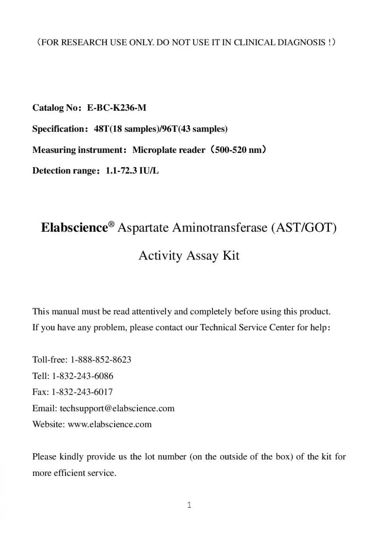
Assay Kit ReitmanFrankel Method Please kindly provide us the lot number on the outside of the box of the kit for more efficient service Catalog No EBCK236M Method Colorimetric method Speci ID: 936658

min sample tissue reagent sample min reagent tissue standard add wells microplate fully reader 200 mix detection unit supernatant

Share:

Link:

Embed:

Download Presentation from below link

Download Pdf The PPT/PDF document "Aspartate Aminotransferase ASTGOT Activi..." is the property of its rightful owner. Permission is granted to download and print the materials on this web site for personal, non-commercial use only, and to display it on your personal computer provided you do not modify the materials and that you retain all copyright notices contained in the materials. By downloading content from our website, you accept the terms of this agreement.


Presentation Transcript

Aspartate Aminotransferase (AST/GOT) Activity Assay Kit (Reitman-Frankel Method) Please kindly provide us the lot number (on the outside of the box) of the kit for more efficient service. Catalog No: E-BC-K236-M Method: Colorimetric method Specification: 96T (Can detect 43 samples without duplication) Instrument: Microplate reader Sensitivity: 1.1 IU/L Detection range: 1.1-72.3 IU/L 1 General information Intended use Background This kit can measure Aspartate Aminotransferase (AST/GOT) activity in animal serum (plasma), tissue, culture cells and cell culture supernatant, etc. AST/GOT is a key enzyme in nitrogen metabolism, which is widely found in plasma and body tissues, including liver, heart, skeletal muscle, kidney, brain, pancreas, lung and erythrocyte. Changes in AST/GOT activity were found in acute pancreatitis, ischemic stroke, severe burns, periodontitis, acute renal disease and motor neuron disease. Detection prin

ciple AST/GOT enables alpha-ketoglutaric acid and aspartic acid to displace amino and keto groups to form glutamic acid and oxaloacetic acid. Oxaloacetic acid can decarboxylate itself to form Pyroracemic acid during the reaction. Pyroracemic acid reacted with 2,4-dinitrophenylhydrazine(DNPH) to form 2,4, dinitrophenylhydrazone showing reddish brown in alkaline solution. Measure the OD values and calculate the enzyme activity. Focus on your research Service for life science 2 Kit components & storage Item Component Reagent 1 Buffer Solution 0.5 mL × 1 vial 2-8 , 6 months Reagent 2 2 mmol/L Sodium Pyruvate 0.5 mL × 1 vial 2-8 , 6 months Reagent 3 Substrate Solution 5 mL × 1 vial 2-8 , 6 months Reagent 4 Chromogenic Agent 5 mL × 1 vial 2-8 , 6 months, shading light Reagent 5 Alkali Reagent 5 mL × 1 vial 2-8 , 6 months Microplate 96 wells No requirement Plate Sealer 2 pieces Note: The reagents must be stored stric

tly according to the preservation conditions in the above table. The reagents in different kits cannot be mixed with each other. Reagents Double distilled water, Normal saline (0.9% NaCl), PBS (0.01 M, pH 7.4) Instruments Microplate reader (500-520 nm), Micropipettor, Vortex mixer, Incubator, Multichannel pipette Materials prepared by users Consumptive material Tips (10 μL, 200 μL, 1000 μL), EP tubes (1.5 mL, 2 mL) 3 Safety data Precautions Some of the reagents in the kit contain dangerous substances. It should be avoided to touch the skin and clothing. Wash immediately with plenty of water if touching it carelessly. All the samples and waste material should be treated according to the relevant rules of laboratory’s biosafety. Before the experiment, please read the instructions carefully, and wear gloves and work clothes. The key points of the assay 1.It is recommended to use multi-channel pipette to add reage

nt 5 working solution to reduce the difference between wells. Detect the sample as soon as possible after collection. The serum sample can be store at 2-8 for 7 days and -20 for 20 days. Pre-assay preparation Reagent preparation 1. Preparation of Reagent 5 working solution: Dilute the Reagent 5 with double distilled water at the ratio of 1: 9 and mix fully. Prepare the fresh solution before use. 2. Incubate reagent 3 at 37 °C for 10 min. The samples should be prepared as conventional methods. Also please refer to appendix II. Sample preparation 4 Dilution of sample It is recommended to take 2~3 samples with expected large difference to do pre-experiment before formal experiment and dilute the sample according to the result of the pre-experiment and the detection range (1.1-72.3 IU/L). Assay protocol Ambient temperature 25-30 Optimum detection wavelength 510 nm Instructions for the use of transferpettor: (1) Equilibrate

the pipette tip in that reagent before pipetting each reagent. (2) Don’t add the liquid outside the tips into the reaction system when pipetting each reagent. Note: The diluent is normal saline (0.9% NaCl) or PBS (0.01 M, pH 7.4). Sample type Dilution factor Human serum 1 Human plasma 1 Porcine serum 1 Rat serum 1 HC-60 cellular supernatant 1 Calu-3 cellular supernatant 1 10% Rat liver tissue homogenization 15-30 10% Rat lung tissue homogenization 2-8 The recommended dilution factor for different samples is as follows (for reference only) 5 Assay protocol Plate set up Note: A-E, standard wells; S1-S43, sample wells; S1'-S43, control wells. 1 2 3 4 5 6 7 8 9 10 11 12 A A A S4 S4' S12 S12' S20 S20' S28 S28' S36 S36' B B B S5 S5' S13 S13' S21 S21' S29 S29' S37 S37' C C C S6 S6' S14 S14' S22 S22' S30 S30' S38 S38' D D D S7 S7' S15 S15' S23 S23' S31 S31' S39 S39' E E E S8 S8' S16 S16' S24 S24' S32 S32' S40 S40' F S1 S1' S9

S9' S17 S17' S25 S25' S33 S33' S41 S41' G S2 S2' S10 S10' S18 S18' S26 S26' S34 S34' S42 S42' H S3 S3' S11 S11' S19 S19' S27 S27' S35 S35' S43 S43' Operating steps (1) Standard wells: Add 5 μL of regent 1 to the standard wells respectively (the pipettes should touch the bottom of the plate). Add 20, 18, 16, 14, 12 μL of reagent 3 to the standard wells from A to E, respectively. Add 0, 2, 4, 6, 8 μL of reagent 2 to the standard wells from A to E, respectively. Sample wells: Add 20 μL of reagent 3 (pre-heated at 37 for 10 min) and 5 μL of sample. Control wells: Add 20 μL of reagent 3 (pre-heated at 37 for 10 min). (2) Mix fully (this is very important), then incubate at 37 for 30 min. (3) Add 20 μL of reagent 4 to each well. 6 Operation table A B C D E 5 5 5 5 5 20 18 16 14 12 0 2 4 6 8 Mix fully (this is very important), then incubate at 37 for 30 min. 20 20 20 20 20 Mix fully with microplate reader for 10

s and incubate at 37 for 20 min. Reagent 5 working 200 200 200 200 200 Mix fully with microplate reader for 10 s, stand for 15 min at room temperature and measure the OD value of each well with microplate reader at 510 nm. (4) Control wells: Add 5 μL of sample to Control wells. (5) Mix fully with microplate reader for 10 s, incubate at 37 for 20 min. (6) Add 200 μL of reagent 5 working solution to each well (the multi-channel pipette is recommended). (7) Mix fully with microplate reader for 10 s, stand for 15 min at room temperature and measure the OD value of each well with microplate reader at 510 nm. Set 5 wells of micro-plate for standard and operate according to the following operating table. The preparation of standard curve Control well Sample well for 10 min) 20 20 5 Mix fully (this is very important), then incubate at 37 for 30 min. 20 20 5 Mix fully with microplate reader for 10 s and incubate at 37 for 2

0 min. 200 200 Mix fully with microplate reader for 10 s, stand for 15 min at room temperature and measure the OD value of each well with microplate reader at 510 nm. The measurement of samples Calculation Definition of international unit: The enzyme amount of 1 μmol of NADH consumed in reaction system (1 mL sample or 1 g tissue protein, 25 ) per minute is defined as 1 unit (wavelength is 340 nm, optical path is 1 cm). Definition of Carmen unit: 1 mL of sample, the total volume of reaction is 3 mL, wavelength is 340 nm, optical path is 1 cm, react at 25 for 1 min, the amount of generated pyruvic acid which oxidize NADH to NAD+ and cause absorbance decreasing 0.001 is as 1 unit. (1 Carmen unit = 0.482 IU/L, 25 ). 8 Note: y: Carmen unit. x: OD standard –OD blank OD blank is the OD value when the carmen unit is 0 510 : OD sample -OD control a, b, c: the constant of standard curve. f: dilution factor of sample before

tested. Cpr: concentration of protein in sample (gprot/L) 1. This kit is for research use only. 2. Instructions should be followed strictly, changes of operation may result in unreliable results. 3. The validity of kit is 6 months. 4. Do not use components from different batches of kit. Notes (1) Serum/plasma AST/GOT activity (IU/L)=[a×(ΔA 510 ) 2 510 +c]×0.482×f (2) Tissue and Cells AST/GOT activity (IU/gprot)=[a×(ΔA 510 ) 2 510 +c]×0.482×f÷Cpr 3.Plot the standard curve by using OD value of standard and correspondent Carmen unit (0, 24, 61, 114, 190 Carmen unit) as x-axis and y-axis respectively. Create the standard curve with graph software (or EXCEL). The Carmen unit of the sample can be calculated according to the formula based on the OD value of sample. The standard curve is y=ax 2 +bx+c. Detection range 1.1-72.3 IU/L Average intra-assay CV (%) 5.3 Sensitivity 1.1 IU/L Averag

e inter-assay CV (%) 6.8 Appendix I Performance characteristics Take 5 μL of human serum, carry the assay according to the operation table. The results are as follows: standard curve: y=2517.55x 2 +74.50x+1.8995, the average OD value of the sample is 0.259, the average OD value of the control is 0.233, and the calculation result is: AST activity (IU/L)= [2517.55 ×(0.259-0.233) 2 +74.5 × ( 0.259 - 0.233 )+1.8995] ×0.482 =2.67 IU/L Example analysis Appendix II Sample preparation Collect fresh blood and stand at 25 for 30 min to clot the blood. Then centrifuge at 2000 g for 15 min at 4 . Take the serum (which is the upper light yellow clarified liquid layer) to preserve it on ice for detection. Take fresh blood into the tube which has anticoagulant (heparin is recommended), centrifuge at 700-1000 g for 10 min at 4 . Take the plasma (which is the upper light yellow clarified liq

uid layer, don't take white blood cells and platelets in the middle layer) to preserve it on ice for detection. Collect fresh urine and centrifuge at 10000 g for 15 min at 4 . Take the supernatant to preserve it on ice for detection. Serum Plasma Urine The following sample pretreatment methods are for reference only. Take 0.02-1g fresh tissue to wash with homogenization medium at 2-8 . Absorb the water with filter paper and weigh. Homogenize at the ratio of the volume of homogenized medium (2-8 ) (mL): the weight of the tissue (g) =9:1, then centrifuge the tissue homogenate for 10 min at 1500 g at 4 . Take the supernatant to preserve it on ice for detection. Meanwhile, determine the protein concentration of supernatant (E-BC-K318-M, E-BC-K168-S, E-BC- K165-S). Tissue sample 11 Collect the cells and wash the cells with homogenization medium for 1~2 times. Centrifuge at 1000 g for 10 min and then discard the supernat

ant and keep the cell sediment. Add homogenization medium at a ratio of cell number (10 6 operated in ice water bath. Centrifuge at 10000 g for 10 min, then take the supernatant and preserve it on ice for detection. Meanwhile, determine the protein concentration of supernatant (E-BC-K318-M, E-BC-K168-S, E-BC- K165-S). Cells Note: 1. Homogenized medium: PBS (0.01 M, pH 7.4) including 0.1 mM EDTA. 2. Homogenized method: (1) Hand-operated: Weigh the tissue and mince to small pieces (1 mm 3 ), then put the tissues pieces to glass homogenized tube. Add homogenized medium into homogenized tube, place the tube into the ice bath with left hand, and insert the glass tamping rod vertically into the homogenized tube with the right hand to grind up and down for 6-8 min. Or put the tissue into the mortar, and add liquid nitrogen to grind fully. Then add the homogenized medium to homogenize. (2) Mechanical homogenate: Weigh

the tissue to EP tube, add the homogenized medium to homogenize the tissue with homogenizer instrument (60 Hz, 90s) in the ice bath. (For samples of skin, muscle and plant tissue, the time of homogenization can be properly prolonged.) (3) Ultrasonication: Treat the cells with ultrasonic cell disruptor (200 W, 2 s/ time, interval for 3 s, the total time is 5 min). 12 1. Please predict the concentration before assaying. If the sample concentration is not within the range of the standard curve, users must determine the optimal sample dilutions for their particular experiments. 2. If the sample type is not included in the manual, a preliminary experiment is suggested to verify the validity. 3. If a lysis buffer is used to prepare tissue or cell homogenates, there is a possibility of causing a deviation due to the introduced chemical substance. Notes for sample www.elabscience.com Focus on your research Service for life s