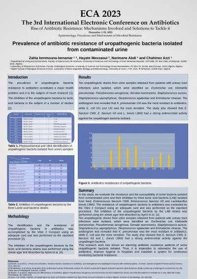
/
A case report of isolated A case report of isolated

A case report of isolated - PowerPoint Presentation

reagan
reagan . @reagan
Follow
342 views
Uploaded On 2022-06-15

A case report of isolated - PPT Presentation

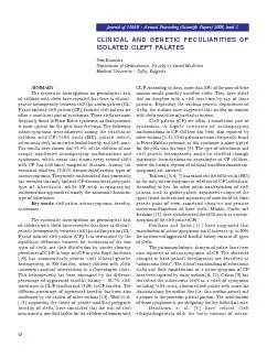
pigmentosus atrophic lichen planus on the supramaxillary area a new variant Summary Slide Why report this case Lichen planus LP is a chronic inflammatory and selflimiting skin condition with phenotypic heterogeneity The two subtypes of LP include lichen planus ID: 918995

planus lichen atrophy case lichen planus case atrophy epidermal clinical pigmentosus atrophic lpp independent phase pathological report

Share:

Link:

Embed:

Download Presentation from below link

Download Presentation The PPT/PDF document "A case report of isolated" is the property of its rightful owner. Permission is granted to download and print the materials on this web site for personal, non-commercial use only, and to display it on your personal computer provided you do not modify the materials and that you retain all copyright notices contained in the materials. By downloading content from our website, you accept the terms of this agreement.


Presentation Transcript

Slide1

A case report of isolated pigmentosus atrophic lichen planus on the supramaxillary area: a new variant?

Summary Slide

Slide2

Why report this case?Lichen planus (LP) is a chronic inflammatory and self-limiting skin condition with phenotypic heterogeneity. The two subtypes of LP include lichen planus pigmentosus (LPP) and atrophic lichen planus (ALP).We herein reported a girl with a complaint of a sharply edged brown plaque beside alae nasi. Both clinical and pathological findings revealed marked pigmentation and epidermal atrophy.

It was diagnosed as atrophic Lichen planus

pigmentosus

(ALPP), and this might be an independent variant of LP or LPP that causes significant epidermal atrophy in the degenerative phase.

Slide3

What was learned from the case? There are few reports on LPP as an independent subtype of LP, and we herein summarized the clinical and/or pathological features of 12 cases, concluding that it might be a catagen phase of LP.This case suggested a pathogenic association between basal cell injury, pigment incontinence, and epidermal atrophy, providing clinical reference for the treatment of special types of LP.