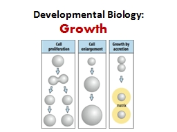
LEC2 Most benign tumors grow slowly amp most of cancers grow faster amp eventually spread locally amp to distant sites metastasis amp causing death In some exception some benign tumors grow more rapidly than some cancers eg leiomyoma uterine fibroid which is benign smooth mus ID: 911273
Download Presentation The PPT/PDF document "II. Rate of growth DR.AYSER HAMEED" is the property of its rightful owner. Permission is granted to download and print the materials on this web site for personal, non-commercial use only, and to display it on your personal computer provided you do not modify the materials and that you retain all copyright notices contained in the materials. By downloading content from our website, you accept the terms of this agreement.
Slide1
II.
Rate of growth
DR.AYSER HAMEED
LEC.2
Slide2Most benign tumors grow slowly
& most of cancers grow faster
& eventually spread locally & to distant sites (metastasis) & causing death.
In some exception, some benign tumors grow more rapidly than some cancers e.g. leiomyoma (uterine fibroid), which is benign smooth muscle tumor influence by estrogen & so increase in size during pregnancy.
Slide3In malignant tumors, rate of growth is inversely related to degree of differentiation
(well differentiated cancer is slowly growing while undifferentiated cancer are grow rapidly).
Some of malignant tumors undergo spontaneous regression, regress the whole primary cancers & leave only their metastasis e.g.
Choriocarcinoma
.
Rapidly growing malignant tumors usually have central area of necrosis (ischemic) because inadequate blood supply to provide the rapid growth of cancer.
Slide4III. Local invasion
:
Benign tumors are:
1. Localized at the site of origin.
2. Not invade the surrounding tissue.
3. Not metastasized.
4. Most of benign tumors have surrounding capsule (derived from the original normal tissue) as a result of atrophy of parenchymal cells & by the pressure of benign tumors.
e.g. leiomyoma of uterus
, fibro adenoma of the breast.
Important not:
Not all benign tumors are capsulated.
Slide5Slide6Slide7Malignant tumors are
:
1. Rapid progressive growth.
2. Infiltration, invasion & penetration of the surrounding.
3. Malignant tumors not develop well defined capsule.
4. Always metastasized.
Local invasion is the second most reliable feature (after metastasis) that distinguishes malignant from benign tumors.
Slide8Slide9Slide10Carcinoma lung invasion
Malignant neoplasms are characterized by the tendency to invade surrounding tissues. Here, a lung cancer is seen to be spreading along the bronchi into the surrounding lung tissues & pleura.
Slide11IV. Metastasis
:
It means development of secondary implants discontinuous with primary tumor, possibly in remote tissues.
Metastasis is the most important characteristic of malignancy.
Not all cancers have equal ability of development metastasis, e.g. Basal cell carcinoma of skin & most of CNS malignancies are highly locally invasive but rarely metastasis.
Slide12Basal cell carcinoma lower lip
These rarely metastasize, but are slow-growing and progressive over time. Leaving them to get larger just makes the plastic surgeon's job much harder, with more disability to the patient, so early detection and excision is a must. Most basal cell carcinomas occur in the head and neck area of adults with prolonged sun exposure. Biopsy & microscopic examination are necessary steps to establish the diagnosis.
Slide13While osteosarcoma is usually metastasize to lung at the time of initial diagnosis
.
In general, the more anaplastic & the larger the primary neoplasm, the more likely is metastatic spread, with some exception extremely small cancers have been known to metastasize.
Slide14Pathways of Metastasis
:
Seeding within body cavities
: These occur when cancers
invade the body cavities (pleura, peritoneum, pericardium & meninges in CNS tumors).
e.g
.
carcinoma of colon, carcinoma of stomach invade the peritoneal cavity, in female extend to both ovaries (
Krukenberg
tumors)
,
carcinoma of lung invade the pleura
&
malignancy of ovary invade the peritoneum.
Slide15Pseudomyxoma peritonei
The entire peritoneal cavity is occupied by a multinodular mucinous mass
Slide16Pseudomyxoma peritonei
Clusters of well-differentiated mucin-producing glandular cells are seen floating in a sea of mucin
.
Slide172. Lymphatic spread
:
This is characteristically seen in carcinomas, while sarcomas are more spread by vascular system.
There are numerous interconnections between lymphatic & vascular systems,
so all forms of cancer may spread through either or both systems
.
The pattern of involvement of lymph node depends principally
on the site of the primary neoplasm & lymphatic pathways of this site e.g. lung carcinoma metastasize first to the regional bronchial lymph nodes, then to the
hilar
lymph nodes.
Slide18Cancer cells arranged themselves inside the lymphatic's either as Emboli(commonest), or as continuous growth(permeation).
In some cases, the cancer cells seem to traverse the lymphatic channels within the immediately proximate nodes to be trapped in subsequent lymph nodes, this is called (
Skip lesions),e.g. cancer of stomach, cancer of prostate involve the cervical lymph nodes.
Slide19It should be remembered, that
lymph nodes enlargement with cancers, not always means cancerous nodal involvement,
it may be due to necrotic debris of the cancer & tumor antigens
, which may induce nodal enlargement in form of lymphadenitis & sinus
histiocytosis
.
Slide20Microscopically, metastatic adenocarcinoma is seen in a lymph node here. It is common for carcinomas to metastasize to lymph nodes. The first nodes involved are those draining the site of the primary.
Lymph node: metastatic adenocarcinoma
Slide213.
Hematogenous
spread
:
is the
most feared consequence of a cancer
; it is the favored pathway for sarcomas.
Arteries are penetrated less readily than veins
; because of the wall thickness is more in arteries.
Source of these malignant cells is from emboli within the lymphatic's
.
The liver & lungs are the most frequently involved secondary sites in such
hematogenous
spread (all portal area drainage flows to the liver & all
caval blood flows to the lungs).
Slide22Pulmonary metastases
Slide23Metastatic adenocarcinoma lung
Slide24Cancers arising near the vertebral column
often
embolize
through the paravertebral venous plexus to set into vertebrae
(e.g.
carcinoma of thyroid & prostate).
4. Intraepithelial cancer spread:
This form of spread may occur
where cancer develop in gland or its duct
e.g.
carcinoma of breast spread to the areolar skin (Paget’s disease of breast).