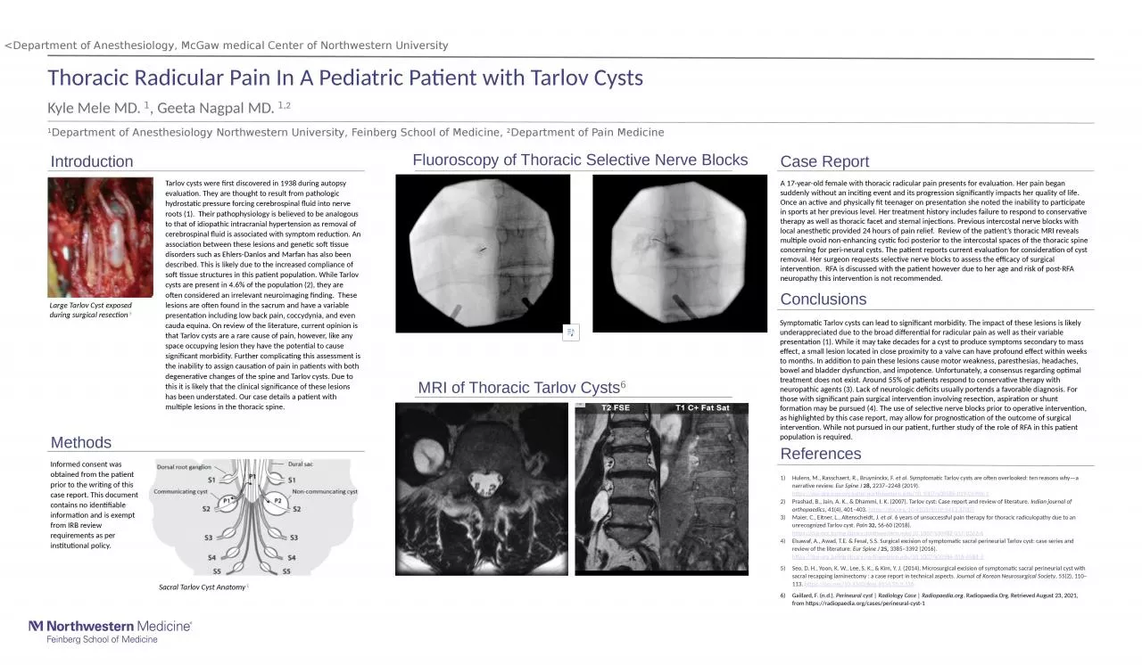
/
1 Department of Anesthesiology Northwestern University, Feinberg School of Medicine, 1 Department of Anesthesiology Northwestern University, Feinberg School of Medicine,

1 Department of Anesthesiology Northwestern University, Feinberg School of Medicine, - PowerPoint Presentation

rosemary
rosemary . @rosemary
Follow
27 views
Uploaded On 2024-02-02

1 Department of Anesthesiology Northwestern University, Feinberg School of Medicine, - PPT Presentation

2 Department of Pain Medicine Introduction A 17yearold female with thoracic radicular pain presents for evaluation Her pain began suddenly without an inciting event and its progression significantly impacts her quality of life ID: 1044091

tarlov pain org cyst pain tarlov cyst org cysts patient thoracic case review lesions https due doi intervention surgical

Share:

Link:

Embed:

Download Presentation from below link

Download Presentation The PPT/PDF document "1 Department of Anesthesiology Northwest..." is the property of its rightful owner. Permission is granted to download and print the materials on this web site for personal, non-commercial use only, and to display it on your personal computer provided you do not modify the materials and that you retain all copyright notices contained in the materials. By downloading content from our website, you accept the terms of this agreement.


Presentation Transcript

1. 1Department of Anesthesiology Northwestern University, Feinberg School of Medicine, 2Department of Pain MedicineIntroduction A 17-year-old female with thoracic radicular pain presents for evaluation. Her pain began suddenly without an inciting event and its progression significantly impacts her quality of life. Once an active and physically fit teenager on presentation she noted the inability to participate in sports at her previous level. Her treatment history includes failure to respond to conservative therapy as well as thoracic facet and sternal injections. Previous intercostal nerve blocks with local anesthetic provided 24 hours of pain relief. Review of the patient’s thoracic MRI reveals multiple ovoid non-enhancing cystic foci posterior to the intercostal spaces of the thoracic spine concerning for peri-neural cysts. The patient reports current evaluation for consideration of cyst removal. Her surgeon requests selective nerve blocks to assess the efficacy of surgical intervention. RFA is discussed with the patient however due to her age and risk of post-RFA neuropathy this intervention is not recommended. Symptomatic Tarlov cysts can lead to significant morbidity. The impact of these lesions is likely underappreciated due to the broad differential for radicular pain as well as their variable presentation (1). While it may take decades for a cyst to produce symptoms secondary to mass effect, a small lesion located in close proximity to a valve can have profound effect within weeks to months. In addition to pain these lesions cause motor weakness, paresthesias, headaches, bowel and bladder dysfunction, and impotence. Unfortunately, a consensus regarding optimal treatment does not exist. Around 55% of patients respond to conservative therapy with neuropathic agents (3). Lack of neurologic deficits usually portends a favorable diagnosis. For those with significant pain surgical intervention involving resection, aspiration or shunt formation may be pursued (4). The use of selective nerve blocks prior to operative intervention, as highlighted by this case report, may allow for prognostication of the outcome of surgical intervention. While not pursued in our patient, further study of the role of RFA in this patient population is required. MethodsFluoroscopy of Thoracic Selective Nerve Blocks MRI of Thoracic Tarlov Cysts6Case Report ConclusionsReferencesThoracic Radicular Pain In A Pediatric Patient with Tarlov Cysts Kyle Mele MD. 1, Geeta Nagpal MD. 1,2<Department of Anesthesiology, McGaw medical Center of Northwestern UniversityTarlov cysts were first discovered in 1938 during autopsy evaluation. They are thought to result from pathologic hydrostatic pressure forcing cerebrospinal fluid into nerve roots (1). Their pathophysiology is believed to be analogous to that of idiopathic intracranial hypertension as removal of cerebrospinal fluid is associated with symptom reduction. An association between these lesions and genetic soft tissue disorders such as Ehlers-Danlos and Marfan has also been described. This is likely due to the increased compliance of soft tissue structures in this patient population. While Tarlov cysts are present in 4.6% of the population (2), they are often considered an irrelevant neuroimaging finding. These lesions are often found in the sacrum and have a variable presentation including low back pain, coccydynia, and even cauda equina. On review of the literature, current opinion is that Tarlov cysts are a rare cause of pain, however, like any space occupying lesion they have the potential to cause significant morbidity. Further complicating this assessment is the inability to assign causation of pain in patients with both degenerative changes of the spine and Tarlov cysts. Due to this it is likely that the clinical significance of these lesions has been understated. Our case details a patient with multiple lesions in the thoracic spine. Chart description can go here. Informed consent was obtained from the patient prior to the writing of this case report. This document contains no identifiable information and is exempt from IRB review requirements as per institutional policy.Hulens, M., Rasschaert, R., Bruyninckx, F. et al. Symptomatic Tarlov cysts are often overlooked: ten reasons why—a narrative review. Eur Spine J 28, 2237–2248 (2019). https://doi-org.ezproxy.galter.northwestern.edu/10.1007/s00586-019-05996-1Prashad, B., Jain, A. K., & Dhammi, I. K. (2007). Tarlov cyst: Case report and review of literature. Indian journal of orthopaedics, 41(4), 401–403. https://doi.org/10.4103/0019-5413.37007Maier, C., Eitner, L., Altenscheidt, J. et al. 6 years of unsuccessful pain therapy for thoracic radiculopathy due to an unrecognized Tarlov cyst. Pain 32, 56-60 (2018). https://doi-org.turing.library.northwestern.edu/10.1007/s00482-017-0262-6Elsawaf, A., Awad, T.E. & Fesal, S.S. Surgical excision of symptomatic sacral perineurial Tarlov cyst: case series and review of the literature. Eur Spine J 25, 3385–3392 (2016). https://doi-org.turing.library.northwestern.edu/10.1007/s00586-016-4584-3Seo, D. H., Yoon, K. W., Lee, S. K., & Kim, Y. J. (2014). Microsurgical excision of symptomatic sacral perineurial cyst with sacral recapping laminectomy : a case report in technical aspects. Journal of Korean Neurosurgical Society, 55(2), 110–113. https://doi.org/10.3340/jkns.2014.55.2.110Gaillard, F. (n.d.). Perineural cyst | Radiology Case | Radiopaedia.org. Radiopaedia.Org. Retrieved August 23, 2021, from https://radiopaedia.org/cases/perineural-cyst-1Large Tarlov Cyst exposed during surgical resection 5Sacral Tarlov Cyst Anatomy 1