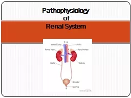
Dr Mohd Aslam Anatomy Basic Renal Physiology Nephron is the functional unit of the kidney Capable of forming urine has two major components Glomerulus Tubule proximal ID: 915087
Download Presentation The PPT/PDF document "Acute Renal Failure- Etiology and Pathop..." is the property of its rightful owner. Permission is granted to download and print the materials on this web site for personal, non-commercial use only, and to display it on your personal computer provided you do not modify the materials and that you retain all copyright notices contained in the materials. By downloading content from our website, you accept the terms of this agreement.
Slide1
Acute Renal Failure- Etiology and Pathophysiology
Dr. Mohd. Aslam
Slide2Anatomy
Slide3Basic Renal Physiology
Nephron
is the functional unit of the kidney
Capable of forming urine, has two major
components:– Glomerulus– Tubule:• proximal• loop of Henle• Distal• collecting
Slide4Outline
Definition of ARF
Epidemiology
Etiology of ARF
Pathophysiology
Management of ARF– Diagnosis of ARF– Treatment of ARF
Slide5DEFINITION:
AKI is a sudden and usually reversible decrease in the
glomerular
filtration rate (GFR) occurring over a period of hours to days
The term “Acute Kidney Injury” now replaces the term ARF; the term Acute Renal Failure should now be restricted to patients who have AKI and “need renal replacement therapy”The kidneys receives 20% of the cardiac output
Slide6‘ACUTE KIDNEY INJURY’
Abrupt reduction [<48 hrs] in kidney function, defined as an absolute increase in
S.creatinine
of ≥0.3 mg/
dL.A percentage increase in S.creatinine of ≥ 50% [1.5 fold from baseline] or a reduction in urine output– documented oliguria of < 0.5 ml/kg/hr, for more than six hours.
Slide7Epidemiology
Epidemiology:
It occurs in
5%of all hospitalized patients and
35% of those in intensive care units
Mortality is high:up to 75–90% in patients with sepsis35–45% in those without
Slide8AKI: Urine Volume
•
Anuria
(< 100 ml/24h)
– Acute bilateral arterial or venous occlusion
– Bilateral cortical necrosis– Acute necrotizing glomerulonephritis– Obstruction (complete)• Oliguria (100-500 ml/24h)
– Pre-renal
azotemia
– ATN
• Non-
Oliguria
(> 500 ml/24h)
– ATN
– Obstruction (partial)
Slide9Classification provides:
– Information on etiology of ARF
– Information on prognosis
– Directs appropriate therapy
Slide10Staging
Slide11Staging
Slide12RISK FACTORS
Advanced age, older than 75 years
Atherosclerosis
Diabetes
HTN
Heart failureLiver diseasePersons with Kidney diseases e.g. preexisting chronic kidney diseasesepsis.Exposed to contrast agents or undergoing cardiac surgery.
Slide13CAUSES OF ACUTE KIDNEY INJURY
Slide14Pre-renal causes of AKI
Volume depletion
• Renal losses (diuretics,
polyuria
)
• GI losses (vomiting, diarrhea)• Hemorrhage• PancreatitisDecreased cardiac output• Heart failure• Pulmonary embolus• Acute myocardial infarction• Severe valvular
heart disease
Slide15Post-renal causes of AKI
Ureteric
obstruction
• Stone disease
• Tumor
• Ligation during pelvic surgeryBladder neck obstruction• BPH• Cancer of the prostate• Drugs (Tricyclic antidepressants, ganglion blockers)• Bladder tumor
• Stone disease, hemorrhage/clot
Urethral obstruction (strictures, tumor)
Slide16Renal Causes of AKI
:
• Large Vascular
• Small vascular and
Glomerular• Interstitial nephritis• Acute tubular necrosis
Slide17Renal Causes of AKI
Large Vascular
:
•
Renovascular disease + ACEI• Renal artery thrombosis/dissection• Cholesterol emboli (recent Cardiac cath/aortic surgery)• Renal vein Thrombosis (hypercoagulable
,
Nephrotic
)
Slide18Acute Glomerulonephritis (GN) / Small
vascular
•
Anti–GBM disease (
Goodpasture
syndrome)• ANCA-associated GN (Wegener granulomatosis, microscopic polyangiitis)• Immune
complex
GN (lupus,
cryoglobulinemia
, primary MPGN)
•
IgA
nephropathy
• HUS/TTP
• Malignant hypertension
Slide19Tubular
• Ischemic
• Toxic
–
Heme
pigment (rhabdomyolysis, intravascular hemolysis)– Crystals (tumor lysis syndrome, ethylene glycol poisoning, methotrexate)
– Drugs (
aminoglycosides
, lithium,
amphotericin
B,
radiocontrast
agents)
Slide20Interstitial
•
Drugs (
penicillins
, NSAIDs,
allopurinol, rifampin, sulfonamides)• Infection (pyelonephritis, viral nephritis)• Systemic disease (Sjogren syndrome,
sarcoid
,
lupus, lymphoma )
Slide21Pathophysiology
•
Increase in
NaCl
delivered to macula
densa. Damage to proximal tubule cells increases NaCl delivery to distal nephron. This causes disruption of feedback mechanism.• Obstruction of tubular lumen.
Casts clog the lumen. This will increase the tubular pressure and then GFR will fall.
•
Backleak
of fluid through the tubular basement membrane.
Slide22Pathophysiology
It includes one of these 3 major patterns:
1)
Syndrome of acute nephritis
2)
Syndrome accompanying tubular pathologyOliguric phaseDiuretic phaseRecovery phase3) Pre – renal syndrome
Slide231)
Syndrome of acute nephritis
This is associated with
glomerulonephritis
Results in increase in
glomerular permeability and decrease in GFRCharacteristic features: mild proteinuria, haematuria,Oedema
Slide242)
Syndrome accompanying tubular pathology
ARF is caused by destruction of the tubular cells of the
nephron
Disease progresses in 3 stages:
a) Oliguria phaseLasts for 7-10 days with urine output less than 400 ml/dayLeads to azotaemia, metabolic acidosis, hyperkalaemia, hypernatraemia
and
hypervolaemia
Slide25b
)
Diuretic phase
Healing of tubules results in improving urinary output
Effects: Dehydration & electrolyte imbalance
c) Recovery phaseFull recovery of tubular cells occurs in half cases while others terminate in deathTime period for recovery – Variable
Slide263)
Pre – renal syndrome
It occurs because of secondary disorders like
ischaemia
& not due to
glomerular or tubular damage.Causes of ischaemia: renal arterial obstruction, hypovolaemia, hypotension or cardiac insufficiency.Due to depressed renal blood flow, there is decrease in GFR causing oliguria, azotemia and edema
Slide27Pre-renal AKI
• History
-- blood or volume loss
-- cardiac failure, arrhythmia,
(
cardiogenic shock)-- sepsis• Physical exam – focus on volume status– Vital signs – current and preceding the development of AKI– Neck veins, lungs, heart, mucous membranes and edema
Slide28Sustained
prerenal
azotemia
is the main factor that predisposes patients to ischemia- induced ATN
Prerenal azotemia and ischemic tubular necrosis represent a continuum. Azotemia progresses to necrosis when blood flow is sufficiently compromised Most cases of ischemic AKI are reversible if the underlying cause is corrected.
Slide29Renal or Intrinsic AKI
–
Glomerular
• History – systemic or primary kidney
• P/E – BP (usually hypertensive), edema
• BUN: creatinine ratio: preserved• Urine analysis : + protein, RBCs +• Often require kidney biopsy
Slide30Renal or Intrinsic AKI
– Interstitial
History – exposure to medications usually 7- 14 days earlier-
penicillins
,
cephalosporins, dilantinP/E – maculopapular erythematous skin rash, 1/3 have fever,
arthralgias
BUN:creatinine
ratio: 10-20:1
•Urine analysis: + protein
WBCs, WBC casts,
eosinophils
Slide31Renal or Intrinsic
ATN – Acute Tubular Necrosis
• The most common type of hospital-acquired ARF
• May be
1) ischemic or
2) nephrotoxic • Most common ATN is ischemic, most often due to a prolonged pre-renal state (prolonged reduced renal perfusion)
Slide32Acute Tubular Necrosis
History – prolonged pre-renal state
Exposure to
nephrotoxin
Aminoglycoside
antibioticsEthylene glycolPigments (myoglobin, hemoglobin)• P/E – assess volume status (to exclude prerenal AKI)
Slide33Contrast-Induced AKI
Prevalence:
• Less than 1% in patients with normal renal function
• Increases significantly with renal insufficiency
Risk factors:
• Renal insufficiency• Diabetes mellitus• Multiple myeloma• High osmolar (ionic) contrast media• Contrast medium volume
Slide34Contrast-induced AKI
Clinical Characteristics:
• Onset - 24 to 48 hrs after exposure
• Duration - 5 to 7 days
• Non-
oliguric (majority)• Dialysis - rarely needed• Urinary sediment - variable• FENa: Low
Slide35Post-Renal AKI
•
History – symptoms (frequency, hesitancy, etc)
- carcinoma, DM, stones, medications
• P/E – distended bladder, prostatic enlargement, pelvic masses , lymph nodes
• Laboratory studies-- elevated BUN:creat ratio -- unremarkable urine sediment -- variable urine chemistries• Renal ultrasound – hydronephrosis
Slide36Rhabdomyolytic
ARF
•
Diagnose with serum CPK (usu. > 10,000), urine dipstick (+) for blood, without RBCs on microscopy, pigmented granular casts
• Common after trauma (“crush injuries”), seizures, burns, limb ischemia occasionally after IABP or cardiopulmonary bypass
• Treatment is largely supportive care.• Alkalinization of urine
Slide37Atheroembolic
ARF
•
Associated with emboli of fragments of atherosclerotic plaque from aorta and other large arteries
• Diagnose by history, physical findings (evidence of other embolic phenomena- CVA, ischemic digits), low serum C3 and C4, peripheral
eosinophilia, eosinophiluria• Commonly occur after intravascular procedures or cannulation ( CABG, AAA repair, etc.)
Slide38Clinical features
•
Signs and symptoms resulting from loss of kidney function:
– decreased or no urine output, flank pain, edema, hypertension, or discolored urine
• Asymptomatic
– elevations in the plasma creatinine– abnormalities on urinalysis
Slide39Clinical features
•
Symptoms and/or signs of renal failure:
– weakness and easy
fatiguability
– Anorexia, vomiting– Seizures, mental status changes – Edema• Systemic symptoms and findings:– Fever, Shortness of breath –
Arthralgias
, Anorexia,
Pruritus
Slide40Complications
Hyperuricemia
,
Hyperkalemia
, Metabolic acidosis
Infection- pneumonia, sepsisGIT- nausea, vomiting, malnutrition, GIT bleedingCNS- asterixis, mental changes, seizuresAnaemia, bleedingHyperphosphatemia
,
hypocalcemia
Cardio- Pulmonary complications
Slide41Acute Renal Failure- Diagnosis and Management
Slide42Differential diagnosis of acute renal failure
Slide43Pre-renal AKI
•
Laboratory studies:
–
BUN:creatinine
ratio – elevated in pre-renal; >20:1– Urine sediment: hyaline and fine granular casts– Urine Specific gravity: high– Urine dipstick: negative (no blood or protein)– Urinary to plasma creatinine ratio: high– Urinary Na: low– FENa: low
Slide44Renal or Intrinsic
ATN – Acute Tubular Necrosis
•
BUN:creatinine
ratio: preserved (10-20:1)
• Urine analysis : negative protein, blood-- granular casts (dirty brown casts)-- renal tubular epithelial cells• Urine chemistries – Urinary Na: high (>40 meq/L)
FENa
: high (>2%)
Urinary to plasma creatinine ratio: low
Slide45Post-Renal AKI
• Laboratory studies--
elevated
BUN:creat
ratio
-- unremarkable urine sediment-- variable urine chemistries• Renal ultrasound – hydronephrosis• Treatment is to relieve the obstruction– Bladder catheterization– Nephrostomy tubes
Slide46INVESTIGATIONS
Biochemistry:
Blood urea,
creatinine,
electrolytes,
Blood gas analysis.Urine osmolality/sodium/creatinine
Slide47Serum Creatinine as a marker
for AKI and GFR
Normal
S.Creatinine
is 0.6-1.2mg/dl and is the most commonly used parameter to assess renal function.
Unfortunately the correlation between S.Creatinine concentration and GFR may be confounded by several factors.
Slide48Slide49Creatinine is not an ideal marker
1.Creatinine excretion is dependent on renal factors independent of function:
– Certain medications such as
cimetidine
and
trimethoprim interfere with proximal tubular creatinine secretion and may cause rise in S. creatinine without fall in GFR.
Slide502.S.Creatinine is dependent on
nonrenal
factors independent of renal function:
S.Creatinine
is dependent on muscle mass, infection, volume of distribution, age, gender, race, body
habitus, diet, presence of amputations.Eg. S. Creatinine of 1.2mg/dl in a 40kg elderly signifies severe reduction of GFR while the same value in a 100kg represents a normal renal function3.Creatinine production and excretion must be in a steady state before creatinine may be used in any formula for the estimation of GFR.
Slide51Fractional Excretion of Na
Since urinary indices depend on urine sodium concentration, they should be interpreted cautiously if the patient has received diuretic
Spot urine Na may be affected (raised) by diuretic use and baseline impaired kidney function (CKD where maximum urine Na
reabsorption
is impaired)
Fractional excretion of Na accounts for this by including creatinine: FeNa = (urine [Na] ÷ plasma [Na]) ÷ (urine creatinine ÷ plasma creatinine) X 100
RENAL INDICES
Renal Failure Index (RFI):
RFI = urine [Na] ÷ urine creatinine /serum creatinine
Slide53Slide54Slide55URINE ANALYSIS
•
Dipstick for blood, protein – Suggests a renal inflammatory process
• Microscopy may show cells, casts, crystals
Slide56RBCs in Urine
Present in
glomerulonephritis
,
vasculitis
,HUS TTPscleroderma crisis
Slide57URINARY CASTS
Hyaline –
prerenal
ARF
Granular –ATN (muddy brown)
Red blood cell casts –glomerulonephritis,vasculitis malignant hypertensionWBC casts- AIN, pyelonephritis ,leukemic or lymphomatous infiltrates
Slide58Crystals
•
Urate
crystals – acute
urate
nephropathy• Oxalate crystals –ethylene glycol ingestion /acyclovir/ indinavir• Eosinophiluria --> > 5 % of WBC s AIN ,atherothrombotic disease
Slide59Haematology
•
Full blood count, blood film:
–
Eosinophilia
may be present in acute interstitial nephritis, cholesterol embolization, or vasculitis (CSS)– Thrombocytopenia and red cell fragments suggest thrombotic microangiopathy
–TTP, HUS
•
Coagulation studies
– Disseminated intravascular coagulation associated with sepsis
Slide60Immunology
Antinuclear antibody (ANA) , Anti-double stranded (
ds
) antibody -
ANA positive in SLE and other autoimmune
disorders;DNA antibodies anti-ds DNA antibodies more specific for SLEC3 & C4 complement concentrations-Low in SLE, acute post infectious glomerulonephritis,
Cryoglobulinemia
ASO and anti-
DNAse
B
titres
High after streptococcal infection
Slide61Immunology
ANCA:
• p-ANCA - Anti PR3 antibodies
• c-ANCA - Anti MPO antibodies
– Associated with systemic
vasculitis - Wegener’s granulomatosis; Microscopic polyangiitis.AntiGBM antibodies:– Present in
Goodpasture’s
disease
Slide62Serology
•
Hepatitis B and C, HIV serology
Slide63Radiology
•
Renal
ultrasonography
: For renal size, symmetry, evidence of obstruction
• Pyelography : localization• MRA/ Doppler US : arterial /venous obstruction
Slide64NEW MARKER
Cystatin
C –protein
Produced by nucleated cells
Filtered and completely reabsorbed
Changes in serum levels occur sooner
Slide65NOVEL BIOMARKERS
1.
IL- 18
2. KIM-1
3.
Gro α/KC4. NGAL-neutrophil gelatinase associated lipocalin5. NHE-3 -Sodium–hydrogen
antiporter
3
Slide66AKI: PREVENTION
Recognize patients at risk (postoperative states, cardiac surgery, septic shock)
Prevent progression from
prerenal
to renal
Preserve renal perfusion(isovolemia, cardiac output, normal blood pressure)Avoid nephrotoxins (aminoglycosides, NSAIDS, amphotericin)
Slide67GENERAL PROTOCOL FOR
MANAGEMENT OF AKI
•
Treat the underlying disease
• Strictly monitor intake and output (weight, urine output, insensible losses, IVF)
• Monitor serum electrolytes• Adjust medication dosages according to GFR• Avoid highly nephrotoxic drugs• Attempt to convert
oliguric
to non-
oliguric
renal failure (
furosemide
)
Slide68FLUID THERAPY
If patient is fluid overloaded:
• Fluid restriction (insensible losses)
• Attempt
furosemide
1-2 mg/kg• Renal replacement therapyIf patient is dehydrated:• Restore intravascular volume first• Then treat as euvolemic
If patient is
euvolemic
:
• Restrict to insensible losses (30-35 ml/100kcal/24 hours) +
other
losses
(urine,
chest
tubes,
etc
)
Slide69SODIUM
•
Most patients have
dilutional
hyponatremia which should be treated with fluid restriction• Severe hyponatremia (Na< 125 mEq/L) : dialysis or hemofiltration
Slide70POTASSIUM
•
Oliguric
renal failure is often complicated by
hyperkalemia
, increasing the risk of cardiac arrhythmias• Treatment of hyperkalemia:– sodium bicarbonate (1-2 mEq/kg)– insulin + hypertonic dextrose– sodium polystyrene : 1 gm/kg (
Hypernatremia
and hypertension are potential complications)
– dialysis
Slide71Slide72MANAGEMENT OF AKI
Metabolic acidosis – soda
bicarb
., if < 15
meq
Hyperphosphatemia – PO4 binders –sevalamerHypocalcemia –calcium carbonateNutrition –restriction of dietary protein < 0.8 g/kg /d, calories – 25-30 kcal /kg/d, enteral nutrition preferred
Slide73Contrast-induced AKI
Prophylatic
Strategies:
• Use I.V. contrast only when necessary
• Hydration
• Minimize contrast volume• Low-osmolar (nonionic) contrast media
Slide74Acute Tubular Necrosis
•
Diagnose by history,
FENa
(>2%)
• sediment with coarse granular casts, RTE cells• Treatment is supportive care.– Maintenance of euvolemia (with judicious use of diuretics, IVF, as necessary)– Avoidance of hypotension– Avoidance of nephrotoxic
medications (including NSAIDs and ACE-I) when possible
– Dialysis, if necessary
• 80% will recover, if initial insult can be reversed
Slide75Hemoglobinuria
+
Myoglobinuria
:
Hemoglobinuria
: Transfusion reactions, HUS (hemolytic uremic syndrome), ECMO (extracorporeal membrane oxygenation)Myoglobinuria: Crush injuries, rhabdomyolysisurine (+) blood but (-) red blood cells
Increase CPK and K+
Treatment:
Aggressive hydration + urine
alkalinization
Mannitol
/
furosemide
Slide76NURSING MANAGEMENT
Excess fluid volume related to decreased
Glomerular
filtration rate and sodium retention
Risk for infection related to alterations in the immune system and host defenses
Imbalanced nutrition: less than body requirementsRisk for injury related to GI bleedingSleep pattern disturbances related to disease conditionProviding skin careProviding support
Slide77Criteria for Initiation of RRT
Anuria
Oliguria
Pulmonary edema
Hyperkalemia
>6.5mmol/LSevere acidemia <7.2Uremic encephalopathyUremic pericarditis
Slide78Therapy
Renal replacement therapy
Furosemide
Dopamine
Fenoldapam
hANP (Anaritide)
Slide79Diagnosis
History collection.
Physical examination.
1.
Asterixis
and myoclonus2. Peripheral edema (if volume overload is present)3. Pulmonary rales (if volume overload is present)4. Elevated right atrial pressure (if volume overload is present)
Slide80Identification of precipitating cause.
Serum creatinine and BUN level .(n 7-18mg/dl)
Serum electrolytes.
Urine analysis.
Renal bladder ultra sound.
Renal scan.CT scans and MRI scan (to identify lesion and masses)The urine will be examined under a microscope.Biopsy.
Slide81GENERAL PROTOCOL FOR
MANAGEMENT
Treat the underlying disease
strictly monitor intake and output (weight, urine
output, insensible losses, IVF)
Monitor serum electrolytes Adjust medication dosages according to GFR Avoid highly nephrotoxic drugs Attempt to convert oliguric to non-
oliguric
renal failure (
furosemide
x 3)
Slide82Acute Renal Failure:
Treatment
•
Water and sodium restriction
• Protein restriction
• Potassium and phosphate restriction• Adjust medication dosages• Avoidance of further insults– BP support–
Nephrotoxins
Slide83Treatment of ARF
Based on type/etiology of AKI (acute kidney injury) i.e., pre-renal, post-renal, or intrinsic renal initially
– Pre-renal – volume, improve renal perfusion
– Post-renal – relieve obstruction
– Intrinsic –
glomerular, tubular, interstitial, vascular (depends on type)• Follow clinically; attention to volume status, avoid additional insults; treat complications of ARF
Slide84Acute Renal Failure
Diagnostic Tools
History and Physical examination
• Blood tests : CBC, BUN/creatinine, electrolytes, uric acid, PT/PTT, CK
• Urinary sediment
• Urinary indices– Urine volume– Urine electrolytes• Radiologic studies– Renal ultrasound (useful for obstructive forms)
– Doppler (to assess renal blood flow)
• Nuclear Medicine Scans
– DMSA: anatomy
– DTPA and MAG3: renal function, urinary
– Excretion and upper tract outflow
Slide85Prevention
A careful history(
nephrotoxic
antibiotic agent
aminoglycosides
, gentamycin, tobramycin, etc.)blood tests and urinalysisDrink enough fluidsDifficulties urinating or blood in the urine should prompt a visitTreat hypotension promptly.
Prevent and treat infections promptly.
Pay special attention to wound, burns and other precursors of sepsis.
Slide86ARF -- Summary
Dx
AKI by falling GFR [rising BUN and creatinine]
Classify into pre-renal, renal, or
postrenal
by history, PE, BUN:creat ratio, urine analysis
Slide87ARF – Summary (cont…)
•
Sometimes also need urine chemistries [urine Na,
FxExNa
and/or
FxExurea] to distinguish pre-renal from ATN• Sometimes need renal ultrasound to verify obstruction [post-renal ARF]• Rarely need other studies, esp to dx type of intrinsic ARF [e.g., kidney biopsy: GN
vs
interstitial nephritis
vs
ATN]
Slide88ARF – Summary (cont…)
•
Distinguishing the types of intrinsic ARF usually depends on history, PE,
urine analysis
– Vascular – large or small vessels
– Tubular = ATN– Interstitial = AIN– Glomerular = acute GN
Slide89THANK YOU