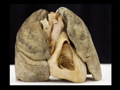
/
Emphysema:  It is characterized by loss of lung elasticity and abnormal enlargement of Emphysema:  It is characterized by loss of lung elasticity and abnormal enlargement of

Emphysema: It is characterized by loss of lung elasticity and abnormal enlargement of - PowerPoint Presentation

scarlett
scarlett . @scarlett
Follow
27 views
Uploaded On 2024-02-03

Emphysema: It is characterized by loss of lung elasticity and abnormal enlargement of - PPT Presentation

Two of the recognized causes of emphysema are smoking which incites lung injury and inherited deficiency of α1 antitrypsin an antiprotease enzyme that protects the lung from injury Emphysema is thought to result from the breakdown of ID: 1044719

asthma cells respiratory bronchial cells asthma bronchial respiratory airway pulmonary response infection chronic persons inflammatory occurs blood caused epithelial

Share:

Link:

Embed:

Download Presentation from below link

Download Presentation The PPT/PDF document "Emphysema: It is characterized by loss ..." is the property of its rightful owner. Permission is granted to download and print the materials on this web site for personal, non-commercial use only, and to display it on your personal computer provided you do not modify the materials and that you retain all copyright notices contained in the materials. By downloading content from our website, you accept the terms of this agreement.


Presentation Transcript

1. Emphysema:

2. It is characterized by loss of lung elasticity and abnormal enlargement of the air spaces distal to the terminal bronchioles with destruction of the alveolar walls and capillary beds . Enlargement of the air spaces leads to hyperinflation of the lungs . Two of the recognized causes of emphysema are smoking which incites lung injury and inherited deficiency of α1- antitrypsin; an antiprotease enzyme that protects the lung from injury.Emphysema is thought to result from the breakdown of elastin and other alveolar wall components by enzyme called proteases which digest proteins.The proteases particularly elastase; which is an enzyme that digest elastin are released from polymorphnuclear leukocytes (e.g neutrophils), macrophages and other inflammatory cells.

3. Cigarette smoking and other irritant stimulate the movement of inflammatory cells into the lungs resulting in an increase release of elastase.In smokers; COPD develops and antiproteases production decrease. Alpha 1 antitrypsin deficiency is a hereditary disease determined by a pair of dominant genes referred to as protein inhibitor (p1 )gene.Laboratory methods are available for measuring α1 – antitrypsin level. Human α1-antitrypsin is available for replacement therapy.

4. There are two commonly recognized types of emphysema; centriacinar or centrilobular and panacinar.The centracinar type affects the bronchioles in the central part of the respiratory lobule, with initial preservation of the alveolar ducts and sacs.It is the most common type and seen predominantly in male smokers.The panacinar type produces initial involvement of the peripheral alveoli and later extends to involve the more central bronchioles.This type is more common in persons with α1- antitrypsin deficiency and in smokers with centriacinar emphysema.

5. Bronchiectasis:It is characterized by permanent dilatation of bronchi caused by destruction of the bronchial muscle wall and elastic supporting tissue. It is not a primary disease but secondary occurs to a number of abnormalities including diffuse bronchitis , cystic fibrosis, pneumonia and T.B.

6. Etiology and pathogenesis:Two processes are critical; obstruction and chronic persistent infection both cause damage to the bronchial walls, leading to weakening and dilatation.Bronchial dilatation is classified as; saccular, cylinderical or varicose.Saccular bronchiectasis involves the proximal third to fourth generation of bronchi → severe dilatation of bronchi with collapse and fibrosis of the more distal lung tissues.

7. Cylinderical → involves 6-8 generations, milder and leads to fewer symptoms.Varicose → involves the 2nd through 8 branching.Localized bronchiectasis: caused by tumor, foreign bodies, mucous plug due to obstructive drainage of mucous secretions.Generalized bronchiectasis: bilateral and affects the lower lobes due to inherited impairment of host mechanism or acquired disorders that permits introduction of infectious organism.

8. Manifestations:Manifested by a chronic productive cough, with several hundred milliliters of foul smelling, purulent sputum, hemoptysis, dyspnea and wheezing. Obstructive pulmonary dysfunction with hypoxemia is seen in moderate to severe cases.

9. Cystic fibrosis:Is a major cause of severe chronic respiratory disease in children and young adults, is an inherited disorder involving fluid secretion by the exocrine glands in the epithelial lining of the respiratory, gastrointestinal and reproductive tracts.Cystic fibrosis is manifested by pancreatic exocrine deficiency and elevation of sodium chloride in sweat. Excessive loss of sodium in the sweat, predisposes young children to salt depletion episodes.Most males with CF had congenital bilateral absence of vas deferens with azospermia. Cystic fibrosis is inherited as autosomal recessive trait.

10. Etiology and pathogenesis:It is caused by mutations in a single gene on the large arm of chromosome 7 that encodes for the cystic fibrosis transmembrane regulator (CFTR), which functions as a Cl – channel in epithelial cell membrane.Mutations in CFTR gene renders the epithelial membrane relatively impermeable to the Cl ions.In sweat glands; concentration of sodium and chloride secreted in the lumen remain unaffected.In persons with CF, the transport of chloride into the airway lumen is impaired → ↑ absorption of sodium and water from the airway into the blood → ↓ water content of the mucocilliary coating of the respiratory tract → dehydration of the mucous layer →viscid secretions → recurrent pulmonary infection.

11.

12. Manifestations:Accumulation of viscid mucous in the bronchi, impaired mucocilliary clearance and lung infection.Chronic bronchiolitis and bronchitis → structural changes in bronchial wall →bronchiectasis → airway obstruction. CF predisposes to chronic infection by pseudomonas aeruginosa , staphylocococcus aureus, haemophilius influenzae.The airway secretions in persons with CF provide a favorable environment for harboring these organisms.Exocrine pancreatic function is abnormal in more than 85% of affected children. Steatorrhea, diarrhea and abdominal pain are common

13. Pulmonary embolism:Develops when a blood-borne substance lodges in a branch of the pulmonary artery and obstructs blood flow.The embolism may consist of a thrombus, air that has been injected, drug intravenous infusion, fat that has been mobilized from the bone marrow after a fracture or amniotic fluid that has entered the maternal circulation during childbirth.

14. Pathogenesis:Most of pulmonary emboli arise from deep vein thrombosis (DVT) in the large veins of the lower legs.Among the physiological factors that contribute to venous thrombosis are venous stasis and venous endothelial injury resulted from prolonged bed rest, severe trauma, surgery, myocardial infarction (MI) and chronic heart failure. Also, from hypercoagubility states.Cancer cells produce thrombin and synthesize procoagulation factors that cause increase thromboembolism.

15. Pregnancy and hormone replacement therapy (oral contraceptive) →↑ resistance to endogenous anticoagulants.The thrombophilias e.g antithrombin III, protein S and C and factor V are groups of inherited disorders affecting coagulation.Obstruction of pulmonary blood flow causes reflex bronchoconstriction in the affected area of the lung, impaired gas exchange and loss of alveolar surfactant, pulmonary hypertension may develop.

16. Manifestations:The clinical manifestations depends on the size and location of obstruction.Small emboli lodged in peripheral branches of pulmonary artery are silent and unrecognized. Moderate size emboli present with breathlessness, pleuritic pain, slight fever and cough of blood sputum. Tachycardia occurs to compensate decreased oxygenation. Massive emboli causes sudden collapse, chest pain, shock, loss of consciousness, rapid pulse, decreased blood pressure , cyanosis and death.

17. Bronchial asthma: It is a chronic inflammatory disease of the airways involving recurring symptoms of air flow obstruction and bronchial hyper responsiveness.Airway obstruction is characterized by episodic wheezing , difficult breathing, feeling of chest tightness and a cough that often is worse at night and in the early morning.

18. Etiology and pathogenesis:Asthma is commonly categorized into 2 types: extrinsic or allergic (atopic) ; due to type I hypersensitivity reaction.And intrinsic or non-atopic that occurs without an allergic component.In either types; episodes of bronchospasm can be triggered by respiratory tract infection, exercise, aspirin ingestion, emotional upset and exposure to bronchial irritant such as cigarette smoking.Asthma may also be classified according to the agents that trigger an attack, these include seasonal, exercise- induced, drug –induced (e.g aspirin).

19. The common denominator underlying all forms of asthma is an exaggerated hypersensitivity response.After exposure to an inciting factor; inflammatory mediators released by activated macrophages, eosinophils, mast cells and basophils induce bronchoconstriction, increased vascular permeability and mucous production. In some persons; persistent changes in airway structure occurs including injury to epithelial cells, smooth muscle hypertrophy and blood- vessel proliferation.

20. T- lymphocytes are involved in the pathogenesis of asthma. It is known that there are 2 subsets of T- helper cells (TH1 and TH2) that develop from the same precursor CD4+ T- lymphocytes.TH1 cells differentiate in response to microbes and stimulate the differentiation of B- cells into immunoglobulins IgM and IgG producing plasma cells.

21. TH2 cells respond to allergens by stimulating B- cells to differentiate into IgE producing plasma cells that bind to mucosal mast cells.Subsequent IgE- mediated reaction to inhaled allergens elicits an asthmatic allergic attack.Atopic asthma : it is typically initiated by a type I hypersensitivity reaction induced by exposure to an extrinsic antigen or allergen.Its onset in childhood or adolescence and seen in persons with a family history of atopic allergy. Persons with atopic asthma often have other disorders such as hay- fever, urticaria and eczema. The mechanism of response to allergens in atopic asthma can be described in terms of the early –phase and late- phase response.

22. The symptoms of the early- phase response ( also called the acute phase response). Which usually develops within (10-20) minutes of exposure to allergens, are caused by the release of chemical mediators from IgE- coated mast cells.The reaction occurs when an antigen binds to a previously sensitized mast cells on the mucosal surface. Mediators release result in the infilteration of inflammatory cells, opening of mucosal intercellular junction and increase access of antigen to the submucosal mast cells. In addition; there is a bronchospasm caused by stimulation of parasympathetic receptor mucosal edema and increased mucous secretions.

23. The late-phase response; develops 4- 8 hrs. after exposure to an asthmatic trigger involves inflammation and increased airway responsiveness. Typically; the response reaches maximum within a few hours and may persist 12-24 hrs.An initial trigger causes release of inflammatory mediators from mast cells, macrophages and epithelial cells. These substances induce the migration and activation of other inflammatory cells ( e.g basophils, eosinophils, neutrophils) which produce epithelial injury and edema, changes in mucocilliary function, reduce clearance of respiratory tract secretion and increased airway responsiveness.

24.

25. Nonatopic asthma (intrinsic): its triggers include respiratory tract infection, exercise, hyperventillation, cold air, drugs and chemicals, hormonal changes and emotional upsets, airborn pollutants and gastroesohageal reflux.Respiratory tract infection caused by viruses, may produce their effects by causing epithelial damage and stimulating the production of IgE antibodies directed toward the viral antigens.

26. Exercise- induced asthma occurs in a number of persons with bronchial asthma. Although the cause is unclear, hyperventilation and corresponding changes in airway physiology may play a role.Increased ventilation rate required to meet higher O2 demands during exercise challenges the ability of airways to condition the inhaled air to correct moist and heat levels.

27. Tobacco smoke and strong odors, induce bronchospasm by irritation of receptors and vagal reflux.Occupational asthma caused by irritants in the workplace such as irritant gases such as SO2, NO2 and ozone.Aspirin and NSAIDs causes bronchial asthma; the mechanism of hypersensitivity pointed to an abnormality in arachidonic acid AA metabolism in which aspirin inhibit the bronchodiltators cyclooxygenase pathway without affecting the lipooxygenase pathway thereby shifting balance toward the bronchoconstrictors leukotriens.Emotional factors trigger asthma by vagal reflux.

28. Manifestations:Wheezing, chest tightness to acute immobilizing attacks. Asthma is often worse at night, studies suggest that there is a circadian and sleep related variation in hormones and respiratory functions.The greater decrease occurs at about 4:00 Am at which time cortisol levels are low, melatonin levels are high and eosinophils activity increased.