Uploads
Contact
/
Login
Upload
Search Results for 'Figure 2 Figure 2 Transmission Electron Microscopy Of Microsporidia Identified In Allograft Sample'
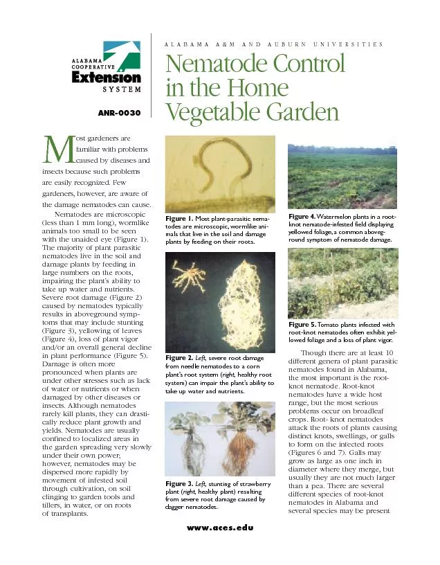
Figure 6. Distinct knots, swellings, or galls visible on the root syst
olivia-moreira
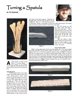
Turning a Spatulaby Tim Kennedyuseful project to make on thelathe is a
phoebe-click
Photosynthesis & Respiration
myesha-ticknor
Figure a
debby-jeon
Modern Truck Electrical Systems
mitsue-stanley
Preliminary electron microscopy studies of bacterial
natalia-silvester
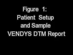
Figure 1: Patient Setup and Sample VENDYS DTM Report
briana-ranney
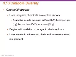
3.13 Catabolic Diversity
alida-meadow
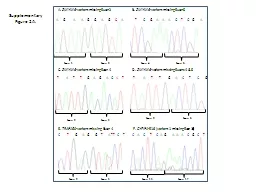
Supplementary Figure 2A.
cheryl-pisano
College Physics Chapter 24 ELECTROMAGNETIC WAVES
marina-yarberry
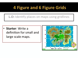
4 Figure and 6 Figure Grids
karlyn-bohler
Figure 7
alida-meadow
Figure 9-1
giovanna-bartolotta
1 Intro
briana-ranney
1 Intro
marina-yarberry
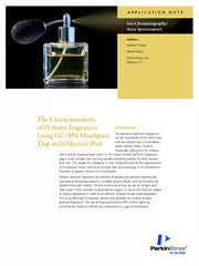
APPLICATION NOTE
myesha-ticknor
Electron Microscopy Lab
mitsue-stanley
Electron Microscopy Lab
trish-goza
ATOMIC FORCE MICROSCOPY Introduction and theoretical background
mitsue-stanley
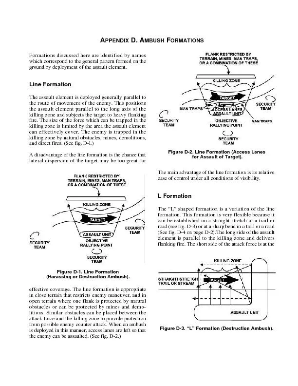
APED D. AMU FOFormations discussed here are identified by nameswhich c
cheryl-pisano
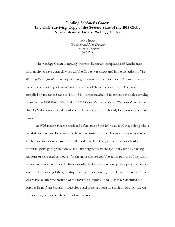
Second State of the 1515 Globe Newly Identified in the Wolfegg Codex
celsa-spraggs
UNIT
danika-pritchard
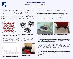
Preparation of a Ferrofluid
natalia-silvester
Sample layout for 80x120cm poster:
tatiana-dople
1
2
3
4
5
6
7