Uploads
Contact
/
Login
Upload
Search Results for 'Figure 4 Figure 4 Brain Images Showing Contrast Enhanced Lesions In The Right Occipital And Left P'
HSC BELONGING, 2013 PAPER1
pasty-toler
Whole Brain/Left Brain/Right Brain
phoebe-click
Figure 2. Soil Removal vs. Stain Type (35-lb. washer/extractor)Figure
sherrill-nordquist
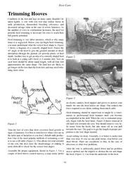
Trimming HoovesCondition of the feet and legs on dairy cattle shouldnt
ellena-manuel
Trimming HoovesCondition of the feet and legs on dairy cattle shouldnt
alida-meadow
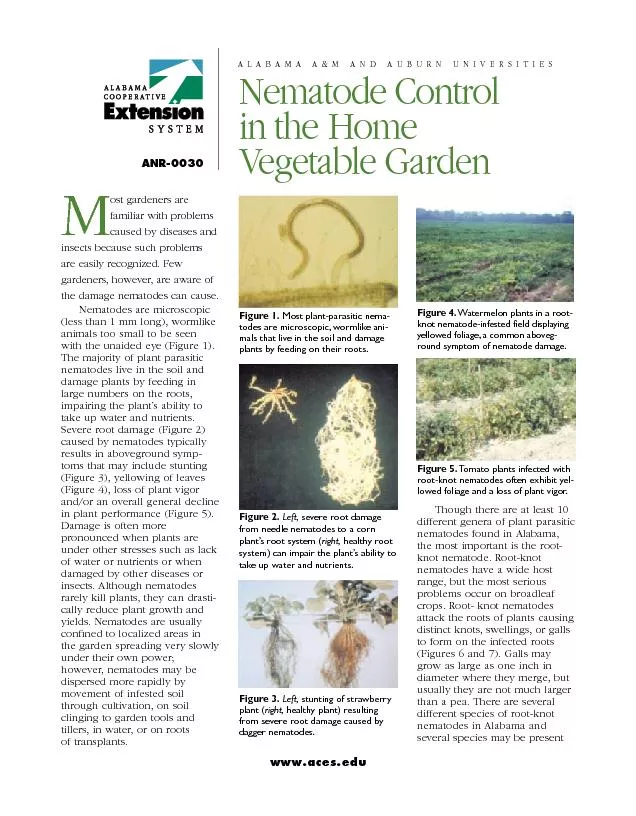
Figure 6. Distinct knots, swellings, or galls visible on the root syst
olivia-moreira
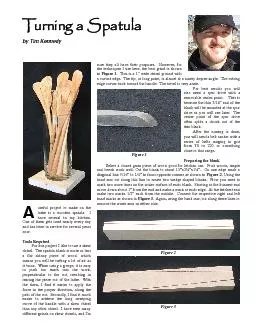
Turning a Spatulaby Tim Kennedyuseful project to make on thelathe is a
phoebe-click
Biofeedback System for Improved Athletic Training
conchita-marotz
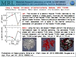
Publications on these systems:
alida-meadow
Radiological approach to
stefany-barnette
1 Introduction: Traumatic Brain Injury
min-jolicoeur
First… Introduction…
tatiana-dople
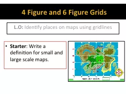
4 Figure and 6 Figure Grids
karlyn-bohler
DO NOW WORK
pamella-moone
Figure 7
alida-meadow
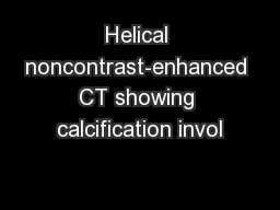
Helical noncontrast-enhanced CT showing calcification invol
test
In this class we are going to learn read papers critically.
lindy-dunigan
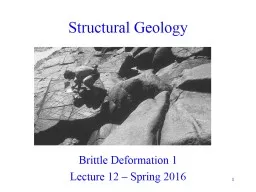
1 Structural Geology
lindy-dunigan
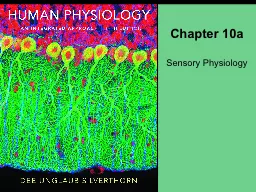
Chapter 10a
tatyana-admore
Figure 9-1
giovanna-bartolotta
Horse. Opening of the Left
pamella-moone
The Language of Space & Time
myesha-ticknor
12 The Central Nervous System
celsa-spraggs
The previous picture “moves” because of tiny muscular movements of your eyes.
calandra-battersby
1
2
3
4
5
6
7