Uploads
Contact
/
Login
Upload
Search Results for 'Figure 4 Figure 4 Brain Images Showing Contrast Enhanced Lesions In The Right Occipital And Left P'
Object Recognition and Feature Detection Using MATLAB
luanne-stotts
1 Topic 2
giovanna-bartolotta
Radiology Case Presentation
tawny-fly
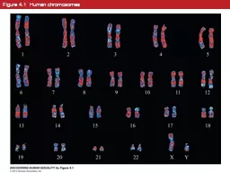
© 2014 Pearson Education, Inc.
pasty-toler
A cerebral aneurysm is a weak area in a blood vessel, w
danika-pritchard
Six-figure grid references
aaron
Enhancing Lesions in the CNS
kittie-lecroy
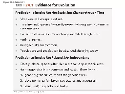
Figure 26.0-1 Why Reproduction Matters
phoebe-click
Muscle Diagrams Figure 10.10a
giovanna-bartolotta
Review Unit 3 and 4 Figure 6.8a
olivia-moreira
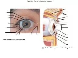
Figure 17-5b The Sectional Anatomy of the Eye.
olivia-moreira
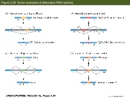
Figure 2.26 Some examples of alternative RNA splicing
test
Figure
calandra-battersby
Figure 15.1 The eye and accessory structures.
luanne-stotts
Figure 23.1
danika-pritchard
Figure 24-9-Table 24-1
danika-pritchard
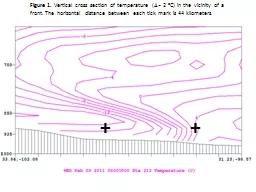
Figure 1.
trish-goza
Figure 4.1 Human chromosomes
debby-jeon
Figure 7.16 Acetals and ketals can be formed from hemiacetals and hemiketals, respectively.
pasty-toler
CEREBRAL LATERALITY: RIGHT BRAIN/LEFT BRAIN
briana-ranney
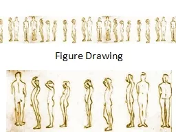
Figure Drawing
celsa-spraggs
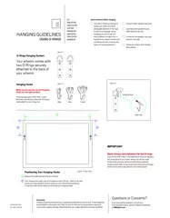
Measure the width between the two D-Rings. Then measure the heig
kittie-lecroy
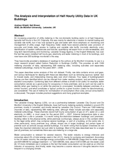
The Analysis and Interpretation of Half Hourly Utility Data in UK Andr
test
Left Brain / Right Brain:
kittie-lecroy
1
2
3
4
5
6
7
8
9