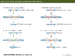
/
Figure 2.26  Some examples of alternative RNA splicing Figure 2.26  Some examples of alternative RNA splicing

Figure 2.26 Some examples of alternative RNA splicing - PowerPoint Presentation

test
test . @test
Follow
404 views
Uploaded On 2017-05-20

Figure 2.26 Some examples of alternative RNA splicing - PPT Presentation

Figure 231 Degradation of casein mRNA in the presence and absence of prolactin Figure 234 Hypothetical model of the regulation of lin14 mRNA translation by lin4 RNAs Figure 235 Current model for the formation and use of microRNAs Part 1 ID: 550301

induction figure part cadherin figure induction cadherin part cell importance model correct morphogenesis sorting amphibian cells micrornas formation current

Share:

Link:

Embed:

Download Presentation from below link

Download Presentation The PPT/PDF document "Figure 2.26 Some examples of alternativ..." is the property of its rightful owner. Permission is granted to download and print the materials on this web site for personal, non-commercial use only, and to display it on your personal computer provided you do not modify the materials and that you retain all copyright notices contained in the materials. By downloading content from our website, you accept the terms of this agreement.


Presentation Transcript

Slide1

Figure 2.26 Some examples of alternative RNA splicing Slide2

Figure 2.31 Degradation of casein mRNA in the presence and absence of prolactinSlide3

Figure 2.34 Hypothetical model of the regulation of lin-14

mRNA translation by lin-4 RNAsSlide4

Figure 2.35 Current model for the formation and use of microRNAs (Part 1)Slide5

Figure 2.35 Current model for the formation and use of microRNAs (Part 2)Slide6

Figure 3.1 Reaggregation of cells from amphibian neurulaeSlide7

Figure 3.2 Sorting out and reconstruction of spatial relationships in aggregates of embryonic amphibian cellsSlide8

Figure 3.4 Hierarchy of cell sorting in order of decreasing surface tensionsSlide9

Figure 3.5 Cadherin-mediated cell adhesion (Part 1)Slide10

Figure 3.6 Importance of the amount of cadherin for correct morphogenesisSlide11

Figure 3.7 Importance of the types of cadherin for correct morphogenesisSlide12

Figure 3.8 Importance of cadherin in cell adhesion and morphogenetic movementsSlide13

Figure 3.13 Ectodermal competence and the ability to respond to the optic vesicle inducer in

XenopusSlide14

Figure 3.14 Lens induction in amphibiansSlide15

Figure 3.15 Schematic diagram of induction of the mouse lensSlide16

Figure 3.16 Feather induction in the chickSlide17

Figure 3.17 Regional specificity of induction in the chickSlide18
Slide19

Figure 3.18 Genetic specificity of induction in amphibians