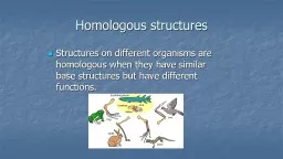
/
Figure 15.1  The eye and accessory structures. Figure 15.1  The eye and accessory structures.

Figure 15.1 The eye and accessory structures. - PowerPoint Presentation

luanne-stotts
luanne-stotts . @luanne-stotts
Follow
385 views
Uploaded On 2019-11-07

Figure 15.1 The eye and accessory structures. - PPT Presentation

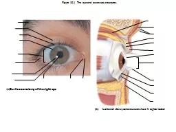
Figure 151 The eye and accessory structures Surface anatomy of the right eye Lateral view some structures shown in sagittal section Figure 152 The lacrimal apparatus Figure 153 Extrinsic eye muscles ID: 764367

eye figure retina view figure eye view retina structures action visual posterior pathway layer neural eyeball humor nerve cranial

Share:

Link:

Embed:

Download Presentation from below link

Download Presentation The PPT/PDF document "Figure 15.1 The eye and accessory struc..." is the property of its rightful owner. Permission is granted to download and print the materials on this web site for personal, non-commercial use only, and to display it on your personal computer provided you do not modify the materials and that you retain all copyright notices contained in the materials. By downloading content from our website, you accept the terms of this agreement.


Presentation Transcript

Figure 15.1 The eye and accessory structures. Surface anatomy of the right eye Lateral view; some structures shown in sagittal section

Figure 15.2 The lacrimal apparatus.

Figure 15.3 Extrinsic eye muscles. Lateral view of the right eye Superior view of the right eye Muscle Action Controlling cranial nerve Summary of muscle actions and innervating cranial nerves

Figure 15.4a Internal structure of the eye (sagittal section). Diagrammatic view. The vitreous humor is illustrated only in the bottom part of the eyeball.

Figure 15.5 Pupil constriction and dilation, anterior view. Label the muscles. Which nerve is responsible for each action and describe the situation for each.

Figure 15.6 Microscopic anatomy of the retina. Neural layer of retina Pathway of light Posterior aspect of the eyeball Cells of the neural layer of the retina Photomicrograph of retina

Figure 15.7 Part of the posterior wall (fundus) of the right eye as seen with an ophthalmoscope . ID structures & functions of each.

Figure 15.8 Circulation of aqueous humor. Lens Cornea 1 2 3 1 2 3

Figure 15.19 Visual pathway to the brain and visual fields, inferior view.