Explore
Featured
Recent
Articles
Topics
Login
Upload
Featured
Recent
Articles
Topics
Login
Upload
Search Results for 'micrograph'
micrograph published presentations and documents on DocSlides.
MICROGRAPH ANALYSIS LAB
by min-jolicoeur
In lab book. Create 5 tables based on the followi...
Figure C53. SEM micrograph of compressed mature yellow-poplar (4mm), T
by alida-meadow
Figure C54. SEM micrograph of compressed mature ye...
Figure C27. SEM micrograph of uncompressed mature yellow-poplar (4mm),
by alida-meadow
Figure C28. SEM micrograph of uncompressed mature ...
2015A Wet Mount Challenge
by celsa-spraggs
Instructions. Wet Mount PT: 2015A. Micrographs 1...
Figure C79. SEM micrograph of compressed mature southern pine, TRT1. .
by calandra-battersby
Figure C80. SEM micrograph of compressed mature so...
Phase transitions andexsolution phenomena in pyroxenes
by min-jolicoeur
001 110 110 Cleavage in the pyroxenes Optical micr...
Electron Micrograph Images and Review
by alida-meadow
Prokaryotic. Eukaryotic Animal Cell. Mitochondria...
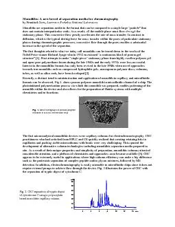
Monoliths: A new breed of separation media for chromatography by Frant
by tatiana-dople
SEM micrograph of porous polymer monolith in a COC...
Measuring from SEM
by myesha-ticknor
Micrographs By Tom Fink • A Micrograph is ...
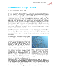
Science Highlight
by myesha-ticknor
Figure 1: Optical micrograph of the giant bacteriu...
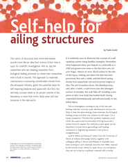
by Paula Gould Image above is an optical micrograph of
by olivia-moreira
Microcapsules containing a red healing agent are ...
Biology 1110
by celsa-spraggs
Principles of Biology. Biology 1110 Laboratory. L...
1. Explain how this animal’s cells will transform starch
by tatyana-admore
organelles. that are involved and the . processe...
Electron Micrograph Images and Review
by sherrill-nordquist
Prokaryotic. Pili. Eukaryotic Animal Cell. Mitoch...
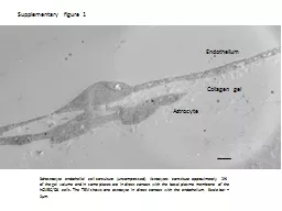
Supplementary figure 1 3d-astrocyte endothelial cell co-culture (uncompressed). Astrocytes constitu
by lucy
hCMEC. /D3 cells. The TEM shows one astrocyte in d...
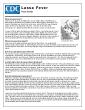
Lassa virus electron micrograph
by dorothy
Image courtesy, C.S. Goldsmith and M. Bowen (CDC)....
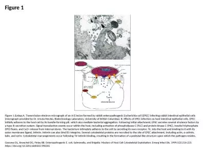
Figure 1 Figure 1. A. Transmission electron micrograph of an A/E lesion formed by rabbit e
by paige
Goosney DL, Knoechel DG, Finlay BB. Enteropathogen...
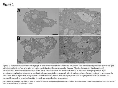
Figure 1 Figure 1. Transmission electron micrograph of amebae isolated from the home hot tub of a a
by joanne
Dey R, Mount H, Ensminger AW, Tyrrell GJ, Ward LP,...
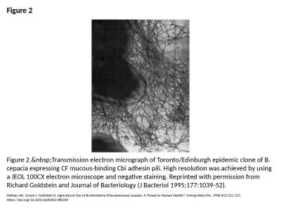
Figure 2 Figure 2. Transmission electron micrograph of Toronto/Edinburgh epidemic clone of
by danya
Holmes AH, Govan J, Goldstein R. Agricultural Use ...
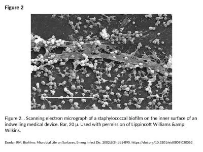
Figure 2 Figure 2. . Scanning electron micrograph of a staphylococcal biofilm on the inner surface
by patricia
Donlan RM. Biofilms: Microbial Life on Surfaces. E...
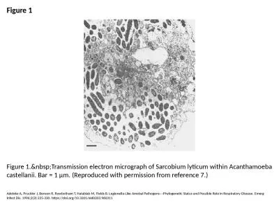
Figure 1 Figure 1. Transmission electron micrograph of Sarcobium lyticum within Acanthamoe
by elise
Adeleke A, Pruckler J, Benson R, Rowbotham T, Hala...
Students Please spend a little time becoming familiar with PowerPoint operations. Running the Po
by oneill
1. To begin left click the slide show screen icon...
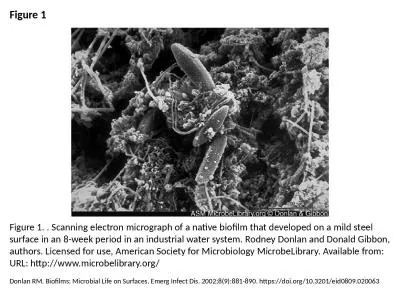
Figure 1 Figure 1. . Scanning electron micrograph of a native biofilm that developed on a mild stee
by victoria
Donlan RM. Biofilms: Microbial Life on Surfaces. E...
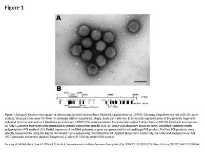
Figure 1 Figure 1. A) Electron micrograph of adenovirus particles isolated from Pipistrell
by lucy
Sonntag M, Mühldorfer K, Speck S, Wibbelt G, Kurt...
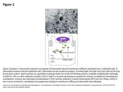
Figure 2 Figure 2. A. Transmission electron micrograph of Salmonella-induced membrane ruff
by victoria
Goosney DL, Knoechel DG, Finlay BB. Enteropathogen...
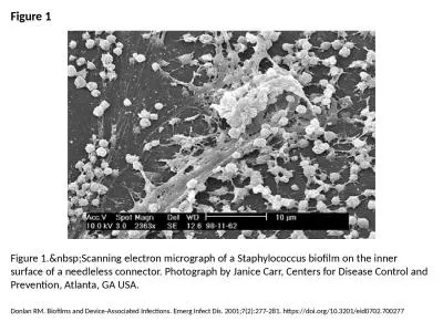
Figure 1 Figure 1. Scanning electron micrograph of a Staphylococcus biofilm on the inner s
by freya
Donlan RM. Biofilms and Device-Associated Infectio...
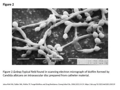
Figure 2 Figure 2. Typical field found in scanning electron micrograph of biofilm formed b
by melody
Jabra-Rizk MA, Falkler WA, Meiller TF. Fungal Biof...
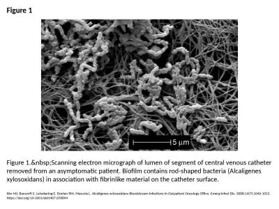
Figure 1 Figure 1. Scanning electron micrograph of lumen of segment of central venous cath
by lydia
Kim MJ, Bancroft E, Lehnkering E, Donlan RM, Masco...
Load More...