Explore
Featured
Recent
Articles
Topics
Login
Upload
Featured
Recent
Articles
Topics
Login
Upload
Search Results for 'Microscope-Cells'
Microscope-Cells published presentations and documents on DocSlides.
Microscope Review The diagram represents a cell in the field of view of a compound light microscope. In which direction should the slide be moved on the microscope stage to center the cell in the field of view
by phoebe-click
Microscope Review The diagram represents a cell i...
Microscope Basics The Compound Microscope
by aaron
Compound Light Microscope. The compound light mic...
Thomas Jefferson National Accelerator Facility Office of Science Education httpeducation
by calandra-battersby
jlaborg Microscopes Microscopes Microscopes Micros...
Microscope Mechanical Stage
by amscope
This model comes with color corrected infinity op...
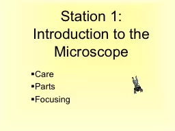
Station 1: Introduction to the Microscope
by sadie
Care. Parts. Focusing. Always carry with 2 hands. ...
The Microscope and Cell Theory
by min-jolicoeur
The Microscope. An understanding of cells and the...
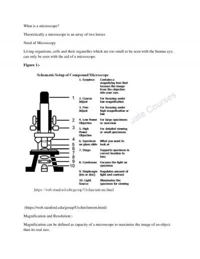
What is a microscope
by elizabeth
Theoretically a microscope is an array of two lens...
Microscopy Microscope is an instrument used to see objects that are too small for the naked eye.
by paisley
. Function of microscope. :. Magnification: . to ....
Parts of the microscope
by pamella-moone
. 2. Compound Light Microscope. Used to observe ...
Microscope
by giovanna-bartolotta
Instrument used to make very small objects appear...
Topic 1: Cells The Cell 1.1 Cell Theory
by dandy
•All living things are made of cells.. •Cells ...
Unit 3: cells Theory, types, structures, movement
by ellena-manuel
Unit 3 Topic 1: The cell theory . By the end of t...
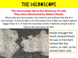
The microscope The microscope led to the discovery of cells.
by tawny-fly
They were discovered by Robert Hooke.. Most . cel...
Discovering Cells
by giovanna-bartolotta
Introduction to cells. Cells form the parts of an...
Cells and Microscopy
by debby-jeon
The Big Questions. How do we even know cells are ...
THE MICROSCOPE A Practical Guide
by madison
Microscopes used in clinical practice are light mi...
Using the fluorescence microscope
by ani
The inverted scope. Objectives are beneath the spe...
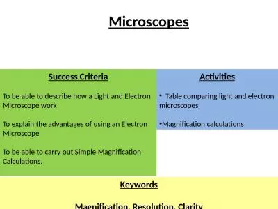
Success Criteria To be able to describe how a Light and Electron Microscope work
by heavin
To explain the advantages of using an Electron Mic...
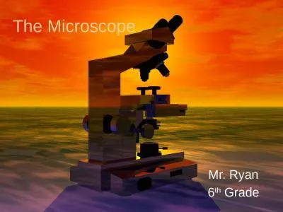
The Microscope Mr. Ryan 6
by blanko
th. Grade. The History. Many people experimented ...
Microscope quiz Review Mrs. Barnes
by mary
Biology I Honors. Which statement BEST describes a...
HW # 39 - Watch “Microscope”
by roxanne
video (link on the website). http://www.youtube.co...
the Microscope Types of Microscopes
by iris
- Compound microscope. - Dissecting microscope. - ...
Seiler ENT Microscopes
by isabella2
(Ear, Nose, and Throat). 2023. Business Overview ....
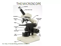
Microscope Basics Parts of the Microscope
by isabella
1 . Eyepiece : the part th. a. t you look through ...
MICROSCOPE
by williams
DEPARTMENT OF MICROBIOLOGY MTN COLLEGE MADURAI. ...
IS A MICROSCOPE
by erica
M I C R O S C O P E CONTENTS: • WHAT ? • HIST...
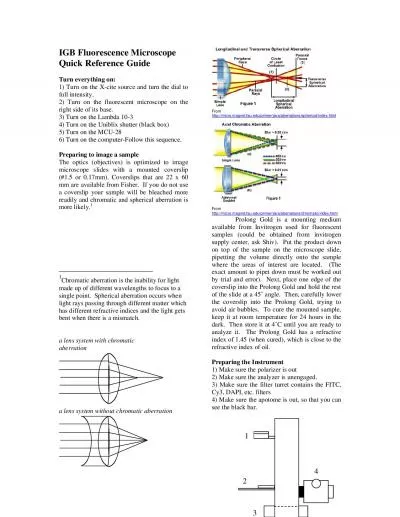
IGB Fluorescence Microscope Quick Reference Guide Turn everything on
by genevieve
Chromatic aberration is the inability for light ma...
The Microscope Learning Objectives
by SmilingSunshine
By the end of this topic, you will be able to:. Na...
Electron Microscope (EM)
by melody
. Electron microscopy (EM) . is an electron beam w...
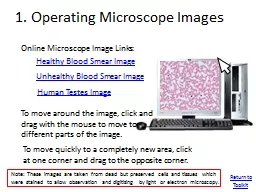
1. Operating Microscope Images
by adah
Online Microscope Image Links:. Healthy Blood Smea...
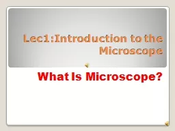
Lec1: Introduction to the Microscope
by eliza
What Is Microscope?. It is an instrument which dea...
Use and Care of a Compound Light Microscope
by daniella
Animal and Food Science Department. Brigham Young ...
THE MICROSCOPE http://biology.unm.edu/ccouncil/Biology_203/Summaries/Microscopes.htm
by dora
1. When moving your microscope, always carry it wi...
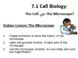
Todays Lesson: The Microscope
by anderson
Learning Objectives…... Create a timeline to sho...
The Compound Microscope 2
by lucinda
nd. stage /Physiology Lab-1. Pharmacology and tox...
Microscopes History of the Microscope
by mary
1590. –first compound microscope. History of th...
Microscopy Microscope DEFINE:
by sophie
instrument that produces . enlarged . image of . o...
See Us at M&M 2020 in Milwaukee
by Semtechsolutions
See Us at M&M 2020 in Milwaukee
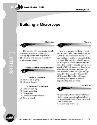
Building a Microscope
by cora
57Level Grades 912Activity 16ABObjectiveScience an...
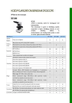
KOZO POLARIZATION SERIES MICROSCOPE XP Series microscope
by ani
XP-100 XP100 is extremely useful for Iatrologyan...
Load More...