Explore
Featured
Recent
Articles
Topics
Login
Upload
Featured
Recent
Articles
Topics
Login
Upload
Search Results for 'Microscope-Stage'
Microscope-Stage published presentations and documents on DocSlides.
Microscope Review The diagram represents a cell in the field of view of a compound light microscope. In which direction should the slide be moved on the microscope stage to center the cell in the field of view
by phoebe-click
Microscope Review The diagram represents a cell i...
Microscope Basics The Compound Microscope
by aaron
Compound Light Microscope. The compound light mic...
Microscope Mechanical Stage
by amscope
This model comes with color corrected infinity op...
Thomas Jefferson National Accelerator Facility Office of Science Education httpeducation
by calandra-battersby
jlaborg Microscopes Microscopes Microscopes Micros...
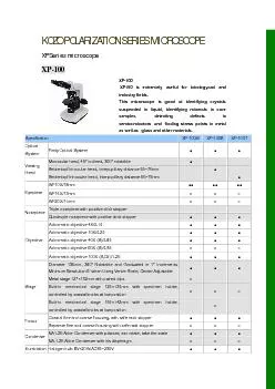
KOZO POLARIZATION SERIES MICROSCOPE XP Series microscope
by ani
XP-100 XP100 is extremely useful for Iatrologyan...
What are the parts of a microscope?
by patchick
Parts of a microscope. Can you guess the parts of ...
Microscope
by stefany-barnette
Basics. By: Kaitlyn Schroeder; ETEAMS 2014. Modif...
DWP SUB LITTLE THEATER ADRFOLEY STAGES STAGE SCORING STAGE STAGE STAGE STAGE AVENUE OF THE STARS GALAXY WAY Pump House Avenue A STAGE STAGE STAGE FOX NETWORK CENTER WM
by myesha-ticknor
FOX BUILDING 89 New York Street NEW EXECUTIVE BUI...
The Microscope Mr. Ryan 6
by blanko
th. Grade. The History. Many people experimented ...
the Microscope Types of Microscopes
by iris
- Compound microscope. - Dissecting microscope. - ...
MICROSCOPE
by williams
DEPARTMENT OF MICROBIOLOGY MTN COLLEGE MADURAI. ...
IS A MICROSCOPE
by erica
M I C R O S C O P E CONTENTS: • WHAT ? • HIST...
The Microscope Learning Objectives
by SmilingSunshine
By the end of this topic, you will be able to:. Na...
Microscopy Microscope is an instrument used to see objects that are too small for the naked eye.
by paisley
. Function of microscope. :. Magnification: . to ....
Use and Care of a Compound Light Microscope
by daniella
Animal and Food Science Department. Brigham Young ...
THE MICROSCOPE http://biology.unm.edu/ccouncil/Biology_203/Summaries/Microscopes.htm
by dora
1. When moving your microscope, always carry it wi...
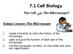
Todays Lesson: The Microscope
by anderson
Learning Objectives…... Create a timeline to sho...
The Compound Microscope 2
by lucinda
nd. stage /Physiology Lab-1. Pharmacology and tox...
Microscopes Care for your microscope
by karlyn-bohler
Always carry your scope with 2 hands (One around ...
The Compound Microscope Physiology Lab-1
by jane-oiler
October , 2017. Background Information. The micro...
Parts and Functions of a Compound Microscope
by lindy-dunigan
Light Microscope. Simple – uses a single lens. ...
Parts and Functions of a Compound Microscope
by luanne-stotts
Light Microscope. Simple – uses a single lens. ...
THE MICROSCOPE
by pasty-toler
http://biology.unm.edu/ccouncil/Biology_203/Summa...
Introduction to the Microscope
by alida-meadow
History. Types. Care. Parts. Focusing. Diggs?. Ha...
POWERPOINT PRESENTATION ON POLARISED MICROSCOPE
by briana-ranney
POLARISED LIGHT MICROSCOPE . Designed to observe ...
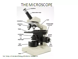
MICROSCOPE PARTS
by pasty-toler
CONCEPTS EXPLORED IN THIS LESSON. ocular lens / e...
Microscope
by tatiana-dople
Basics. Nosepiece. Objectives. Stage Clips. Light...
Parts of the microscope
by pamella-moone
. 2. Compound Light Microscope. Used to observe ...
Microscope Structure
by lindy-dunigan
From its humble beginnings the microscope has und...
THE MICROSCOPE A Practical Guide
by madison
Microscopes used in clinical practice are light mi...
Using the fluorescence microscope
by ani
The inverted scope. Objectives are beneath the spe...
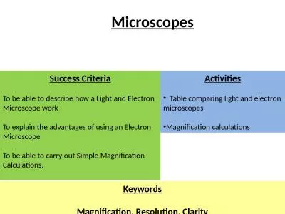
Success Criteria To be able to describe how a Light and Electron Microscope work
by heavin
To explain the advantages of using an Electron Mic...
Microscope quiz Review Mrs. Barnes
by mary
Biology I Honors. Which statement BEST describes a...
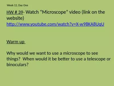
HW # 39 - Watch “Microscope”
by roxanne
video (link on the website). http://www.youtube.co...
Seiler ENT Microscopes
by isabella2
(Ear, Nose, and Throat). 2023. Business Overview ....
Microscope Basics Parts of the Microscope
by isabella
1 . Eyepiece : the part th. a. t you look through ...
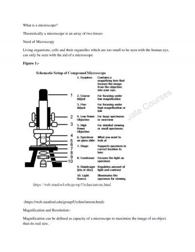
What is a microscope
by elizabeth
Theoretically a microscope is an array of two lens...
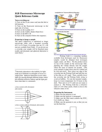
IGB Fluorescence Microscope Quick Reference Guide Turn everything on
by genevieve
Chromatic aberration is the inability for light ma...
Electron Microscope (EM)
by melody
. Electron microscopy (EM) . is an electron beam w...
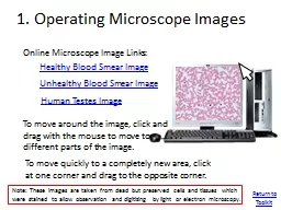
1. Operating Microscope Images
by adah
Online Microscope Image Links:. Healthy Blood Smea...
Load More...