/
Blood Components, Physical Characteristics, and Volume Blood Components, Physical Characteristics, and Volume

Blood Components, Physical Characteristics, and Volume - PowerPoint Presentation

sherrill-nordquist
sherrill-nordquist . @sherrill-nordquist
Follow
435 views
Uploaded On 2018-03-16

Blood Components, Physical Characteristics, and Volume - PPT Presentation

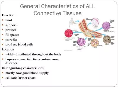
Blood transports everything nutrients wastes and body heat Blood is a complex fluid connective tissue with both solid and liquid components Solid living blood cells formed elements Erythrocytes red blood cells RBCS ID: 653433

rbcs blood rbc cells blood rbcs cells rbc oxygen body platelets clotting

Share:

Link:

Embed:

Download Presentation from below link

Download Presentation The PPT/PDF document "Blood Components, Physical Characteristi..." is the property of its rightful owner. Permission is granted to download and print the materials on this web site for personal, non-commercial use only, and to display it on your personal computer provided you do not modify the materials and that you retain all copyright notices contained in the materials. By downloading content from our website, you accept the terms of this agreement.


Presentation Transcript

Slide1

BloodSlide2

Components, Physical Characteristics, and Volume

Blood transports everything (nutrients, wastes, and body heat)

Blood is a complex fluid connective tissue with both solid and liquid components.

Solid – living blood cells (formed elements)Erythrocytes (red blood cells –RBCS)Leukocytes (white blood cells –WBCS)Platelets (function in the blood-clotting process)Liquid – nonliving fluid matrix (plasma)Slide3

An average blood sample (

hematocrit

) contains (by volume):

55% plasma45% RBCsLess than 1% WBCsLess than 1% plateletsSlide4
Slide5

Blood color varies from scarlet (oxygen-rich) to a dull red (oxygen-poor)

Whole blood is slightly alkaline (pH 7.35 – 7.45)

8% of body weight

Total adult blood volume is about 6 quartsSlide6
Slide7

Plasma

Straw-colored fluid

90% water

Helps to distribute body heat evenly throughout the bodyOver 100 different solutes(plasma proteins, nutrients, respiratory gases, hormones, wastes, and products of cell metabolism)Slide8

Plasma proteins are most abundant solutes

Most are made by the liver

Not used for cellular metabolism

Composition varies continuously as cells remove or add substances to the bloodBody organs make dozens of adjustments daily to maintain solutes at life-sustaining levels.Slide9

Formed Elements

Erythrocytes

Ferries oxygen in blood to all cells

AnucleateContain very few organellesContains iron-containing hemoglobin (Hb) protein to transport oxygenLack mitochondria and make ATP by anaerobic mechanismsSmall biconcave cells that provide a large surface area relative to volume

Outnumber WBCs by about 1000 to 1Slide10

About 5 million RBC/mm³ (as the RBC volume increases, blood viscosity increases)

The more

Hb

a RBC contains, the more oxygen it will carryAverage of 12-18 hemoglobin per 100 ml bloodA single RBC contains about 250 million Hb moleculesEach Hb can carry 4 molecules of oxygen

So each RBC carry about 1 billion molecules of oxygenSlide11
Slide12

Leukocytes

Far less numerous than RBCs

4,000 – 11,000 WBC/mm³

The only complete cells in blood (contain nuclei and usual organelles)Helps defend the body against damage by bacteria, viruses, parasites, and tumor cellsAble to slip into and out of the blood vessels (diapedesis) to cause inflammatory or immune responsesLeukopenia is an abnormally low WBC countSlide13

Can locate areas of tissue damage and infection in the body

Respond to certain chemicals that diffuse from the damaged cells (positive

chemotaxis

)Following the diffusion gradient to pinpoint areas of tissue damageGather in large numbers to destroy foreign substances or dead cellsThe body speeds up their productionTotal WBC count above 11,000 cells/mm³is referred to as leukocytosis

Indicates the presence of a bacterial or viral infection in the bodySlide14
Slide15

Platelets

Not cells in the strict sense

Fragments of very large multinucleate cells called

megakaryocytes that ruptureNormal platelet count in blood is about 300,000/mm³Needed for the clotting processSlide16
Slide17

Write a paragraph/overview of what we learned about blood.

Draw a picture and thoroughly describe each of the solid/cellular components of blood (RBC, WBC and platelets)

THIS IS DUE AT THE END OF THE PERIOD!!!Slide18

Blood Cell Formation

Occurs is red bone marrow

Flat bones of the skull and pelvis, ribs, sternum, and proximal epiphysis of the

humerus and femur.Each type of blood cell is produced in different numbers in response to body needs and stimuliAll formed elements arise from a common type of stem cell ( hemocytoblast) in the red bone marrowTwo types of descendents

Lymphoid stem cell – produces lymphocytesMyeloid stem cell – produces all other classes of formed elementsSlide19

RBCs are unable to divide (

anucleate

) and have a limited life span of 100 to 120 days

They begin to fragment and their remains are eliminated by the phagocytes in the spleen and liverLost cells are replaced more or less continuously Developing RBCs divide many times and then begin synthesizing huge amounts of hemoglobinWhen hemoglobin has been accumulated, nucleus and most organelles are ejected and the cell collapsesSlide20

The young RBC (

reticulocyte

) still contains some rough ER but begins transporting oxygen

Within two days they have ejected the remaining ER and have become fully functional erythrocytesThe process from hemocytoblast to mature RBC takes 3 to 5 daysPlatelet production is accelerated by thrombopoietinSlide21

Rate of erythrocyte production is controlled by a hormone called erythropoietin from liver and kidneys

When blood levels of oxygen begin to decline, the kidneys increase release of erythropoietin

Bone marrow is targeted and is stimulated to make more RBCs

Excessive amount of oxygen in the bloodstream depresses erythropoietin release and RBC productionColony stimulating factors (a hormone) trigger formation of leukocytes and platelets and also enhance the ability of mature leukocytes to protect the bodySlide22
Slide23

Hemostasis

Stoppage of blood flow is fast and localized

Blood loss at the site is permanently prevented when fibrous tissue grows into the clot and seals the hole in the blood vessel.

Three phases occur in rapid sequenceSlide24

Platelet plug formation

Membrane is broken so endothelium and collagen fibers are exposed to oxygen

Platelets become “sticky” and cling to the damaged site

Platelets release chemicals that attract more plateletsPlatelet plug is formedSlide25

Vascular spasms

Platelets release serotonin that causes the blood vessel to go into spasms

Blood loss decreases until clotting can occurSlide26

Coagulation

Injured tissues release

thromboplastin

(helps the clotting process)A platelet phospholipid interacts with thromboplastin and Ca⁺ to form a

prothrombin activatorThe activator converts prothrombin in the plasma into the enzyme thrombinThrombin joins soluble fibrinogen proteins into long hair-like molecules of insoluble webbingSlide27

Coagulation continued

Forms a meshwork that traps the RBCs and forms the basis of the clot

Within the hour, the clot begins to retract, squeezing serum (plasma minus the clotting proteins) from the mass and pulling the ruptured edges of the blood vessel closer togetherSlide28

Normally, blood clots within 3 to 6 minutes

Once the clotting cascade has started, triggering factors are rapidly inactivated to prevent widespread clotting

Eventually the endothelium regenerates and the clot is broken downSlide29
Slide30

Create a comic strip of

hemostasis

including pictures and thoroughly descriptive captions.Slide31

Disorders of Hemostasis

Undesirable Clotting

Usually occurs in the legs

Clot that develops and persists in an unbroken blood vessel is a thrombosisMay prevent blood flow if large enoughA thrombosis that breaks away from the vessel wall and floats freely in the bloodstream is an embolusUsually no problem unless/until it lodges in a blood vesselCaused by anything that roughens the blood vessel endothelium and encourages clinging of platelets

Slowly flowing blood or blood pooling is another risk factor (especially in immobilized patients)Slide32

Bleeding disorders

Common causes are platelet deficiency, deficits of clotting factors and genetic conditions

Thrombocytopenia results from an insufficient number of circulating platelets

Normal movement causes spontaneous bleeding from small blood vessels (petechia)Hemophilia is a hereditary bleeding disorder Lack of the factors needed for clottingMinor tissue trauma results in prolonged bleeding and can be life-threateningSlide33
Slide34

Human Blood Groups

Plasma membranes of RBCs have genetically determined proteins (antigens)

Each of us tolerates our own cellular (self) antigens

RBC proteins will be recognized as foreign if transfused into another person with different RBC antigens Antibodies present in the plasma attach to “foreign” RBCs causing RBCs to clump (agglutination)Slide35

This leads to the clogging of small blood vessels throughout the body

Foreign RBCs are

lysed

and Hb is unable to deliver oxygenClogged vessels affect kidney tubules to cause kidney failure, fever, chills, nausea, and vomiting may occur.Slide36

Over 30 common RBC antigens

ABO blood groups are based on which of the two antigens (type A or B) a person inherits

Absence of both antigens results in blood type O

Presence of both antigens results in blood type ABPossession of either A or B antigen yields type A or B blood, respectivelySlide37

Rh

blood groups are based on

agglutinogen

D (originally identified in Rhesus monkeys)Most Americans are Rh⁺ (RBCs carry the Rh antigen)If an Rh⁺ person receives Rh⁺ blood their immune system begins producing anti-

Rh⁺ antibodies Hemolysis does not occur with the first transfusionIt takes time for the body to react and start making antibodies Slide38

In subsequent transfusions, a typical reaction occurs (patient’s antibodies attack/rupture donor RBCs)

First time pregnant

Rh

⁺ women who carry Rh⁺ babies usually results in the delivery of a healthy babyMother is sensitized by Rh⁺ antigens and forms anti- Rh⁺ antibodiesIf she becomes pregnant again with an

Rh⁺ baby, her antibodies will cross the placenta and destroy the baby’s RBCsSlide39
Slide40

Developmental Aspects of Blood

Embryonic development of the entire circulatory system occurs within 28 days

Embryonic blood cells are circulating in the newly formed blood vessels around day 28

Fetal Hb has a greater ability to pick up oxygen (fetal blood is less oxygen-rich than that of the mother)Fetal blood cells are gradually replaced by RBCs that contain the more typical HbFetal RBCs are destroyed and the products are released in the bile

If this happens at a rate faster that the immature liver can handle, the infant becomes jaundicedSlide41

Write a journal entry as if you are a person suffering from a

hemostatic

disorder. You must explain your disorder and how you feel about having that disorder. You may also portray a doctor explain what to expect while telling a patient that they have that disorder, are pregnant with a child who has the disorder. This is due at the end of the period!