/
A 40 YEAR OLD COMATOSE MALE WITH PALATAL MYOCLONUS A 40 YEAR OLD COMATOSE MALE WITH PALATAL MYOCLONUS

A 40 YEAR OLD COMATOSE MALE WITH PALATAL MYOCLONUS - PowerPoint Presentation

shoesxbox
shoesxbox . @shoesxbox
Follow
342 views
Uploaded On 2020-06-15

A 40 YEAR OLD COMATOSE MALE WITH PALATAL MYOCLONUS - PPT Presentation

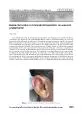
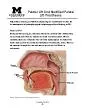
Teaching Neuro Images Neurology Resident and Fellow Section 2017 American Academy of Neurology History of posterior circulation stroke 4 months earlier due to hypertension Spastic quadriplegia ID: 777402

academy neurology american 2017 neurology academy 2017 american venkatesh degeneration olivary nucleus hypertrophic imaging bilateral circulation posterior stroke palatal

Share:

Link:

Embed:

Download Presentation from below link

Download The PPT/PDF document "A 40 YEAR OLD COMATOSE MALE WITH PALATAL..." is the property of its rightful owner. Permission is granted to download and print the materials on this web site for personal, non-commercial use only, and to display it on your personal computer provided you do not modify the materials and that you retain all copyright notices contained in the materials. By downloading content from our website, you accept the terms of this agreement.


Presentation Transcript

Slide1

A 40 YEAR OLD COMATOSE MALE WITH PALATAL MYOCLONUS

Teaching NeuroImages

NeurologyResident and Fellow Section

© 2017 American Academy of Neurology

Slide2

History of posterior circulation stroke 4 months earlier due to hypertensionSpastic quadriplegia

Bilateral extensor plantar reflex and Palatal myoclonus Vignette

Venkatesh et al.

© 2017 American Academy of Neurology

Slide3

Venkatesh et al.

Imaging

© 2017 American Academy of Neurology

Slide4

Venkatesh et al.

Imaging

© 2017 American Academy of Neurology

Slide5

Bilateral hypertrophic olivary degeneration following posterior circulation stroke

Hypertrophic olivary degeneration is a transsynaptic degeneration involving interconnecting fibers of inferior olivary nucleus, red nucleus and contralateral dentate nucleus forming three corners of Guillian-Mollaret triangle.1,2 Symmetrical enlargement and T2 hyperintense olives helps in differentiating this entity from demyelination, tumors and inflammatory processes.

© 2017 American Academy of Neurology Venkatesh et al.