/
 Functions of Major Brain Regions  Functions of Major Brain Regions

Functions of Major Brain Regions - PowerPoint Presentation

stefany-barnette
stefany-barnette . @stefany-barnette
Follow
342 views
Uploaded On 2020-04-03

Functions of Major Brain Regions - PPT Presentation

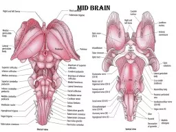
Pages 239252 Regions of the Brain Cerebral hemispheres cerebrum Diencephalon Brain stem Cerebellum 2015 Pearson Education Inc CEREBRUM Functions by Lobes Frontal Primarily motor function ID: 775210

ventricle cerebral brain education ventricle cerebral brain education pearson 2015 area stem choroid plexus diencephalon gland body cerebellum hemisphere

Share:

Link:

Embed:

Download Presentation from below link

Download Presentation The PPT/PDF document " Functions of Major Brain Regions" is the property of its rightful owner. Permission is granted to download and print the materials on this web site for personal, non-commercial use only, and to display it on your personal computer provided you do not modify the materials and that you retain all copyright notices contained in the materials. By downloading content from our website, you accept the terms of this agreement.


Presentation Transcript

Slide1

Functions of Major Brain Regions

Pages 239-252

Slide2

Regions of the Brain

Cerebral hemispheres (cerebrum)DiencephalonBrain stemCerebellum

© 2015 Pearson Education, Inc.

Slide3

CEREBRUM: Functions by Lobes

Frontal

:

Primarily motor function

Prefrontal Cortex

(anterior association)

attention, memory, decisions

Parietal

: primarily sensory stimuli integration

Touch/tactile, spatial information, body position

Temporal

: Auditory and Olfactory

stimuli integration

Broca’s

area

: motor component of speech- saying words

Occipital

: Visual

stimuli integration

Slide4

Figure 7.13c Left lateral view of the brain.

Primary motor area

Premotor area

Anterior

association area

Working

memory andjudgment

Broca’s area(motor speech)

Olfactoryarea

Central sulcus

Primary somaticsensory area

Gustatory area(taste)

Speech/language(outlined by dashes)

Posteriorassociationarea

Visual area

Auditory area

Problemsolving

Languagecomprehension

(c)

Slide5

Diencephalon

An anatomical location made up of the:thalamushypothalamusepithalamus: roof of third ventricle and pineal gland

© 2015 Pearson Education, Inc.

Slide6

Figure 7.16a Diencephalon and brain stem structures.

Third ventricle

Anterior

commissure

Hypothalamus

Optic chiasma

Pituitary gland

Mammillary body

Pons

Medulla oblongata

Spinal cord

(a)

Cerebral hemisphere

Corpus callosum

Choroid plexus of third

ventricle

Occipital lobe of

cerebral hemisphere

Thalamus

(encloses third ventricle)

Pineal gland

(part of

epithalamus)

Corpora quadrigemina

Cerebral aqueduct

Cerebral peduncle

Fourth ventricle

Choroid plexus

Cerebellum

Midbrain

Slide7

Diencephalon- Thalamus

Helps shape third ventricle wallsthe Gateway structureRelay station for sensory impulsesSends impulses to cortex for interpretationAll parts of the brain communicating with the cerebral cortex MUST send signals through the thalamus

© 2015 Pearson Education, Inc.

Slide8

Diencephalon- Hypothalamus

Helps shape third ventricle wallsExerts control over the autonomic nervous system Regulates: body temperature, water balance/thirst, metabolism/hunger, heart rate, blood pressureHouses limbic center (emotional response)emotions and drivesRegulates pituitary glandHouses mammillary bodies (reflex centers for smell)

© 2015 Pearson Education, Inc.

Slide9

Diencephalon- Epithalamus

Helps shape roof of third ventricleHouses the pineal glandproduces hormone melatoninregulates sleep cycles and reproductive behaviorsIncludes the choroid plexus of the 3rd ventricle

© 2015 Pearson Education, Inc.

Slide10

Figure 7.16a Diencephalon and brain stem structures.

Third ventricle

Anterior

commissure

Hypothalamus

Optic chiasma

Pituitary gland

Mammillary body

Pons

Medulla oblongata

Spinal cord

(a)

Cerebral hemisphere

Corpus callosum

Choroid plexus of third

ventricle

Occipital lobe of

cerebral hemisphere

Thalamus

(encloses third ventricle)

Pineal gland

(part of epithalamus)

Corpora

quadrigemina

Cerebral aqueduct

Cerebral peduncle

Fourth ventricle

Choroid plexus

Cerebellum

Midbrain

Structures of the Brain Stem

Slide11

Brain Stem- Midbrain

cerebral peduncles: two bulging fiber tracts that convey ascending and descending impulsesLocated anteriorlycerebral aqueduct: allows passage of CSF corpora quadrigemina: four rounded protrusions that are the visual and auditory reflexAlso called superior and inferior colliculiLocated posteriorly

© 2015 Pearson Education, Inc.

Slide12

Brain Stem- Pons

The anteriorly bulging center part of the brain stemMostly composed of fiber tractsIncludes nuclei involved in the control of respiration

© 2015 Pearson Education, Inc.

Slide13

Brain Stem-

Medulla oblongata

Merges into the spinal cordHouses fourth ventricleConnects projection fiber tracts from the cerebrumHomeostatic control center for autonomic fxs:Heart rate controlBlood pressure regulationRespirationSwallowing/Vomiting

© 2015 Pearson Education, Inc.

Slide14

Brain Stem- Reticular Formation

Gray matter along the entire brain stemPlays a role in motor control of visceral (internal) organsIncludes the Reticular Activating System (RAS) Assists in wake/sleep cycles and consciousnesssensory information filter

© 2015 Pearson Education, Inc.

Slide15

Cerebellum

Controls balance/posture and equilibriumCoordination of body movements and fluid contraction of skeletal muscleAlcohol affects coordination via the cerebellum

© 2015 Pearson Education, Inc.

Slide16

Figure 7.16a Diencephalon and brain stem structures.

Third ventricle

Anterior

commissure

Hypothalamus

Optic chiasma

Pituitary gland

Mammillary body

Pons

Medulla oblongata

Spinal cord

(a)

Cerebral hemisphere

Corpus callosum

Choroid plexus of third

ventricle

Occipital lobe of

cerebral hemisphere

Thalamus

(encloses third ventricle)

Pineal gland

(part of epithalamus)

Corpora quadrigemina

Cerebral aqueduct

Cerebral peduncle

Fourth ventricle

Choroid plexus

Cerebellum

Midbrain

This

midsagittal

section shows the gray matter outlining the arbor vitae (white)

The Cerebellum