/
Thalassemias  and related disorders(cont.): Thalassemias  and related disorders(cont.):

Thalassemias and related disorders(cont.): - PowerPoint Presentation

tabitha
tabitha . @tabitha
Follow
27 views
Uploaded On 2024-02-03

Thalassemias and related disorders(cont.): - PPT Presentation

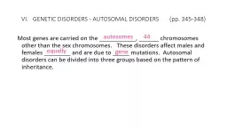
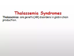
Assist Prof DrMaysem Mouayad Alwash Note The thalassaemias are classified into α β δβ γδβ δ γ and εγδβ thalassaemias according to the type of globin chains ID: 1044510

globin thalassaemia mutations haemoglobin thalassaemia globin haemoglobin mutations diagnosis major severe iron anaemia transfusion red blood production normal chain

Share:

Link:

Embed:

Download Presentation from below link

Download Presentation The PPT/PDF document "Thalassemias and related disorders(cont..." is the property of its rightful owner. Permission is granted to download and print the materials on this web site for personal, non-commercial use only, and to display it on your personal computer provided you do not modify the materials and that you retain all copyright notices contained in the materials. By downloading content from our website, you accept the terms of this agreement.


Presentation Transcript

1. Thalassemias and related disorders(cont.): Assist. Prof. Dr.Maysem Mouayad Alwash

2. Note:The thalassaemias are classified into α-, β-, δβ-, γδβ-, δ-, γ- and εγδβ-thalassaemias, according to the type of globin chain(s)that is produced in reduced amounts .The two major categories are the α- and β-thalassaemias, while the rare formsinclude the γ-, δ- and εγδβ- thalassaemias.

3. Β-Thalassemias

4. Β-ThalassemiasThe most important public health problems because they are common and usually produce severe anaemia in their homozygous and compound heterozygous states.

5. Genetic basis of disease: molecular pathologyThe β-thalassaemias are considered to be autosomal recessive disorders since individuals who have inherited one abnormal β-gene (carrier) are asymptomatic and the inheritance of two abnormal β-globin genes is required to produce a clinically detectable phenotype

6. -300 β-thalassaemia alleles (including deletions) have been characterized. -Only 20 β-thalassaemia alleles account for more than 80% of the β-thalassaemia mutations in the whole world. (This is because in each of the high-frequency areas, only a few (four to six)mutations are common, reflecting local selection due to malaria, with a varying number of rare ones. Each of these populations thus has its own unique group of mutations ).

7. The vast majority of β-thalassaemiamutations are point mutations (i.e. single-base substitutions) and small insertions or deletions of one to two bases.

8. -Mutations completely inactivate the β-gene with no β-globin production resulting inβ0-thalassaemia. -Mutations that allow the production of some β-globin cause β+ or β++ thalassaemia.

9. β‐Thalassaemia major Pathophysiology:Thalassaemia major is a severe, transfusion-dependent, inherited anaemia. There is a profound defect of β chain production. Excess α chains accumulate and precipitate in the red cell precursors in the bone marrow resulting in

10. ineffective erythropoiesis. The few cells that leave the marrow are laden with precipitated α chains and are rapidly removed by the reticuloendothelial system. The constant erythropoietic drive causes massive expansion of bone marrow and extramedullary erythropoiesis. If untreated,80% of children with β thalassaemia major die within thefirst 5 years of life.

11. The pathophysiology of β-thalassaemia.

12. Clinical features1-Severe anaemia becomes apparent at 3–6 months .Typically the infant presents in the first year with failure to thrive, pallor and a swollen abdomen.

13. 2-Enlargement of the liver and spleen :occurs as a result of-excessive red cell destruction, -extramedullary haemopoiesis- iron overload.

14. 3- Expansion of bones caused by intense marrow hyperplasia Leads to a thalassaemic facies and to thinning of the cortex of many bones with a tendency to fractures andbossing of the skull with a ‘hair‐on‐end’ appearance on Xray

15. hair‐on‐end’

16. 4-Thalassaemia major is the disease that most frequently underlies transfusional iron overload.The clinical features (due to hepatic, endocrine and cardiac iron overload)

17. 5-Infections occur frequently. In infancy, without adequate transfusion, anaemia predisposes to bacterial infections.-Pneumococcal, Haemophilus and meningococcal infections.-Yersinia enterocolitica ,Klebsiella, and fungal infection. -Transfusion of viruses by blood transfusion may occur

18. 6-Liver disease -hepatitis C but hepatitis B -Human immunodeficiency virus (HIV).- Iron overload may also cause liver damage.

19. 7-Osteoporosis may occur in well‐transfused patients. It is more common in diabetic patients with endocrine abnormalities

20. 8-Hepatocellular carcinoma incidence is increased in those with iron overload and chronic hepatitis B or C.

21. Laboratory diagnosisThe full blood count:low Hb, low MCV, low MCH. blood film :shows severe hypochromic, microcytic anaemia ,normoblasts, target cells and basophilic stippling and small red cell fragments The reticulocyte count is elevated, but less than expected for the degree of anaemia, due to the ineffective erythropoiesis .

22. Blood film in β‐thalassaemia major post‐splenectomy.There are hypochromic cells, target cells and many nucleated redcells (normoblasts). Howell–Jolly bodies are seen in same redcells.

23. -Renal function is normal.-Liver function tests show elevation of bilirubin, aspartate aminotransferase and lactate dehydrogenase, with a normal alanine aminotransferase.-Erythropoietin levels will be high. -Soluble transferrin receptor levels up to 30 timesgreater than normal. -White cell and platelet counts should be normal unless there is hypersplenism.

24. A bone marrow aspirate is not essential to make the diagnosis, but if performed shows very marked erythroid hyperplasia, with dyserythropoiesis. Many of the erythroid precursors show inclusions after incubation with methyl violet; similar inclusions are found in the peripheral red cells after splenectomy. Immunoelectron microscopy confirms that the inclusions in β-thalassaemia consist of precipitated α- globin chains.

25. -Haemoglobin analysis is needed to confirm the diagnosis .-Electrophoretic or Chromatographic techniques. -The vast majority consisting of HbF; small amounts of HbA may be present depending on the β- globin mutation, the age of the child and whether the child has been transfused.

26. Absence of HbA confirms a diagnosis of β0- thalassaemia, while presence of HbA (pretransfusion sample) confirms β+ -thalassaemia

27. Testing of the parents should confirm the diagnosis, both typically being carriers of β-thalassaemia, with HbA2 levels greater than 3.5% and MCH below 27 pg..

28. These findings are sufficient to make a diagnosis of severe β- thalassaemia, although where DNA analysis is available it isoften used to identify the β-globin mutations and confirm the diagnosis

29. β‐Thalassaemia trait (minor)-This is a common, usually symptomless abnormality -characterized by a hypochromic, microcytic blood picture (MCV and MCH very low) but high red cell count (>5.5 × 10¹²/L) and mild anaemia (haemoglobin 100–120 g/L). -It is usually more severe than α thalassaemiatrait. -A raised Hb A2 (>3.5%) confirms the diagnosis. .

30. The diagnosis allows the possibility of prenatal counselling. If the partner also has β‐thalassaemia trait there is a 25% risk of a thalassaemia major child

31.

32. Thalassaemia intermedia:(Non-transfusion-dependent thalassaemia) :The term thalassaemia intermedia is used to describe patients with the clinical picture of thalassaemia which, although not transfusion dependent, is associated with a much more severe degree of anaemia than is found in carriers for α- or β- thalassaemia. It is increasingly referred to as non-transfusion dependent thalassaemia (NTDT)

33. Non‐transfusion dependent thalassaemia:(-.

34. The patient with thalassaemia intermedia may show bone deformity, enlarged liver and spleen, extramedullary erythropoiesis , leg ulcers, gallstones, osteoporosis andvenous thrombosis.

35. Iron overload is caused by increased iron absorption and occasional transfusions, e.g. during pregnancy or infections or given to reduce bone deformity

36. Hb H disease (three‐gene deletion α‐thalassaemia) is a type of thalassaemia intermedia without iron overloador extramedullary haemopoiesis.

37. 𝛃-Thalassaemia in association with haemoglobinvariantsIn many populations, because there is a high incidence of both β-thalassaemia and other globin gene mutations, it is quite common for an individual to inherit a β-thalassaemia allele from oneparent and a gene for a structural haemoglobin variant from the other.

38. Although numerous interactions of this type havebeen described, only three are common and significant: HbS/β- thalassaemia, HbC/β-thalassaemia and HbE/β-thalassaemia

39. δβ‐ThalassaemiaThis involves failure of production of both β and δ chains.δβ0 or (δβ) 0 (delta beta zero) thalassaemia (sometimes also designated Gγ Aγ (δβ)0 thalassaemia) results from the deletion of both δ andβ genes , but with preservationof the γ genes. Aγδβ0 or (Aγδβ) 0 thalassaemia (sometimes also designated Gγ(Aγδβ )0 thalassaemia) results from deletions of the A γ, δ and β genes.

40. -The phenotype of heterozygotes resembles that of β thalassaemia trait, but the haemoglobin A2 percentage is often normal -Haemoglobin F is consistently elevated, varying from 5% to 20%. The distribution of haemoglobin F( best observed by flow cytometry) is heterocellular.

41. The blood film features are very similar to those of β thalassaemia trait.

42. -Homozygotes and compound heterozygoteswho also have a severe β + or β 0 mutation may have thalassaemia intermedia rather than thalassaemia major. -Homozygotes for δβ0 or Aγδβ0 thalassaemiahave 100% haemoglobin F. In δβ thalassaemia homozygotes, Gγ and Aγ globin chains are present in similar amounts, whereas homozygotes for Aγδβ0 thalassaemiahave only Gγ globin chains

43. Blood film an adult male with δβthalassaemia trait.

44. Hereditary persistence of fetal haemoglobin(HPFH)a range of inhereted disorders characterizedby decreased or absent HbA production and a variable compensatory increase in HbF synthesis.-

45. These are a heterogeneous group of genetic conditions caused by deletions or cross‐overs affecting the production of β and γ chains or, in non‐deletion forms ,by point mutations upstream from the γ‐globin genes or in the BCL11A gene

46. -HPFH heterozygotes have essentiallynormal red cell indices, normal HbA2 levels and HbF levels of 10–35%. - A pancellular (or homogeneous) distributionof HbF in HPFH.

47. -HPFH homozygotes are asymptomatic with slightly reduced MCV and MCH, and compound heterozygotes with β thalassaemia havevery mild disease.

48. HPFH

49. Haemoglobin LeporeThis is an abnormal haemoglobin caused by unequal crossing‐ over of the β and δ genes to produce a polypeptide chain consisting of the δ chain at its amino end and β chain at its carboxyl end.

50. The δβ‐fusion chain is synthesized inefficiently and normal δ‐ and β‐chain production is abolished.It can be considered as a type of δβ-thalassaemia

51. Heterozygotes for Hb Lepore have hypochromic microcytic red cells, normalHbA2 and variably increased HbF.-The homozygotes show thalassaemia intermedia

52.