/
Essentials of Human Anatomy & Physiology Essentials of Human Anatomy & Physiology

Essentials of Human Anatomy & Physiology - PowerPoint Presentation

tatyana-admore
tatyana-admore . @tatyana-admore
Follow
570 views
Uploaded On 2016-06-10

Essentials of Human Anatomy & Physiology - PPT Presentation

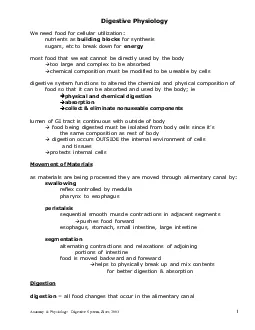
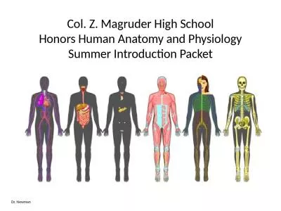
Copyright 2003 Pearson Education Inc publishing as Benjamin Cummings Slides 632 644 Seventh Edition Elaine N Marieb Chapter 6 The Muscular System Lecture Slides in PowerPoint by Jerry L Cook ID: 355847

Share:

Link:

Embed:

Download Presentation from below link

Download Presentation The PPT/PDF document "Essentials of Human Anatomy & Physio..." is the property of its rightful owner. Permission is granted to download and print the materials on this web site for personal, non-commercial use only, and to display it on your personal computer provided you do not modify the materials and that you retain all copyright notices contained in the materials. By downloading content from our website, you accept the terms of this agreement.


Presentation Transcript

Slide1

Essentials of Human Anatomy & Physiology

Copyright © 2003 Pearson Education, Inc. publishing as Benjamin Cummings

Slides 6.32 – 6.44

Seventh Edition

Elaine N. Marieb

Chapter 6

The Muscular System

Lecture Slides in PowerPoint by Jerry L. CookSlide2

Types of Ordinary Body Movements

Slide 6.32

Copyright © 2003 Pearson Education, Inc. publishing as Benjamin Cummings

Flexion

Extension

Rotation

Abduction

CircumductionSlide3

Body Movements

Slide 6.33

Copyright © 2003 Pearson Education, Inc. publishing as Benjamin Cummings

Figure 6.13Slide4

Special Movements

Slide 6.34

Copyright © 2003 Pearson Education, Inc. publishing as Benjamin Cummings

Dorsifelxion

Plantar flexion

Inversion

Eversion

Supination

PronationOppositionSlide5

Types of Muscles

Slide 6.35

Copyright © 2003 Pearson Education, Inc. publishing as Benjamin Cummings

Prime mover – muscle with the major responsibility for a certain movement

Antagonist – muscle that opposes or reverses a prime mover

Synergist – muscle that aids a prime mover in a movement and helps prevent rotation

Fixator – stabilizes the origin of a prime moverSlide6

Naming of Skeletal Muscles

Slide 6.36a

Copyright © 2003 Pearson Education, Inc. publishing as Benjamin Cummings

Direction of muscle fibers

rectus

(straight)oblique (slanted)

Relative size of the muscle

maximus

(largest)

minimus

(smallest)

longus

(long)Slide7

Naming of Skeletal Muscles

Slide 6.36b

Copyright © 2003 Pearson Education, Inc. publishing as Benjamin Cummings

Location of the muscle

many

muscles are named for bones (e.g.,

temporalis)Number of origins

biceps (two heads)

triceps

(three heads)

quadriceps

(four heads)Slide8

Naming of Skeletal Muscles

Slide 6.37

Copyright © 2003 Pearson Education, Inc. publishing as Benjamin Cummings

Location of the muscles origin and insertion

Example:

sterno

(on the sternum)Shape of the muscle

Example: deltoid (triangular)

Action of the muscle

Example: flexor and extensor (flexes or extends a bone)Slide9

Head and Neck Muscles

Slide 6.37

Copyright © 2003 Pearson Education, Inc. publishing as Benjamin Cummings

Facial Muscles

Frontalis

– muscle covering frontal bone; allows for lifting eyebrows and wrinkling forehead

Orbicularis

Oculi – circular muscle around eye; allows closing, squinting, blinking, and winking of eye

Orbicularis

Oris – circular muscle around lips; closes mouth and protrudes lipsSlide10

Head and Neck Muscles

Slide 6.37

Copyright © 2003 Pearson Education, Inc. publishing as Benjamin Cummings

Facial Muscles (cont’d)

Buccinator

– runs horizontally along cheek and inserts into O.

oris; flattens cheek in whistling, blowing, and chewing

Zygomaticus

– runs from side of mouth up to cheekbone; raises corner of mouth upward Slide11

Head and Neck Muscles

Slide 6.37

Copyright © 2003 Pearson Education, Inc. publishing as Benjamin Cummings

Chewing Muscles

Masseter

– covers the angle of the lower jaw, running from

zygomatic process to mandible; closes the jaw

Temporalis – covers temporal bone; acts as a synergist/aids the

masseter in closing the jawSlide12

Head and Neck Muscles

Slide 6.37

Copyright © 2003 Pearson Education, Inc. publishing as Benjamin Cummings

Neck Muscles

Platysma

– covers

anterolateral neck, originates from chest and inserts into area around mouth; pulls corners of mouth downward

Sternocleidomastoid – paired muscles with two heads originating from the sternum and clavicle, then inserting into the mastoid process; bend the head down forward and to the sidesSlide13

Head and Neck Muscles

Slide 6.38

Copyright © 2003 Pearson Education, Inc. publishing as Benjamin Cummings

Figure 6.14Slide14

Trunk Muscles

Slide 6.37

Copyright © 2003 Pearson Education, Inc. publishing as Benjamin Cummings

Anterior Trunk Muscles

Pectoralis

Major – covers upper part of chest, originates from shoulder girdle and first six ribs, inserts on proximal

end of humerus

; adducts and flexes arm

Intercostal Muscles – between the ribs; external intercostals raise rib cage to allow air in, internal intercostals depress cage to force air outSlide15

Trunk Muscles

Slide 6.37

Copyright © 2003 Pearson Education, Inc. publishing as Benjamin Cummings

Anterior Trunk Muscles (cont’d)

Rectus Abdominals – paired muscles running from pubis to rib cage; flex the vertebral column and compress the abdominal contents

External Oblique – paired muscles of the lateral walls of the abdomen, running from the last eight ribs to the

ilium

; flex the vertebral column and rotate the trunk and bend laterallySlide16

Trunk Muscles

Slide 6.37

Copyright © 2003 Pearson Education, Inc. publishing as Benjamin Cummings

Anterior Trunk Muscles (cont’d)

Internal Oblique – paired muscles deep to external

obliques

, originate from iliac crest and insert into the last three ribs; same function as external obliques

Transversus

abdominals – deepest muscle of abdominal wall with fibers running horizontally, originates from lower ribs and iliac crest and inserts into the pubis; compresses the abdominal contentsSlide17

Trunk Muscles

Slide 6.37

Copyright © 2003 Pearson Education, Inc. publishing as Benjamin Cummings

Anterior Arm Muscles

Biceps

Brachii

– originates from the shoulder girdle and inserts into the radial tuberosity; flexes and

supinates the forearm

Brachialis – deep to the bicep; flexes the forearm

Brachioradialis – originates from the humerus

and inserts into the distal forearmSlide18

Trunk Muscles

Slide 6.39

Copyright © 2003 Pearson Education, Inc. publishing as Benjamin Cummings

Figure 6.15Slide19

Trunk Muscles

Slide 6.37

Copyright © 2003 Pearson Education, Inc. publishing as Benjamin Cummings

Posterior Trunk Muscles

Trapezius

– paired muscles with origin running from base of occipital bone down to the end of the thoracic vertebrae, insert on scapular spine and clavicle; extend the head and move the scapula

Latissimus

Dorsi – large paired muscles of lower back, originates from lower back and

ilium then inserts into proximal end of humerus

; extends and adducts humerusSlide20

Trunk Muscles

Slide 6.37

Copyright © 2003 Pearson Education, Inc. publishing as Benjamin Cummings

Posterior Trunk Muscles (cont’d)

Erector

Spinae

– paired set of three muscle columns that extend the length of the vertebral column; extend the vertebral column

Deltoid – originates from the scapular spine and clavicle and inserts into the deltoid tuberosity

of humerus; abduct the armSlide21

Trunk Muscles

Slide 6.37

Copyright © 2003 Pearson Education, Inc. publishing as Benjamin Cummings

Posterior Arm Muscles

Triceps

Brachii

– originates from shoulder girdle and proximal humerus, inserts into

olecranon process of ulna; extends the forearm (antagonist of biceps)Slide22

Deep Trunk and Arm Muscles

Slide 6.40

Copyright © 2003 Pearson Education, Inc. publishing as Benjamin Cummings

Figure 6.16Slide23

Lower Limb Muscles

Slide 6.37

Copyright © 2003 Pearson Education, Inc. publishing as Benjamin Cummings

Pelvis, Hip and Thigh Muscles

Gluteus

Maximus

– originates from sacrum and ilium and inserts on

gluteal tuberosity of femur, forms the buttocks; extends the thigh, especially when climbing and jumping

Gluteus

Medius – originates from ilium and inserts into femur; abducts the thigh with the hipSlide24

Lower Limb Muscles

Slide 6.37

Copyright © 2003 Pearson Education, Inc. publishing as Benjamin Cummings

Pelvis, Hip and Thigh Muscles (cont’d)

Iliopsoas

– originates from the iliac bone and lower vertebrae and inserts on the lesser

trochanter of the femur; flexes the leg at the hip

Adductor Muscles – originate from the pelvis and inserts medially towards the proximal end of the femur; adducts the thighSlide25

Lower Limb Muscles

Slide 6.37

Copyright © 2003 Pearson Education, Inc. publishing as Benjamin Cummings

Pelvis, Hip and Thigh Muscles (cont’d)

Hamstring group – three muscles originating from the

ischial

tuberosity and inserts onto both sides of the proximal end of the tibia; extend the leg

Sartorius – originates from anterior iliac crest and inserts into medial side of tibia; flexes the thigh Slide26

Lower Limb Muscles

Slide 6.37

Copyright © 2003 Pearson Education, Inc. publishing as Benjamin Cummings

Pelvis, Hip and Thigh Muscles (cont’d)

Quadriceps group – four muscles that make the anterior thigh, originating from the femur or pelvis and inserting into the

tibial

tuberosity

; extends the knee and flexes the hipSlide27

Muscles of the Pelvis, Hip, and Thigh

Slide 6.41

Copyright © 2003 Pearson Education, Inc. publishing as Benjamin Cummings

Anterior View

Posterior ViewSlide28

Lower Limb Muscles

Slide 6.37

Copyright © 2003 Pearson Education, Inc. publishing as Benjamin Cummings

Lower Leg Muscles

Tibialis

Anterior – originates from upper tibia and inserts at the

tarsals, running parallel to anterior crest; acts to

dorsiflex and invert the foot

Extensor Digitorum Longus

– originates from lateral tibial condyle

and proximal radius and inserts into toes 2-5, running just lateral to

tibialis

; extends toes and

dorsiflexes

footSlide29

Lower Limb Muscles

Slide 6.37

Copyright © 2003 Pearson Education, Inc. publishing as Benjamin Cummings

Lower Leg Muscles (cont’d)

Gastrocnemius

– forms calf muscle, two head originate from each side of distal femur and inserts onto the heel

bone; main part of plantar flexion

Soleus – originates from tibia and inserts into the

tarsals, just deep to gastrocnemius; aids in plantar flexion of footSlide30

Muscles of the Lower Leg

Slide 6.42

Copyright © 2003 Pearson Education, Inc. publishing as Benjamin Cummings

Figure 6.19Slide31

Superficial Muscles: Anterior

Slide 6.43

Copyright © 2003 Pearson Education, Inc. publishing as Benjamin Cummings

Figure 6.20Slide32

Superficial Muscles: Posterior

Slide 6.44

Copyright © 2003 Pearson Education, Inc. publishing as Benjamin Cummings

Figure 6.21