/
Transport across cell membrane Transport across cell membrane

Transport across cell membrane - PowerPoint Presentation

tatyana-admore
tatyana-admore . @tatyana-admore
Follow
558 views
Uploaded On 2016-07-30

Transport across cell membrane - PPT Presentation

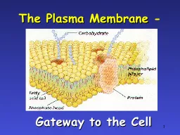
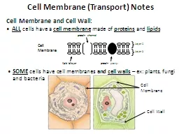
by Vani Gupta Types of cell membrane transport Factors affecting transport Cell membrane Chemical gradient Electrical gradient Rate of transport Passive transport ID: 425643

transport cell diffusion membrane cell transport membrane diffusion concentration proteins protein gradient ions cells water molecules endocytosis active high

Share:

Link:

Embed:

Download Presentation from below link

Download Presentation The PPT/PDF document "Transport across cell membrane" is the property of its rightful owner. Permission is granted to download and print the materials on this web site for personal, non-commercial use only, and to display it on your personal computer provided you do not modify the materials and that you retain all copyright notices contained in the materials. By downloading content from our website, you accept the terms of this agreement.


Presentation Transcript

Slide1

Transport across cell membrane

by

Vani

GuptaSlide2

Types of cell membrane transport

Factors affecting transport

Cell membrane

Chemical gradient

Electrical gradient

Rate of transport

Passive transport

Diffusion

Osmosis

Facilitated diffusion

Active transport

Pumps

phagocytosis

Endocytosis

/

exocytosisSlide3

Factors affecting transport:

cell membrane

The cell needs to absorb and excrete various compounds throughout its life.

These compounds need to pass through the membrane which is made from a

phospholipid bilayerThe phospholipid bilayer is formed by phospholipid molecules bipolar molecule: the fatty acid side is hydrophobic, the phosphoric side is hydrophilicSlide4

The membrane is permeable to:

The membrane is impermeable to:

H2O

Gases (O2, CO2, N2)

LipidsSmall, neutral molecules (such as urea) Small, charged molecules“large molecules” such as amino acids, glucose and larger

These compounds must go through channels present in the membrane in order to enter or exit the cellSlide5

Factors affecting transport:

Chemical gradient

Compound moves from an area of high concentration to low concentration (or concentration gradient)

All compounds permeable to the

phospholipid bilayer will move this waySlide6

Factors affecting transport:

Electrical force

Positive ions are attracted to negative ions and vice versa

Ions are repelled by ions of the same charge (+ against + and – against -)Slide7

Movement across the cell membrane

Both chemical and electrical forces (electrochemical force) drive the movement of compounds across the cell membraneSlide8

Factors affecting the rate of transport

The rate of transport will depend on:

The concentration gradient

The compound permeability to the membrane

The type and number of charges present on the compoundSlide9

Crossing

of cell membrane

fats and oils can pass directly through

inside celloutside cell

waste

lipid

sat

sugar

aa

H

2

OSlide10

Types of Transport Proteins

Channel proteins are embedded in the cell membrane & have a pore for materials to cross

Carrier proteins can change shape to move material from one side of the membrane to the otherSlide11

Cell membrane channels

Need to make “doors” through membrane

protein channels allow substances in &

out

specific channels allow specific material in & outH2O channel, salt channel, sugar channel, etc.

inside cell

outside cellSlide12

Protein channels

Proteins act as doors in the membrane

channels to move specific molecules through cell membrane

HIGH

LOWSlide13

Passive transport

Compounds will move from area of

high concentration toward area of lower concentration

No ATP is needed for this type of transportPassive transport mainly TWO types A-Osmosis B-Diffusion-diffusion again two types a-

simple diffusion- no energy needed

b-

facilitated diffusion-

no energy needed

-

help through a protein channelSlide14

Osmosis

Each compound obeys the law of diffusion

diffusion of water from HIGH concentration of water to LOW concentration of water

across a semi-permeable membraneHowever, some compounds are unable to cross the cell membrane (glucose, electrolytes…)Water can cross  will enter or exit the cell depending its concentration gradient.Slide15

where is osmosis importantSlide16

Cells in Solutions Slide17

PLASMOLYSIS

Isotonic Solution

NO NET MOVEMENT OF H

2

O (equal amounts entering & leaving)Hypotonic SolutionCYTOLYSIS

Hypertonic Solution

PLASMOLYSISSlide18

Diffusion

Simple diffusion

-

no energy needed

Movement across higher to lower concentration gradient. Facilitated diffusion- Some compounds are unable to diffuse through the membrane. They will be allow to cross if the membrane has proteins that can bind these compounds and enable to cross toward the area of lower concentrationSlide19

Simple and facilitated diffusion

inside cell

outside cell

lipid

inside cell

outside cell

H

2

O

simple diffusion

facilitated diffusion

H

2

O

protein channelSlide20

Simple Diffusion

Doesn’t require energy

Moves high to low

concentrationExample: Oxygen or water diffusing into a cell and carbon dioxide diffusing out.Slide21

Simple Diffusion

The rate of diffusion will be increased when there is :

Concentration

: the difference in between two areas (the gradient) causes diffusion. The greater the difference in concentration, the faster the diffusion.

Molecular size

:

smaller substances diffuse more

quickly.

Large molecules (such as starches and proteins) simply cannot diffuse through.

Shape of Ion/Molecule

: a substance’s shape may

prevent

it from diffusing rapidly, where others may have a shape that aids their diffusion.

Viscosity of the Medium

: the lower the viscosity, the more

slowly

molecules can move through it.Slide22

Movement of the Medium

: currents will aid diffusion. Like the wind in air,

cytoplasmic

steaming (constant movement of the cytoplasm) will aid diffusion in the cell.Solubility: lipid - soluble molecules will dissolve through the phospholipid

bilayer

easily,

as will gases like CO

2

and O

2

.

Polarity

:

water

will diffuse, but because of its polarity, it will

not

pass through the non-polar phospholipids. Instead, water passes though specialized

protein ion

channelsSlide23

Facilitated diffusion

Doesn’t require energy

Uses transport proteins to move high to low concentration

Examples: Glucose or amino acids moving from blood into a cell.Slide24

where is facilitated transport importantSlide25

Slide26

Active Transport

-

Pumps

- phagocytosis - Endocytosis/exocytosisSlide27

Active transport

ATP (energy) is needed

 pump

Moves

materials from LOW to HIGH concentrationAGAINST concentration gradientSlide28

Example-1

ATPase

pumps

The most common: Na/K pumps

 reestablish membrane potential. Present in all cells.Two K+ ions are exchanged with 3 Na + ionsSlide29

EXAMPLES OF ACTIVE TRANSPORT

Example 2

: the thyroid gland accumulates iodine as it is needed to manufacture the hormone thyroxin.

The iodine concentration can be as much as 25 times more concentrated in the thyroid than in blood. Slide30

Example 3

: In order to make ATP in the mitochondria, a proton pump (hydrogen ion) is required.Slide31

where is active transport importantSlide32

Endocytosis

Endocytosis

: (“Endo” means “in”). Endocytosis is the taking in of molecules or particles by invagination of the cell membrane forming a vesicle. Integrity of plasma membrane is maintained.

This requires energy.

Endocytosis

is fallowed by

exocytosis

on the other side. –

Transcytosis

, vesicle trafficking, or

cytopempsis

.Slide33

Types

of

endocytosis

1. pinocytosis (cell drinking): small molecules are ingested and a vesicle is immediately formed. This is seen in small intestine cells (villi) 2. phagocytosis

(cell eating):

large

p

articles, (visible with light microscope) are

invaginated

into the cell (

ie

:

white blood cells ‘eat’ bacteriaSlide34
Slide35

Phagocytosis

Used to engulf large particles such as food, bacteria, etc. into vesicles

Called “Cell EatingSlide36

Capture of a Yeast Cell (yellow) by Membrane Extensions of an Immune System Cell (blue)Slide37

Receptor-Mediated

Endocytosis

Some integral proteins have receptors on their surface to recognize & take in hormones, cholesterol, etc.Slide38

Exocytosis

Exocytosis

: (“

Exo” means “out”.) Exocytosis is the reverse

of endocytosis.

This is where a cell

releases the contents

of a vesicle outside of the cell.

These contents may be

wastes, proteins, hormones

, or some other product for secretion.

This also

requires energy.

Example: vesicles from the

Golgi

fuse with the plasma membrane and the proteins are released outside of the cell.Slide39
Slide40

Fusion of vesicle with plasma membrane is mediated by a number of accessory proteins- SNARE protein.

Require stimulus and Ca.

Exception-

Renin

from JG cells and PTH from parathyroid gland by decrease in intracellular Ca.Constitutive Secretion- Immunoglobulin from plasma Cells and collagen from fibroblast.Regulated- endocrine gland, pancreatic acinar cells Slide41

Membrane Transport Proteins

1.

Water Channels or

Aquaporins

(AQPs) – 12 types Amount of water is regulated by No. of AQPs They are known as gated channel although they are pores. Two types a) Aquaporins- only water. b) Aquaglyceroporins- also for small molecules. Slide42

2- Ion Channels-

All cells specially on excitable cells – Neurons and muscle cells

Selective and non selective

Gated – voltage gated and extracellular agonist or antagonist gated ex – acetylcholine gated cationic specific channel at motor end plate of skeletal muscle.

Conductance- 1-2 picosimens and > 100 picosimens. Ex- Na, K, Ca, Cl, Anion , cation.Slide43

3.Solute Carriers-

> 40 types , > 300 transporters.

three gps-1.

Uniporters

- single molecule across the membrane (GLUT ) 2. Symporters- Two or more molecules Ex- Na-k-cl Symporter (Kidney) Na - Glucose Cotransporter. 3. Antiporters- Two or more molecules in opposite directions Ex :Na- H antiporter ( PH regulation) 3Na- Ca , Cl- HCO3 Slide44

4.ATP DEPENDENT TRANSPORTERS

1.

ATPase

Ion Transporters

1. P- Type- gate phosphorylted during transport. Na- K ATP ase. 2. V- Type- Vacuolar H- ATPase – urine acidification on Vacules like endosomes and lysosomes.2. ATP – binding cassette (ABC) transporters – 7 subgroups transport diverse group of ions ex- Cl, Cholesterol, bile acids, drugs, iron and organic anions.EX:- Cystic fibrosis transmembrane regulator. Multidrug Resistance Protein. organic Anions. . Slide45

Molecular Motors:

Kinesin

- over the microtubule

Dynein

- retrogate transportMyosin- over the microfilaments.- 18 types aSlide46

Q-1 all membrane processes, such as pumping and channelling of molecules are carried out by.

a-lipid

b-carbohydrate

c-nucleic

acid d-proteinSlide47

Q-2

Which of the following statement about membrane transport protein is incorrect

a-carrier

proteins are similar to enzymes in that they show saturation

b-carrier protein can facilitate both active and passive transport c-channel protein can facilitate both active and passive transport d-the Na+ /Glucose transport protein carries out secondary active transport.Slide48

Q-3 Diffusion across the plasma membrane is more rapid if a substance is

a-a

protein

b-hydrophilic

c-high in its oil : water partition coefficient d-larger and globular in shapeSlide49

Q-4 the difference between simple diffusion and facilitated transport is that facilitated transport.

a-is

concentration dependent

b-occurs

across plasma membrane c-require membrane protein d-utilize a substance moving with its concentration gradientSlide50

Q-5 Erythrocyte glucose transporter specifically transports glucose down its concentration gradient and exhibit hyperbolic saturation kinetics .This is an example of

a-active

mediated transport

b-passive mediated transport c-non- mediated transport d-group translocation Slide51

Q-6 which one of the following is a correct statement for Na-K

ATPase

.

a-it gives out 3 Na-ions and takes in 2 K-ions b- it gives out 2 Na-ions and takes in 3 K-ions c- it gives out 3 Ca-ions and takes in 2 K-ions d-it gives out 3 Na-ions and takes in 2 Ca-ionsSlide52

Q7-which of the following effects of the steroid digitalis is observed after treatment of congestive heart failure.

a-decrease

in

cytosolic

sodium levels b-inhibition of Na-K ATPase c-decrease in the force of heart muscle contraction d-stimulation of the plasma membrane ion pump.Slide53

Q8-you wish to design a new drug which will act as an

ionophore

to deliver Ca

2+

across the nerve cell membrane .This drug would most likely be a-hydrophobic on the outside and hydrophilic on inside b-insoluble in lipid c-soluble in proteins d-smaller than 0.001 nm in diameterSlide54

Q9- the process by which a cell secretes macro-molecule by fusing a vesicle to the plasma membrane is called

a-

endocytosis

b-

exocytosis c-pinocytosis d-phagocytosisSlide55

Q10- free fatty acids enter cell by

a-passive

diffusion

b-active

diffusion c- through carrier protein d – Active transport Slide56

Q-11

Aquaglyceroporins

transport-

a. Water only b. water and small molecules. c. Water and Glucose d. Water and salt.Slide57

Q-12 Which of the fallowing is responsible for PH

Regulation-

a.

Antiporters

.b. Symportersc. Uniporters.d. Co-porters.Slide58

Q-13 V type – transporters are

a.

ATPase

dependent.

b. Symporters. c. Carrier Proteins. d. Receptor Proteins. Slide59

Q-14 .GLUT is an example of-

a.

Antiporters

.

b. Symportersc. Uniporters.d. Co-porters.Slide60

Q-15 Presence of Ion channels are must on

a. Excitable tissue.

b. Non excitable tissue.

c. Renal tissue

d. Cardiac muscle.Slide61

Q-16.

Na- K

ATPase

transport Na-

a. Towards Concentration gradient. b. Against Concentration gradient. c. Towards electro chemical gradient. d. Against electrochemical gradient.Slide62

Q-17. Rennin secretion from JG cells is an example of-

a.)

Exocytosis

b.) pincocytosis c.) Vacular movement. d.) Transcytosis.Slide63

Q.- 18. PTH secretion fro parathyroid glands require-

a.) low intracellular Ca.

b.) high Intracellular Ca.

c.) Low intracellular K.

d.) high Intracellular K. Slide64

Q-19.

Transcytosis

incudes

- a. Endocytosis and phagocytosis. b. Endocytosis and pincocytosis. c. Endocytosis and exocytosis. d. Endocytosis only.Slide65

Q-20.

Transcytosis

occurs at

a).

Epethelial Cells. b). Endocrine Cells. c). Nerve cells. d). None of the above.